


Rare and Unusual Cases
VICTOR BERGAMASCO, MBBS
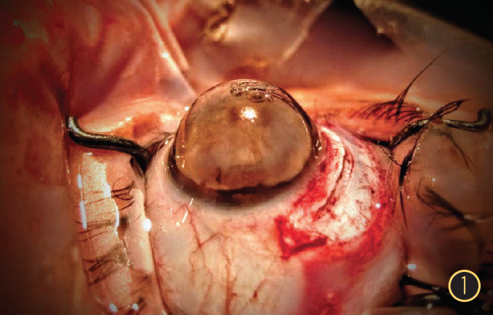
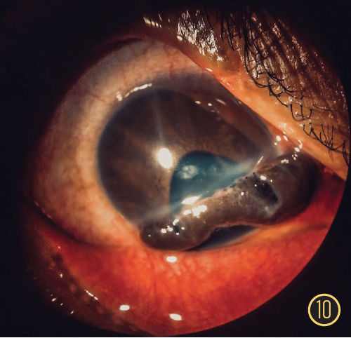
The Globe
This image shows keratoglobus and a total fibrotic cataract in a 45-year-old woman with Ehlers-Danlos syndrome who underwent uneventful extracapsular cataract extraction followed a few months later by limbus-to-limbus penetrating keratoplasty.
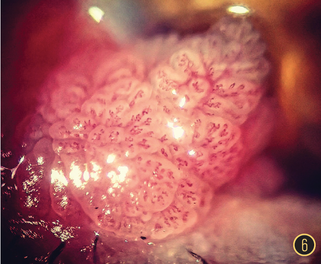
MARCELO MESQUITA MAIA, MD
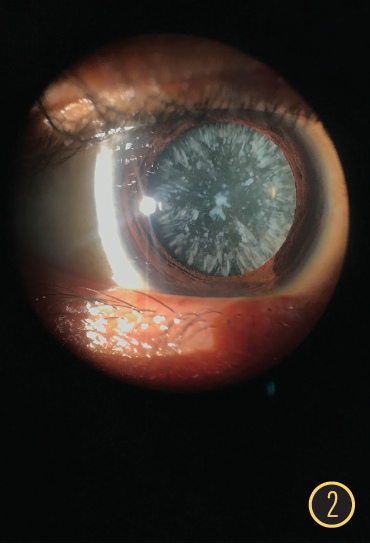
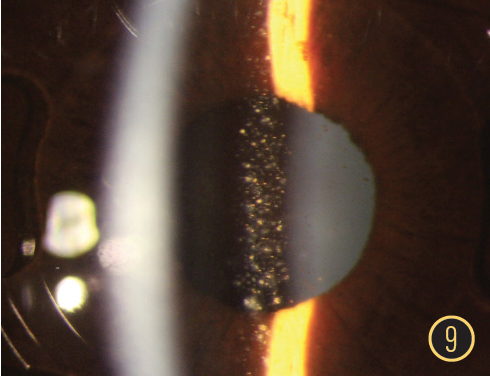
Confetti
This is a photograph of an interesting and unusual type of cortical cataract.
ALEXANDER MARTÍNEZ RUA, MD
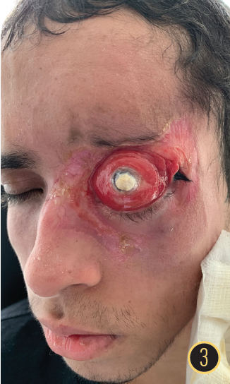
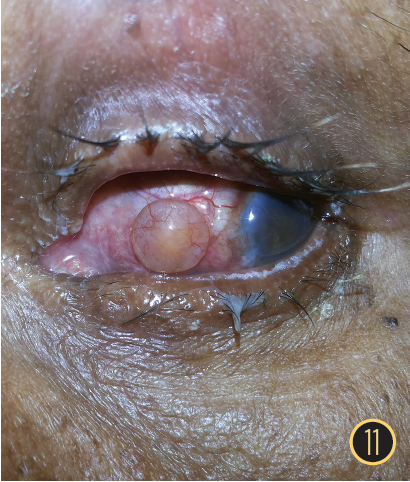
Necrotizing Fasciitis
The image in this photograph is of necrotizing fasciitis in a patient with acute lymphoid leukemia.
Interesting and Artistic Images
MANAS NATH, MBBS, DO,
FAEH, FAICO
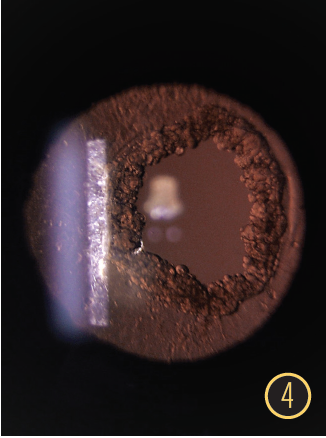
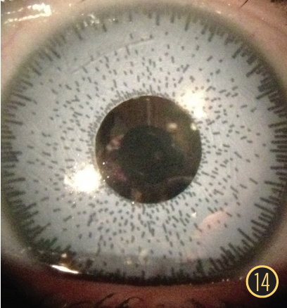
Ring of Pearls
This slit-lamp retroillumination image shows proliferation of Elschnig pearls at the margin of an Nd:YAG laser posterior capsulotomy opening. The posterior capsulotomy was performed more than 2 years prior to presentation.
CELSO DE SOUZA DIAS JÚNIOR, MD
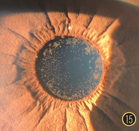
An Encounter With Semicircles
This image of fluorescein semicircles in a nonglaucomatous eye was taken during Goldmann applanation tonometry.
ADNAN BERKAY KISAKÜREK, MD
Conjunctival Squamous Papilloma
A 45-year-old man presented with a mass on the inferior eyelid of his right eye. He reported experiencing foreign body sensation in the eye as well as irritation and itching.
Surgical Complications
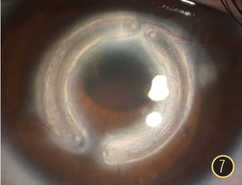
CLAUDIO TRINDADE, MD, PHD
Neovascularization After Corneal Ring Segments
This image was captured with a slit lamp using open-slit illumination and diffuse background illumination; a 35-year-old patient with keratoconus presented with a complex web of neovascularization around an intrastromal corneal ring segment 7 years after ring segment implantation. White deposits along the intrastromal tunnel caused visual and cosmetic problems.
ROBERTO JULE, MD
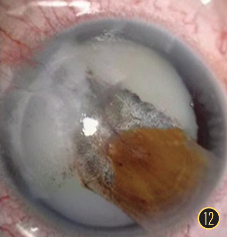
Misplaced
An image of a cataract fragment in the posterior pole after posterior capsular rupture during cataract surgery.
RICARDO STOCK, MD
Artisan Pigmentation
A photograph of iris pigment attached to an Artisan Phakic IOL (Ophtec) after uneventful surgery.
Ocular Trauma
CAROLINA MENDOZA, MD
A Prolapsed Self-Sealing Iris
This image of the right eye of a 29-year-old woman with a history of penetrating corneal laceration shows a corneal and scleral laceration with prolapsed iris sealing the corneal injury, which kept the anterior chamber formed.
KUNAL ASHOK MANDLIK, MBBS, MS, FAEH(CORNEA)
Phacocoele
This is an image of the eye of a patient with a history of trauma who presented with a lens extrusion in the subconjunctival space through a scleral tear.
LEKSEJ MEDIC, MD
Traumatic Cataract With Total Iridodyalisis
An image of a patient with a traumatic cataract with subtotal iridodialysis caused by a penetrating injury of the cornea and sclera.
Slit-Lamp Images
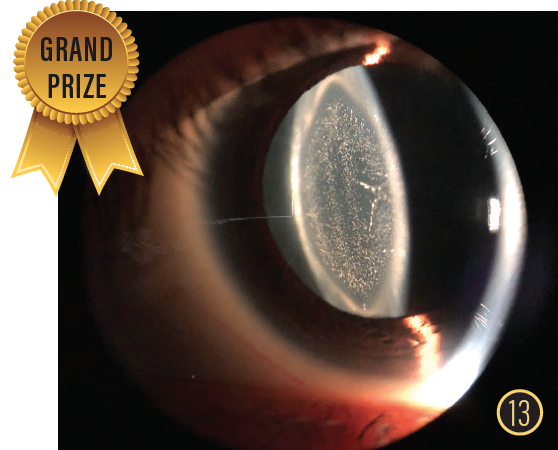
GRAND PRIZE WINNER | JOCELYN RIVERA, MD
Galaxy
This image shows a dense cerulean cataract in a 28-year-old woman with high myopia who presented with a 1-year history of progressive visual loss in her left eye.
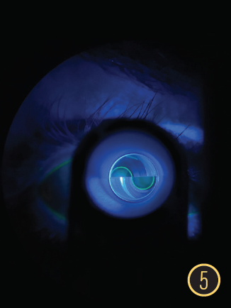
MICHAEL RIZEN, MD
Don’t It Make My Brown Eyes Blue
This is a photo of a patient who received iris implants in South America and presented with uveitis. Removal of the implants was recommended to prevent long-term complications of glaucoma and corneal decompensation.
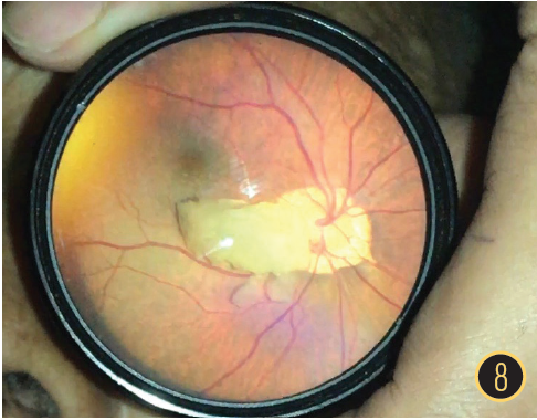
GABRIEL CASTILHO SANDOVAL BARBOSA, MD
Epicapsular Stars
This is a photograph of a remnant of the tunica vasculosa lentis that appears as star-shaped brown pigments in the anterior lens capsule.

