I have long been fascinated by keratoconus, which is usually described as a mysterious corneal dystrophy of unknown origin. Despite our ability to detect the pronounced structural changes and deformations of the corneal wall synonymous with keratoconus on topography, the disease lacks specific genetic and biomolecular markers—at least to the best of our knowledge. For many years now, a large part of my clinical practice has been dedicated to the diagnosis, management, and prevention of keratoconus, and I have worked tirelessly to uncover the true root cause of the disease.
AT A GLANCE
- The author argues that keratoconus is not a dystrophy of unknown genetics and biomolecular substratum but rather a syndrome caused by eye rubbing.
- If the etiology of keratoconus is mechanical, it could be possible that the structural changes and deformations in the cornea are initiated and aggravated by the mechanical force of rubbing the eye.
- Patients should be warned of the deleterious effects of chronic and vigorous eye rubbing.
Finally, I have a simple yet provocative hypothesis to share: eye rubbing causes keratoconus.1
NOT JUST A RISK FACTOR
A few years back, during the examination of a patient with such badly scarred and protruding corneas that I did not need a slit-lamp examination to confirm keratoconus, I found myself asking the following question: How could such a quiescent organ as the cornea undergo such a dramatic morphologic change without so much as a biologic cascade to explain it? It was at that time that I decided to look harder for an answer, one that had not yet been considered.
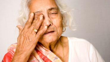
For a while now, the ophthalmic community has acknowledged that eye rubbing is a risk factor for keratoconus and its progression, and many clinical observations and reports support this line of thought.2-6 However, eye rubbing has never been acknowledged as the root cause. Looking beyond the realm of existing evidence has not been routinely attempted—until now. I would argue that keratoconus is not a dystrophy of unknown genetics and biomolecular substratum but rather a syndrome caused by eye rubbing. To put it another way, the progressive deformation and thinning of the corneal wall—the hallmark of keratoconus—are caused by trauma to the cornea that results from chronic and constant eye rubbing.
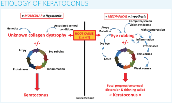
Historically, the main hypothesis in the etiology of keratoconus has been molecular, in that genetics, environmental conditions, and other unknown general factors are thought to be keys to the appearance of the disease. I propose that the etiology of keratoconus is instead mechanical, in that structural changes and deformations in the cornea are initiated and aggravated by the mechanical force of rubbing the eye (see Etiology of Keratoconus).
Additional mechanical factors such as corneal refractive surgery and compression of the cornea at night by the hand, pillow, or mattress may also accelerate distortion of the cornea. In the former, LASIK can cause dry eye disease—once again triggering eye rubbing—which may be more detrimental to a thinned cornea. In the latter, the prolonged contact between the eye and eyelids and the pillow or mattress overnight can cause local irritation, dryness, contamination, and itch—circumstances that can incite eye rubbing and, subsequently, lead to inflammation of the cornea and ocular surface. This may also explain why keratoconus is often worse in one eye, the one on the side of the head on which the person sleeps. The variable way a person rubs his or her eyes may also account for the large spectrum of presentation of keratoconus, especially in early disease.
To further accentuate the correlation between eye rubbing and keratoconus, the effect of the mechanical stress—the release of proteinases in the stroma—triggers progressive thinning in the cornea, making it even more vulnerable to the mechanical trauma induced by the rubbing.
In my mechanical hypothesis, keratoconus cannot occur without a repeated mechanical injury such as rubbing. Once the duration and frequency of that rubbing exceed the capacity of the cornea to uphold native structural and biomechanical resistance, a mechanical imbalance is reached, and corneal deformation results. At this point, topographic findings characteristic of keratoconus are detectable.
GENETICS OR EYE RUBBING?
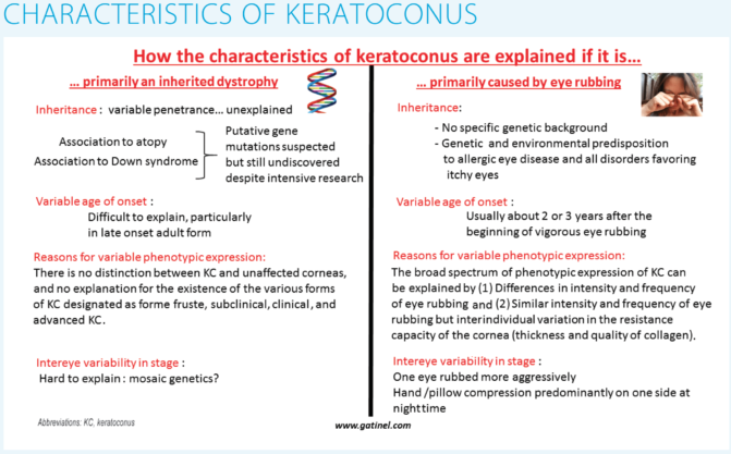
As the two diagrams in Characteristics of Keratoconus show on page 24, the variation in the patient’s age and the laterality, severity, and broad spectrum of phenotypic expression of keratoconus presentation is better explained by excessive eye rubbing than by corneal degeneration caused by an unknown genetic disorder or molecular cascade.
If the same eye rubbing intensity, duration, and frequency are applied to two corneas—one with natively reduced corneal thickness and strength and one thicker and stronger—the one with less biomechanical strength will likely succumb to deformity more readily and more significantly than the other. The repetitive force of the fingers and knuckles rubbing the eye can cause corneal collagen to stretch and become disorganized. Additionally, the acute rise in IOP that results from the sudden reduction in eye volume caused by the fingers can stretch the scleral shell, leading to a refractive shift of axial myopia. Despite contradicting reports, axial length in keratoconus patients seems to be slightly but significantly higher than in nonkeratoconus patients.7
The exact genetics of keratoconus has not yet been elucidated. The frequency of occurrence of keratoconus in close family members is not clearly defined, but the pattern of inheritance is estimated to be less than 20%.8,9 With such variable penetrance, the age of onset is difficult to explain through genetics. It is also difficult to explain through genetics the intereye variability in the stage of this disease.
Yet, genetics can still influence the development of keratoconus if the root cause is the very act of eye rubbing. In this context, the genetic component is related to the predisposition of conditions that lead to increased eye rubbing and to variations in corneal thickness and resistance. These can be, for instance, Down syndrome,10,11 Tourette syndrome,12,13 atopy,14 sleep apnea,15 shift work sleep disorder, pregnancy, and sex (males are more likely to develop keratoconus). With regards to the last, eye makeup worn during the day may preclude women from rubbing their eyes, resulting in a lower incidence of keratoconus in women. Likewise, the recent increase in computer use and, subsequently, the development of computer vision syndrome in some individuals could elicit eye rubbing and account in part for the recent increase in the prevalence of keratoconus.16 The increased sensitivity of the corneal surface after PRK may also induce less eye rubbing postoperatively. Such exacerbated sensitivity does not exist after LASIK, which also induces more dry eye. These factors may partly account for the higher prevalence of ectasia after LASIK than after PRK.
AT A MICROSCOPIC LEVEL
Another question that came to my mind when evaluating the eye rubbing hypothesis is: How are the changes seen in keratoconus at the microscopic level explained by eye rubbing?
This might, indeed, not only be a causal link between the act of eye rubbing and the development of keratoconus, but it might also explain the lack of inflammation in the form of neovascularization and cell infiltration seen in keratoconic eyes. At the microscopic level, it has also been proven that the biomechanical weakening observed in keratoconus is focal, involving the (rubbed) central or paracentral zone of the cornea and sparing the periphery.17
Additionally, the act of eye rubbing can cause progressive distention, disorganization, and severing of corneal fibrils, which can disturb the homogeneity of the fiber matrix, decrease the cornea’s ability to dissipate the mechanical energy evenly, and affect corneal regularity. Thus, chronic eye rubbing could lead to changes in corneal macroscopic regularity and to the presentation of classic topographic features inherent in keratoconus. The mechanical strength and quality of an eye’s collagen fibrils could also influence the susceptibility of one cornea to developing keratoconus over another.
A PERFECT COUNTEREXAMPLE
Marfan syndrome, a disorder with an identifiable genetic mutation of the fibrillin-1 molecule, represents a perfect counterexample to explain the holes in current theories on the pathogenesis of keratoconus. This is because no keratoconic pattern is found in the eyes of Marfan syndrome patients, despite the reduction of collagen strength present in the corneal stroma. Although these corneas are thinner, they tend to be flatter rather than steeper. Because the force resulting from IOP is evenly distributed against the posterior surface of the cornea and the inner surface of the sclera, in the absence of a focal stress, a softer eyeball will undergo progressive distension of its shell. This causes the local radii of curvature of the cornea to increase (and the corneal curvature to decrease), with concomitant progressive thinning.
Talking to Patients
What You Need to Know

- The frequency and duration of eye rubbing may be difficult to document.
- Elicit a history of repetitive eye rubbing in an appropriate manner; often, patients are too embarrassed to admit to the habit and may not readily volunteer or may underreport the habit.
- Some patients may not even be aware of their eye rubbing habit until they are sensitized to it.
- Ask patients to have family, friends, and coworkers survey and observe their habits in an attempt to have patients more accurately portray their eye rubbing during follow-up visits.
- Ask patients to pay attention to specific times of eye rubbing, including upon waking, before going to bed, and after removing their contact lenses or eye makeup.
- Pay attention to any discrepancies in the presentation of keratoconus between eyes; often, the worse eye is the one being rubbed more often or more vigorously.
- Ask patients to change their sleeping position to avoid nocturnal extended ocular compression against objects such as the mattress, the hand, and the pillow.
Other than corneal flatness, displacement of the crystalline lens from increased zonular weakening and zonular loss is one of the ocular diagnostic criteria for Marfan syndrome. Thus, morphologic abnormalities resulting from fibrillin-1 mutations are able to produce displacement of the lens but fail to produce any corneal changes other than flattening and thinning, suggesting that, without an additional factor, altered biomechanics from collagen abnormalities alone are insufficient to account for the steepening and weakening seen in keratoconus.
This leads to another question: In most patients, keratoconus is not associated with a systemic disease, so how can an eye develop keratoconus? The answer is eye rubbing. This would explain how other tissues in the body are preserved, as the repetitive force of the fingers and knuckles rubbing the eye can cause corneal collagen to stretch and become disorganized. Additionally, the acute rise in IOP that results from the sudden reduction in eye volume caused by the fingers can stretch the scleral shell, leading to a refractive shift of axial myopia. Despite contradicting reports, the axial length of keratoconus patients seems to be slightly but significantly greater than in nonkeratoconic patients.7
The biomechanically weaker cornea of a Marfan patient may be more vulnerable to the effects of the eye rubbing. Similarly, corneas that were thinned by laser corneal refractive surgery may also be at increased risk of corneal ectasia if they were weakened by repetitive rubbing pre- and/or postoperatively.
CONCLUSION
It is understandable that there will be skeptics of the theory I propose herein, that the root cause of keratoconus is indeed eye rubbing. Although this theory blatantly defies the widely accepted concept that keratoconus is an inherited corneal dystrophy of unknown origin, I have applied it in the daily management of keratoconus patients with great success. As yet, none of my patients who have completely stopped rubbing their eyes has experienced keratoconus progression. Of course, such observations are ongoing and require longer follow-up; however, I believe my theory on eye rubbing and keratoconus to be compatible with most of what is known about the disease.
It may not be possible currently to prove my hypothesis, but in my opinion, it provides a more comprehensive explanation of what is commonly observed in keratoconus and a better explanatory framework than the current biomolecular and genetic concepts of a dystrophy. I may sound like a heretic, but it is also important to acknowledge that, as long as exact genetic and biomolecular pathways are not established, the current concept that keratoconus is a dystrophy remains an unproven assumption as well.
I believe that no specific gene exists for keratoconus, and therefore, I consider it not a dystrophy but a primary mechanical disease. It should be coined more descriptively by the acronym RICE, for rubbing-induced corneal ectasia. Genetic predispositions to conditions leading to eye rubbing (atopy and immune system pathologies) could, however, coexist with this concept.
If my hypothesis is accurate, we can finally close the chapter on the mystery of the pathogenesis of keratoconus. I sincerely hope that I am correct, and I would urge my peers to educate their patients on the deleterious effects of chronic and vigorous eye rubbing in an attempt to slowly eradicate this disease altogether. For some helpful information, see Talking to Patients: What You Need to Know.
1. Gatinel D. Eye rubbing, a sine qua non for keratoconus? International Journal of Keratoconus and Ectatic Corneal Diseases. 2016;5(1):6-12.
2. Maumenee IH. The eye in the Marfan syndrome. Trans Am Ophthalmol Soc. 1981;79:684-733.
3. McMonnies CW. Mechanisms of rubbing-related corneal trauma in keratoconus. Cornea. 2009;28(6):607-615.
4. McMonnies CW, Boneham GC. Keratoconus, allergy, itch, eye-rubbing, and hand-dominance. Clin Exp Optom. 2003;86(6):376-384.
5. Sugar J, Macsai MS. What causes keratoconus? Cornea. 2012;31(6):716-719.
6. Carlson AN. Expanding our understanding of eye rubbing and keratoconus. Cornea. 2010;29(2):245.
7. Rozema JJ, Zakaria N, Ruiz Hidalgo I, Jongenelen S, Tassignon MJ, Koppen C. How abnormal is the noncorneal biometry of keratoconic eyes? Cornea. 2016;35(6):860-865.
8. Burdon KP, Vincent AL. Insights into keratoconus from a genetic perspective. Clin Exp Optom. 2013;96(2):146-154.
9. Kennedy RH, Bourne WM, Dyer JA. A 48-year clinical and epidemiologic study of keratoconus. Am J Ophthalmol. 1986;101(3):267-273.
10. Scherbenske JM, Benson PM, Rotchford JP, James WD. Cutaneous and ocular manifestations of Down syndrome. J Am Acad Dermatol. 1990;22(5 pt 2):933-938.
11. Van Splunder J, Stilma JS, Bernsen RM, Evenhuis HM. Prevalence of ocular diagnoses found on screening 1539 adults with intellectual disabilities. Ophthalmology. 2004;111(8):1457-1463.
12. Kandarakis A, Karampelas M, Soumplis V, et al. A case of bilateral self-induced keratoconus in a patient with Tourette syndrome associated with compulsive eye rubbing: case report. BMC Ophthalmol. 2011;21(11):28.
13. Mashor RS, Jumar NL, Ritenour RJ, Rootman DS. Keratoconus caused by eye rubbing in patients with Tourette syndrome. Can J Ophthalmol. 2011;46(1):83-86.
14. Sibbald B, Rink E, D’Souza M. Is the prevalence of atopy increasing? Br J Gen Pract. 1990;40(337):338-340.
15. Gupta PK, Stinnett SS, Carlson AN. Prevalence of sleep apnea in patients with keratoconus. Cornea. 2012;31(6):595-599.
16. Klamm J, Tarnow KG. Computer vision syndrome: a review of literature. Medsurg Nurs. 2015;24(2):89-93.
17. Roberts CJ, Dupps WJ Jr. Biomechanics of corneal ectasia and biomechanical treatments. J Cataract Refract Surg. 2014;40(6):991-998.

