CASE PRESENTATION
A 5-year-old girl with no past ocular history presents to the emergency room after being accidently stabbed in the left eye with the sharp end of a pencil. The patient undergoes a corneal laceration repair, during which she is noted to have a 5-mm corneal laceration, iris prolapse, and a large anterior capsular violation superiorly to inferiorly with vitreous prolapse. Postoperatively, the surgeon notes a white cataract and cortical material in the anterior chamber.
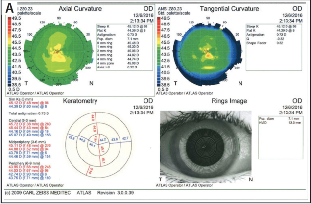
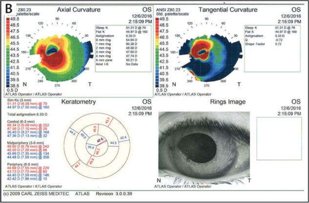
The patient presents 4 weeks after the open globe repair. Her UCVA measures 20/20 OD and 20/400 OS with no improvement with manifest refraction. A slit-lamp examination shows a sealed full-thickness 5-mm central corneal laceration with 10–0 nylon sutures and white cortical material extending from the lens to the cornea in the area of the wound (Figure 1). There is minimal cell in the anterior chamber, and the pupil is irregular but does not dilate well. There is no view to the posterior pole, but the B-scan shows no retinal detachment. Neither the Lenstar LS900 (Haag-Streit USA) nor the IOLMaster (Carl Zeiss Meditec) can capture keratometry (K) readings for the left eye, but measurements with the Atlas 9000 Corneal Topography System (Carl Zeiss Meditec; Figure 2) are shown for both eyes.

Figure 1. Appearance of the patient’s left eye 4 weeks after open globe repair.


Figure 2. Measurements of the right (A) and left (B) eyes with the Atlas 9000 Corneal Topography System.
When would you proceed with cataract extraction? Which K readings would you use to calculate the IOL power, and which lens would you insert, given the lack of sufficient anterior capsular support? Please discuss your surgical approach to this case.
—Case prepared by Zaina Al-Mohtaseb, MD.


SHEETAL BRAR, MS, AND SUMIT “SAM” GARG, MD
Surgery must be undertaken at the earliest opportunity, because the compromised capsule and the cortical matter in the anterior chamber increase the risk of secondary glaucoma and uveitis. We would aspirate the cortical matter with bimanual irrigation and aspiration (I/A) using low infusion pressure, followed by an anterior vitrectomy (pars plana approach) to clear the vitreous. Our preference would be to leave the eye aphakic initially, because accurate keratometry would be difficult, given the corneal sutures and wound edema. If the corneal wound showed adequate healing, we would remove the sutures during the surgery.
After a few weeks, we would have the child fitted for a contact lens for the purposes of visual rehabilitation and amblyopia prevention. Depending on her visual status, secondary IOL implantation could be considered 3 to 6 months later. If the topography remained highly irregular after 6 months, K readings from the right (healthy) eye could be considered, because the child does not have a previous ocular history. We would aim for a myopic outcome to assist with future corneal refractive procedures and to account for possible elongation of the eye.
If the posterior capsule were adequate to support an IOL, a three-piece lens could be implanted in the sulcus (with or without optic capture). If capsular support were inadequate, we would consider scleral glued IOL fixation.
After the secondary IOL implantation, it would be important to assess the patient’s vision, the scar’s impact, and the topography. Visual rehabilitation could be achieved with a rigid or scleral contact lens, topography-guided PRK to regularize corneal shape and reduce aberrations, or lastly, a penetrating keratoplasty if the scar is in the visual axis and impairing vision.
GREGORY S. H. OGAWA, MD, AND DEEPINDER K. DHALIWAL, MD, LAc


We would typically wait 4 to 6 weeks after open globe repair to remove the traumatic cataract. This allows enough time for anterior chamber inflammation to subside. Because this 5-year-old is still in the amblyogenic period, however, expeditious visual rehabilitation is important.
In cases of corneal trauma, K values can be unreliable. We would use the K values from the fellow eye to calculate the IOL power.
We would use bimanual I/A to remove the cataract through two limbal paracenteses. After staining the capsule with trypan blue or indocyanine green dye, we would attempt to convert the anterior capsular tear into a round capsulorhexis. If there were a posterior capsular break, we would “plug” it with Viscoat (Alcon) while removing the rest of the cataract. Then, we would perform a vitrectomy through the same paracentesis used for the cortical aspiration. We find that triamcinolone acetonide (Triesence; Alcon) can be useful for visualizing subtle vitreous strands. If the anterior and posterior capsules split on different meridians, then we would not implant an IOL but would instead wait for the anterior and posterior capsules to fuse, effectively creating a single lens capsule membrane over 2 to 3 months.
We would use a three-piece acrylic IOL and contour the capsular opening (if it were not already the correct size) for sulcus haptics with posterior optic capture. If the posterior capsule were intact, then we would see what the split anterior capsule looked like in terms of whether we could achieve reasonable coverage of the haptics of a one-piece acrylic lens by placing it in the bag. My preference in this case would be a one-piece acrylic IOL, because it would place minimal stress on the capsule and decrease the potential for the anterior split to extend posteriorly. If anterior and posterior capsular support were insufficient, we would consider intrascleral haptic fixation of a three-piece acrylic IOL (Yamane technique1) or Gore-Tex suturing (W.L. Gore & Associates) of an Akreos AO60 lens (Bausch + Lomb).
CATHLEEN M. McCABE, MD

I would proceed with cataract surgery at the earliest opportunity. The goal would be to remove the cataract, repair the iris, insert a stable posterior chamber IOL, and plan for future refractive management. IOL selection is challenging, given the irregular cornea and the likelihood that the K readings will change with removal of the sutures and healing of the laceration. I would use measurements for the fellow eye to select a sulcus-based lens power, while knowing that the patient may undergo further corneal procedures (penetrating keratoplasty) to maximize her vision in the future. I would plan to implant a three-piece IOL with scleral fixation of the haptics. Alternatively, the patient could be left aphakic and could wear a contact lens until the eye stabilized, she was older, and/or she had undergone a corneal transplant.
Intraoperatively, I would stain the anterior capsule with VisionBlue (trypan blue; Dutch Ophthalmic USA) for better visualization. Using a dispersive viscoelastic (Viscoat), I would then reform the anterior chamber while taking care to avoid overinflation. I always have extra viscoelastic on the tray and on hand in the OR for complicated cases like this one. An anterior chamber maintainer would then be placed. Next, I would use a diamond knife to make a paracentesis and place iris hooks as needed to maximize visualization, given the poorly dilating pupil. The nuclear material in a child this age will be easy to aspirate, and a 23- or 25-gauge vitrector could be used so that the incisions could remain small, improving anterior chamber stability.
In adults when the plan involves vitrectomy, I often place 23-gauge trocars at the start of surgery. In this case, however, I would not be able to visualize and confirm correct placement of the trocar during insertion, so I would wait until the cataract was cleared before placing a sclerotomy through the pars plana, if needed. Because the posterior capsule is compromised, it is likely that insufficient support remains for creation of a primary posterior capsulotomy with capture of the optic. Sulcus placement of a three-piece IOL would not be possible without additional support, so I would perform a more complete anterior vitrectomy and consider placing a sutured or sclera-fixated IOL. An Aaris EC-3 PAL three-piece IOL (Carl Zeiss Meditec) can be sclerally fixated by externalizing the haptics through 30-gauge needles placed through the conjunctiva and sclera. A flange at the end of the haptic is created with cautery in a technique described by Yamane.1 An alternative method would be the off-label use of a Gore-Tex suture to sclerally fixate an Akreos AO60 lens. Finally, I would repair the iris defect and reform the pupil using 10–0 nylon sutures (Ethicon) with sliding Siepser knots.
Postoperatively, the patient will need close observation, including amblyopia prevention with glasses or contact lenses and patching or atropine. Future intervention may include a corneal transplant to improve the vision in the affected eye.
ELIZABETH YEU, MD

Complications of the anterior segment from trauma can be challenging regardless of the patient’s age, but pediatric cases carry unique concerns related to ocular immaturity and potential amblyogenesis. The original surgeon in this case was spot-on in performing the corneal repair and leaving the traumatic lens damage alone. I will always default to deferring a traumatic cataract repair during an open globe repair. As demonstrated by this case, although there is obvious lens violation, the eye can remain relatively uninflamed and normotensive. Phacolytic intraocular hypertension is something that needs to be watched for very closely. One major benefit of leaving the damaged lens alone initially is that adequate fibrosis of the injured lens capsule will occur rather quickly, which can isolate the damage and prevent extension of the capsular violation.
This child needs surgery sooner rather than later because of the dense intumescence that is now impeding proper vision—an amblyogenic risk. With an older patient, I would consider waiting several months to allow full healing of the corneal laceration, remove the corneal sutures, and then perform IOL power calculations. The lack of stromal opacification or fibrosis within the laceration line demonstrates that the wound is not ready for suture removal at 4 weeks, so cataract surgery should proceed with the corneal sutures still in place.
I would be cautious about how much pressure I applied posteriorly, because the posterior capsule has been violated. Too much sudden pressure within the anterior chamber from overfilling with viscoelastic or a suddenly deepened anterior chamber with irrigation could drive more lenticular material posteriorly. I would stain the anterior capsule with trypan blue, then carefully aspirate lenticular material manually with a 25-gauge hydrodissection cannula or the like.
Because children develop progressive myopia after cataract surgery,2,3 I would target a result of approximately +3.00 D of hyperopia, as recommended by the American Academy of Ophthalmology.4 For the IOL power calculation, I would use the average K values of about 45.00 D, which is the average value of the central 3-mm zone in the healthy contralateral eye, instead of the approximately 47.00 D erroneous value of the aberrated cornea from the corneal laceration repair and the sutures that are present.
After careful lens removal, the anterior capsule may require some excision and reshaping for an adequate central capsulotomy opening, but I would not be surprised if there were enough support to house a three-piece sulcus IOL, in which case my preference would be a round-edged IOL such as the Sensar AR40e (Johnson & Johnson Vision). If the anterior capsule were inadequate, I would opt for iris versus sulcus fixation using a 9–0 polyprolene suture. Long-term studies have shown that posterior iris fixation of IOLs can provide very good visual and functional outcomes, with low dislocation rates.5,6
Postoperatively, the clinical care of this patient will be a labor of love, with amblyopia therapy starting 1 day after surgery. Once the corneal sutures are removed, she will need to continue amblyopia treatment, potentially necessitating her wearing a rigid gas permeable contact lens to normalize the corneal aberrations and optimize vision.
WHAT I DID: ZAINA AL-MOHTASEB, MD

I discussed at length with the patient’s parents the potential surgical complications, given the capsular violation and the unknown visual potential because of the traumatic nature of the cataract, lack of view to the posterior pole, and amblyogenic age. I also advised them that the patient might need surgery in the future (given the corneal laceration) and that I might not be able to place an IOL during the initial surgery. If a patient with a violated anterior capsule and lenticular material in the anterior chamber does not have uncontrolled IOP or inflammation, then I like to wait at least 3 to 4 weeks after open globe repair to allow fibrosis of the anterior and posterior capsules in addition to healing of the corneal wound. Of course, I did not want to wait longer in this case because of the risk of amblyopia. As usual, I prepared for the worst and made sure I had all the necessary intraocular microinstrumentation, including intraocular forceps and scissors, capsular tension rings, and iris hooks.
Watch It Now
See how Zaina Al-Mohtaseb, MD, managed this case.
To calculate the lens power (for both a one- and a three-piece IOL), I used the corneal power information from the right eye and the axial length from immersion data on the left eye. Because of the corneal scar and irregular K readings, I explained to the parents that the patient’s best vision would be achieved with a hard contact lens regardless of the IOL used.
I administered tropicamide and phenylephrine as well as intracameral 1:4,000 preservative-free epinephrine for pupillary dilation. I stained the capsule with trypan blue to evaluate where the capsular violation was. After filling the anterior chamber with a dispersive viscoelastic, I placed iris hooks to aid pupillary dilation. Luckily, the cataract was soft, so I used bimanual I/A to remove the milky cortical material and remaining cortex. I find that bimanual I/A provides a very stable chamber. I was able to enter the area of the anterior capsular tear with the aspirator to remove the cortical material. I was also able to create an extra paracentesis incision to improve my access to the remaining cortical material. In eyes like this one, it is important not to allow the chamber to collapse at any point during surgery by filling it with viscoelastic before the removal of instruments.
After extracting the lens and cortical material, I could see a large U-shaped anterior capsular tear, with anterior capsular fibrosis in the visual axis, and an intact posterior capsule. There was fibrosis of the posterior capsule with central opacification—most likely in an area of previous violation—and there was no vitreous in the anterior chamber. I used 25-gauge intraocular scissors to cut the anterior capsular fibrosis and to create a half-circle anterior capsulotomy from the remaining anterior capsule. Given the posterior capsular opacification and the patient’s age (she would be unable to remain still for a postoperative YAG capsulotomy), I used a cystotome and Utrata forceps to create a 4.5-mm posterior capsulotomy. Luckily, there was no violation of the anterior hyaloid face and no vitreous prolapse. I was therefore able to insert a three-piece lens, place the haptics in the sulcus, prolapse the optic posteriorly, and reverse optic capture it in the posterior capsulotomy.
After removing the iris hooks and suturing the wound with 10–0 Vicryl sutures (Ethicon), I removed the remaining viscoelastic using I/A with the bimanual anterior vitrector just in case I encountered any vitreous. I instilled triamcinolone acetonide to confirm that there was no vitreous in the anterior chamber and for its anti-inflammatory properties. I injected acetylcholine for miosis to provide coverage of the IOL’s edge and to make sure the pupil was round. In the area of the irregular pupil superonasally, I performed an iris sweep to make sure there were no vitreous strands. I hydrated the incisions and found that the lens was well centered and the chamber deep at the end of surgery.
Three weeks postoperatively, the patient had a BCVA of 20/50, and I sent her for a contact lens fitting.
1. Yamane S, Inoue M, Arakawa A, Kadonosono K. Sutureless 27-gauge needle–guided intrascleral intraocular lens implantation with lamellar scleral dissection. Ophthalmology. 2014;121(1):61-66.
2. Lambert SR. Changes in ocular growth after pediatric cataract surgery. Dev Ophthalmol. 2016;57:29-39.
3. Kora Y, Shimizu K, Inatomi M, et al. Eye growth after cataract extraction and intraocular lens implantation in children. Ophthalmic Surg. 1993;24(7):467-475.
4. Trivedi RH, Wilson ME Jr. Selecting intraocular lens power in children. EyeNet Magazine. January 2006. http://bit.ly/2r76PkO. Accessed June 7, 2017.
5. Kavitha V, Balasubramanian P, Heralgi MM. Posterior iris fixated intraocular lens for pediatric traumatic cataract. Middle East Afr J Ophthalmol. 2016;23(2):215-218.
6. Yen KG, Reddy AK, Weikert MP, et al. Iris-fixated posterior chamber intraocular lenses in children. Am J Ophthalmol. 2009;147(1):121-126.


