Interesting & Artistic Images
Silicone Ballet: Trio of Bubbles in the Eye
By Muhammad Saad, MBBS, FCPS Resident
The photograph shows a unique composition of silicone bubbles within the eye.

The Pearl
Aows AlJammal, MBChB
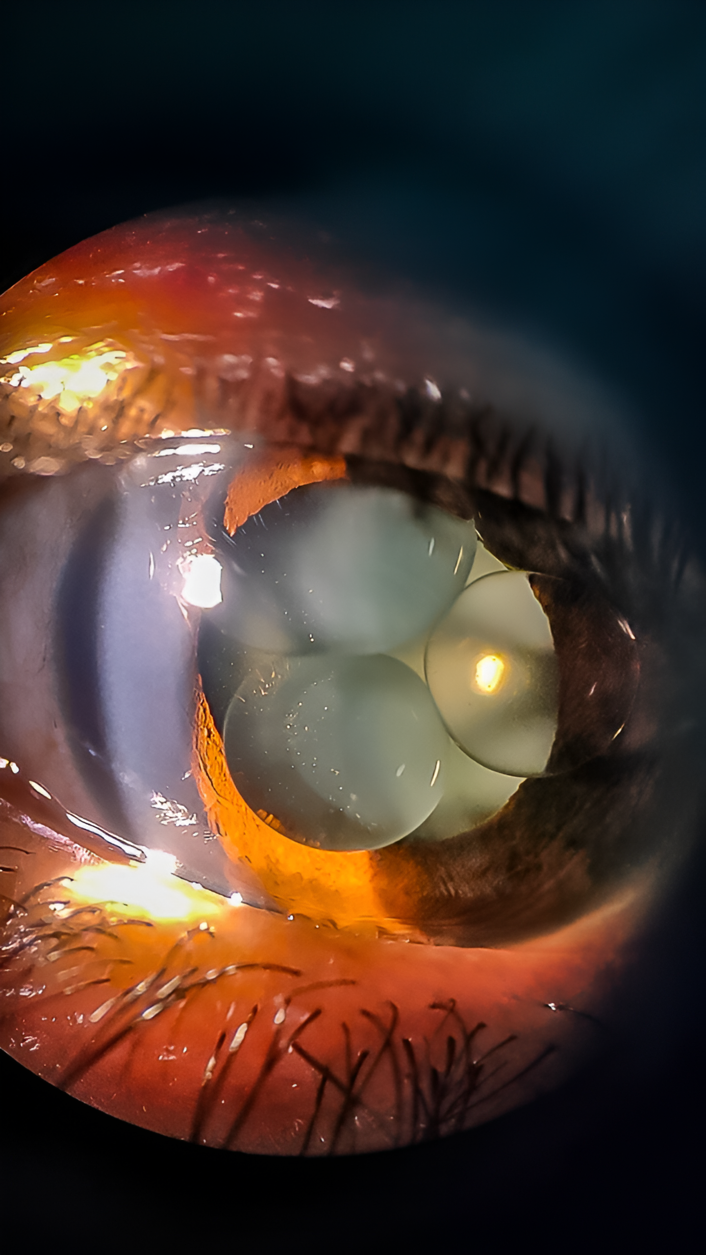
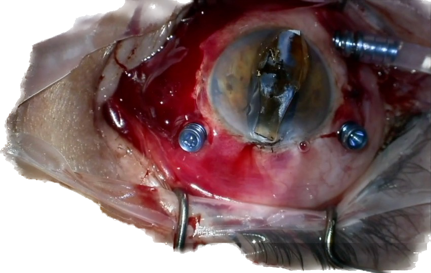
The photograph shows a mature cataract in a patient undergoing simultaneous penetrating keratoplasty and cataract extraction.

The Beehive
Deepanshu Agrawal, MBBS, DO, DNB, FVRS, and Rinal Pandit, MBBS, MS, FMRF, FAICO
Captured at a postoperative visit 1 month following cataract surgery and silicone oil removal, the image reveals a honeycomb pattern of residual silicone oil droplets on a hydrophilic IOL.

Hanging on by a Thread
Grand Prize Winner
Abha Amin, MD
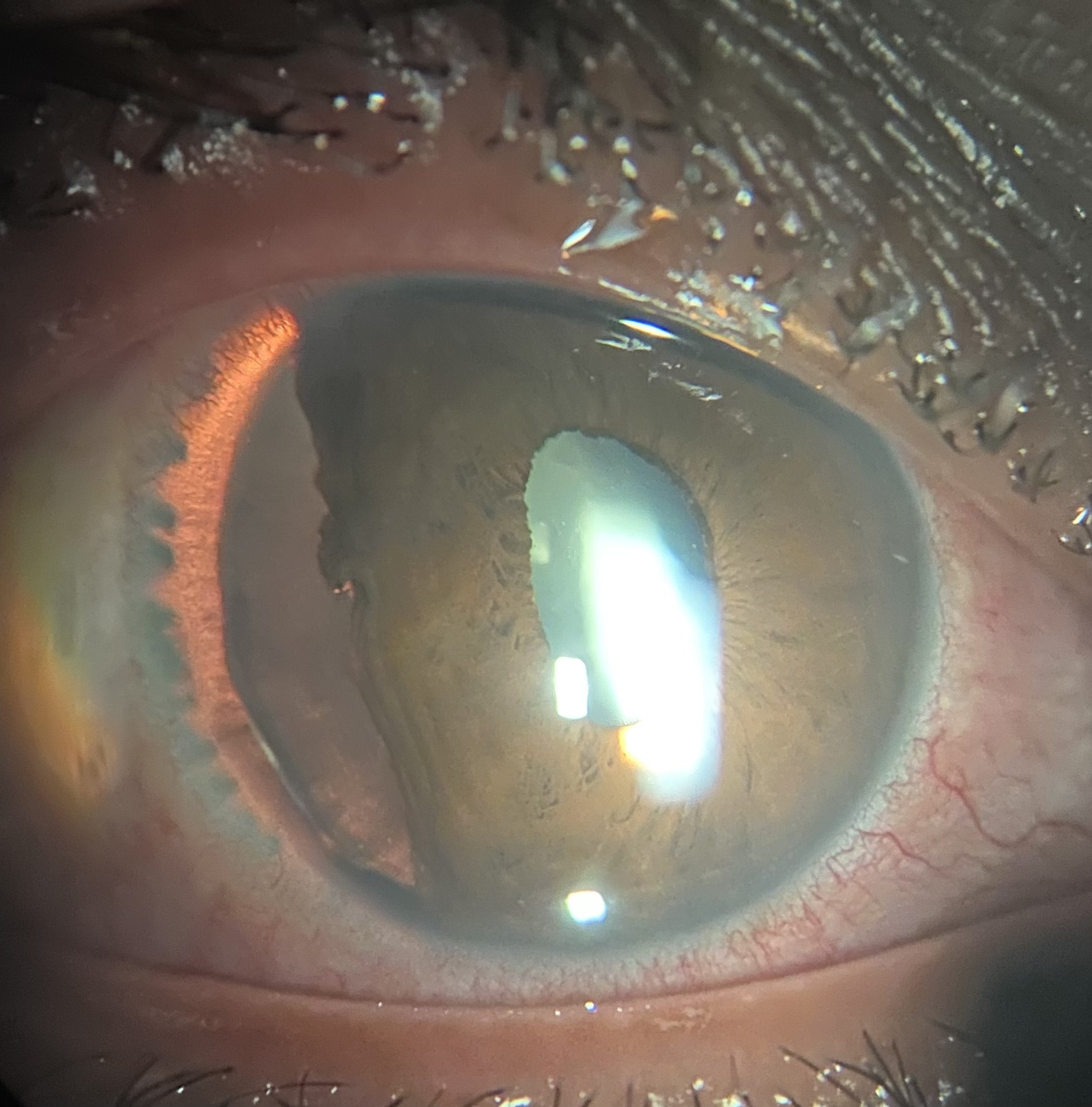
In the photograph, zonulopathy has caused lens subluxation.

Resting Demodex on an Eyelash
Goktug Demirci, MD
The photograph of a patient with ocular rosacea features a resting Demodex folliculorum amid cylindrical dandruff. The image is an unfiltered light microscope capture.
Rare & Unusual Diseases

Corneal Keloid in a Radial Keratotomy Wound
Giuliano Freitas, MD, PhD
A whitish, elevated lesion has risen from a radial keratotomy wound in the eye of a 72-year-old man.

Endophthalmitis in a Patient With Presumed Axenfeld-Rieger Syndrome
Sarishka Desai, BA, and Majida Gaffar, MD
Severe endophthalmitis has occurred in a patient with congenital glaucoma and anterior segment dysgenesis, presumed to be Axenfeld-Rieger syndrome.

Radiant Nevus of Ota Beauty
Akshita Ghiya, MD
In this 14-year-old girl with Nevus of Ota, grey-blue hyperpigmented lesions surround the left eye, and hyperpigmented patches in the sclera can also be seen.

Dead Bag Syndrome
Andres German Alza, MD
The IOL–capsular bag complex has dislocated in the eye of a patient who underwent bilateral cataract surgery.

Wynorrific: a Paradox of Beauty and Horror
Jesus Santiago Vidaurri-Martinez, MD
The photograph captures a rare occurrence of synchysis scintillans in the anterior chamber.
Slit Lamp

Zonular Dehiscence
Darrin Landry, CRA, OCT-C, FOPS
Superior zonular dehiscence led to lens dislocation. The image was captured with sclerotic scatter illumination.

Blood Is Coming, Game of Vision
Mohamed Afifi, MRCSEd, FICO, EBO
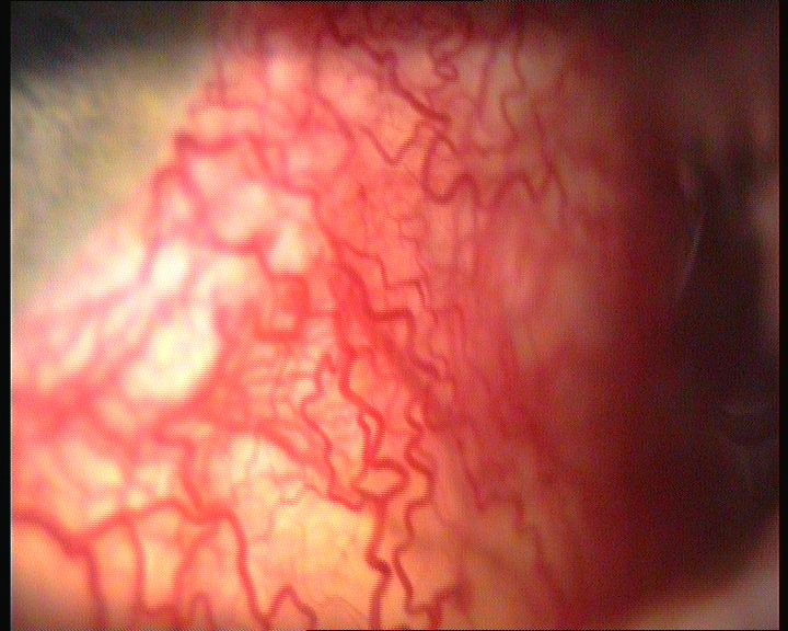
The photograph captures an acute invasion of blood vessels into the cornea from the conjunctiva. The cornea is under siege from all sides.
Carotid Cavernous Fistula
Ana Miguel, MD, PhD, FEBO, GCSRT
A patient sought a second opinion for nontraumatic bilateral hyperemia in the left eye (photo) that had developed 10 months earlier. Clinical signs included chemosis, nonpulsatile proptosis, corkscrew vessels, and paresis of the left fourth cranial nerve. An MRI report confirmed a carotid-cavernous fistula, which was successfully treated with endovascular intervention.

Rivers of Blood
Nayeli Bernal, MPH
A patient exhibits neovascular glaucoma.

Heart-Shaped Pupil
Abu Serhan Hashem, MD
The pupil has a unique heart-shaped appearance.
Surgical Complications

Houston, We Have a Problem!
Rami Shasha, MD, FRCSC, ABO
A patient was referred following an aborted cataract procedure at another facility. A slit-lamp examination revealed a large nuclear fragment occupying nearly 50% of the anterior chamber.

Cataract Surgery on a Vascularized Cornea
Riadh Romdhane, MD
A patient who was blind in both eyes due to vascularized corneas and cataracts underwent extracapsular cataract extraction. Postoperatively, they regained the ability to see up to 3 m.

Ice Finger of Death
Rodolfo Bonatti, MD
When the tube of an Ahmed Glaucoma Valve (New World Medical) is too long, it can make contact with the crystalline lens and cause a localized freezing cataract to form.

Dislocated Sutured Plate Haptic IOL
Steven G. Safran, MD, PA
The patient had a sutured plate haptic IOL. When the suture broke, the lens fell posteriorly onto the retina. The lens was successfully removed, and intrascleral haptic fixation of the replacement IOL was performed with the Yamane technique.

Massive Bleb Elevation
Thomas Tien, MD and Irving M. Raber, MD
A patient presented with bleb dysesthesia resulting from significant bleb elevation following a trabeculectomy.
Trauma
Traumatic Lens Dislocation in an Eye With Anterior Megalophthalmo
Huda Al-Ghadeer, MD, FRCS
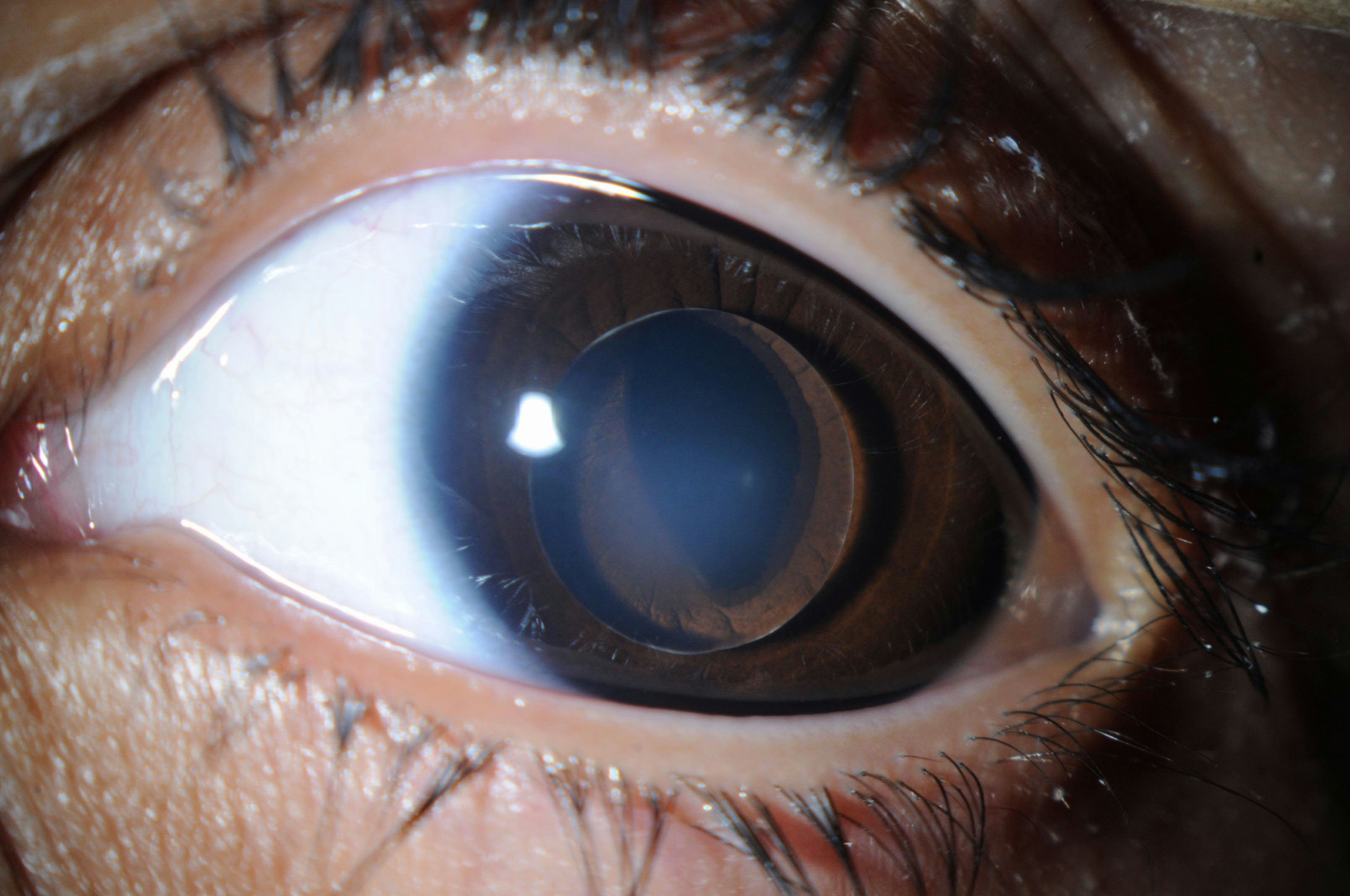
A slit-lamp photograph of the left eye reveals megalocornea, microspherophakia, lens dislocation in the anterior chamber, and ectropion uveae.
Miraculous Extraction
Claudio Brancato, MD
A 62-year-old man presented after sustaining accidental ocular trauma in a domestic setting that damaged his right eye. The foreign body, presumed to be glass or metal, was successfully emulsified and removed via corneal access.
Traumatic Iridodyalisis
Jesus Santiago and Vidaurri-Martinez, MD
The photograph illustrates traumatic iridodialysis secondary to blunt trauma. Both the crystalline lens and zonules remain intact over the area of dialysis, with ciliary processes in situ.

Staple in Cornea
Ashley Perez
A staple causes penetrating corneal trauma.

Traumatic Inclusion Cyst
Mehrnaz Atighehchian, MD
The image shows a traumatic conjunctival-scleral inclusion cyst following a penetrating injury. A 15-year-old male patient exhibited a sizable traumatic conjunctival inclusion cyst 4 years following the initial repair of a corneal-scleral laceration. The size of the cyst has increased since 1 year earlier.