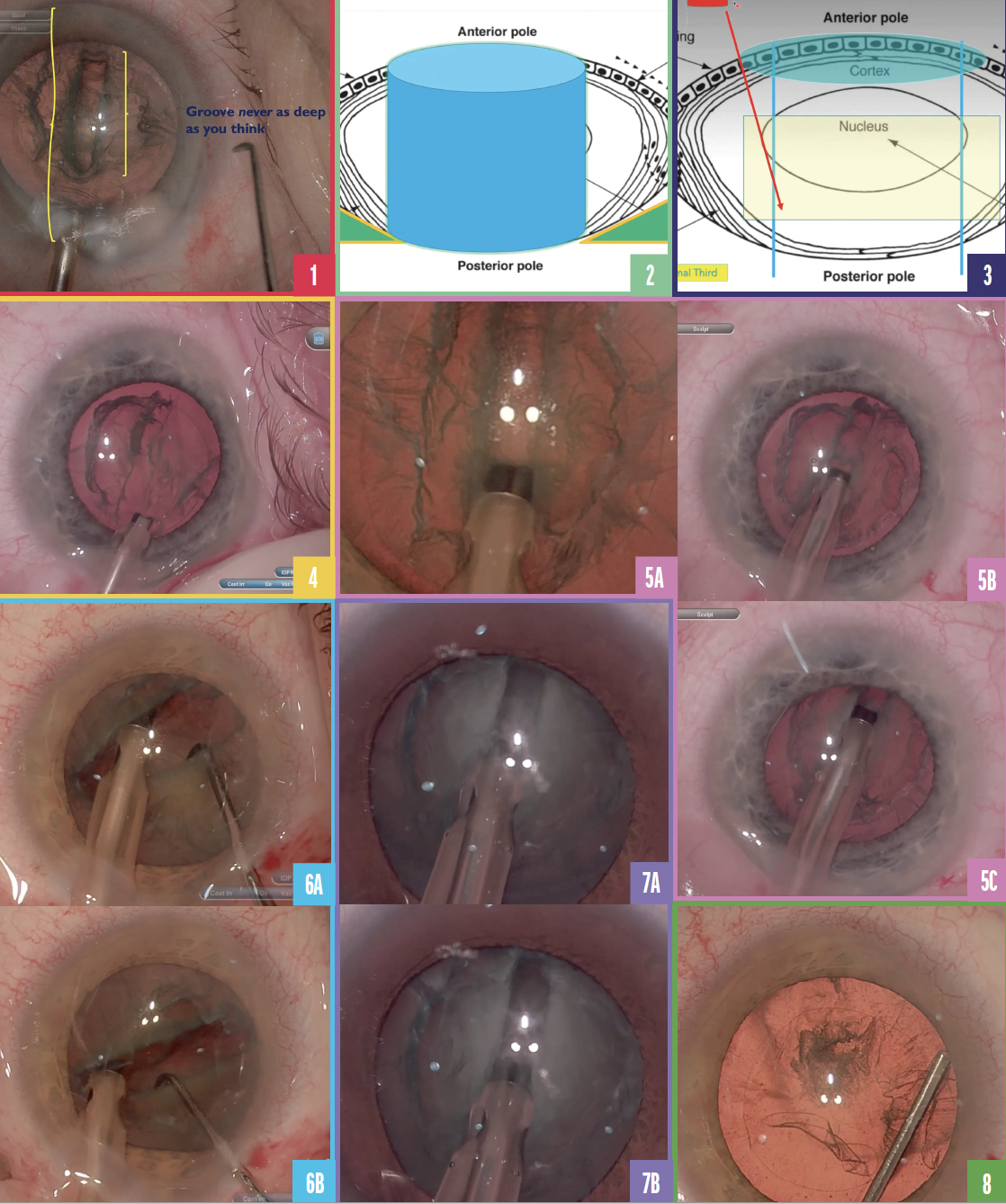
Strategies to create the groove and crack the lens deserve greater attention. The phaco tip is usually angled downward toward the posterior capsule to create the groove without an awareness of the lens shape and natural curvature in the posterior capsule. Ignoring this z-axis concept of the nucleus makes it challenging to gauge proper groove length and depth (Figure 1) and risks piercing the posterior capsule. The surgeon must be constantly weighing the risks with the knowledge that a groove that is too short and shallow can make the lens difficult to crack.

Figure 1. Of the total lens, the groove is only about 50%, making it more difficult to crack.
Figure 2. The 3D effect of the lens and the z-axis.
Figure 3. A phaco tip inserted through a primary incision near the proximal capsulotomy can access only about 40% of the lens and not the proximal end (red arrow). The red cylinder near the top left represents the primary incision, the blue area represents the capsulorhexis, and the yellow rectangle represents the distal third of the lens.
Figure 4. The top 15% of the lens is aspirated.
Figure 5. The phaco tip is positioned at a steep angle and down toward the anterior capsular edge to create the proximal third of the groove (A). The phaco tip is flattened to create the middle third of the groove (B). The phaco tip is angled up to follow the curve of the posterior capsule and create the distal third of the groove (C).
Figure 6. The groove is cracked in the distal third (A). The groove is separated by moving from the distal third to the proximal edge (B).
Figure 7. The needle of the phaco tip is used to scrape into the lens (A,B).
Figure 8. During hydrodissection, the posterior wave can help indicate how deep the groove can be made.
Aggressively sculpting the groove deepest near where the proximal capsulotomy matches with the thickest section of the lens carries less risk of piercing the posterior capsule as the phaco tip is angled sharply down. This creates space to then angle the phaco tip flat, parallel to the posterior capsule, as the groove is then extended distally. Here and in the accompanying video (watch below), I outline my technique for creating the groove.
BACK TO BASICS: ANATOMY OF THE LENS AND THE RULE OF THIRDS
Anatomy. The 3D effect of the lens and z-axis on groove creation should not be overlooked when creating a strategy for the groove in the nucleus (Figure 2). The distal third of the lens has a posterior capsule that is angled up as the lens tapers. The center third of the lens is thickest and therefore the safest area to groove. It is effectively impossible to access the proximal third of the lens when one considers that the phaco instrument is straight and must traverse through a primary incision near the limbus and then the capsulotomy opening with a proximal edge that is typically 2.5 mm from the center of the lens with an average diameter of 10 mm. Typically, just 40% to 50% of the lens is available to create the groove through this approach (Figure 3).
Groove creation may be thought of in thirds—the proximal, middle, and distal thirds. Each third requires a different angle of the phaco tip to match the axial thickness of the lens.
Proximal third. In this third, the phaco tip should be inserted at as steep an angle as possible (ideally 80º to 90º) starting close to the proximal anterior capsular edge.
Middle third. The middle third of the groove has the most lens density. With the space created in the proximal groove, the surgeon can now flatten the tip in this area. Safety and control are optimized when phaco is performed with the tip flat. For dense lenses, if needed, the sharp needle of the metal beveled phaco tip can be used to create depth (see Ice Scraper Technique, ).
Distal third. The distal third is the most dangerous portion of the groove to create. The posterior distal lens curves up, and the lens often becomes softer. There is a risk of vitreous surge through the lens into the bag. The phaco tip should be curved up along the contour of the posterior capsule to allow the groove to become more superficial (see Quick Tip for Hydrodissection).
ICE SCRAPER TECHNIQUE
When I’m nervous about cracking the nucleus in dense cataracts and want to go deeper with the groove, I use the needle of the phaco tip to scrape into the lens and then lift (Figure 7). I call this the ice scraper technique because it is like using a shovel to remove layers of ice after a snowstorm. The phaco tip is kept flat, but more ultrasound energy is used.
MY TECHNIQUE
Remove superficial cortical material. Without ultrasound, the top 15% of the lens is aspirated to help improve red reflex and depth visualization (Figure 4).
Approach the proximal third at a steep angle. The groove is initiated at the edge of proximal capsulotomy. The phaco tip is held at a steep angle because this is the thickest area of the lens (Figure 5A).
Advance the phaco tip to the middle and distal thirds of the lens. Once the phaco tip is engaged in the middle third, the phaco tip is flattened out (Figure 5B). Then, in the distal third, the phaco tip is curved upward to follow the contour of the posterior capsule (Figure 5C).
Crack the groove. The instruments are placed deep into the groove to achieve an effective crack. Most surgeons crack in the middle of the groove. I find it is best, however, to crack the groove at the distal third (Figure 6). When the groove is cracked in the middle, it is harder to separate the two large halves of the lens and creates more stress on the bag and zonules. When the groove is cracked on the distal third—and it is deep—visualization and the red reflex are improved. The vector forces allow enlargement of the groove without putting much stress on the bag.
QUICK TIP FOR HYDRODISSECTION
Watch the posterior wave and pay attention to the depth because it can indicate how deep the groove can be made (Figure 8).
Once the distal crack is created, the groove is unzippered by moving toward the proximal edge and cracking again. By holding the two hemi-nuclei apart, the depth of the plate can be visualized, and the smaller side of the nucleus can be rotated to be removed first.
CONCLUSION
Thinking about the groove as three different sections and strategizing the angle of the phaco tip according to each third allow a deeper and longer groove to be created.
*Eyetube is prowered by BMC, parent company to CRST


