
High-quality visualization has become an invaluable tool for use during clinical procedures, teaching, and training. This is especially true of advanced three-dimensional (3-D) technology available to surgeons and their teams.
The ability to work, record, and display in 3-D versus 2-D provides surgeons a realistic depth of field and helps them navigate through procedures more efficiently. These 3-D systems have revolutionized surgical training in medical fields such as laparoscopic surgery.
APPLICATIONS TO TRAINING
In ophthalmology, lectures with slides have been the gold standard in training for decades—2-D training materials for an inherently 3-D specialty. Residents are exposed to some 2-D video, but it is inadequate for conceptualizing cataract surgery. Trainees do observe procedures in the OR and at times have the opportunity to watch surgery through the assistant's microscope, but even the microscope is almost invariably in 2-D! Some ORs have 2-D monitors, but they are often set up sporadically and usually with low image quality. As a result, senior residents perform their first phacoemulsification never having seen the depth required to sculpt a nucleus. They are left with the trial-and-error—words you never want to use in medicine—method when they begin to operate.
Now, 3-D technology gives the observer the surgeon's view in real time. This technology reduces the learning curve like no other tool in ophthalmologists armamentarium, and it is invaluable for training residents as well as surgeons with years of experience. At the Mackool Eye Institute, we use the new Sony 3-D system. It mounts quickly on the Luxor (Alcon) and Lumera (Carl Zeiss Meditec) microscopes, allowing advanced 3-D image capture and display within minutes (Figure).
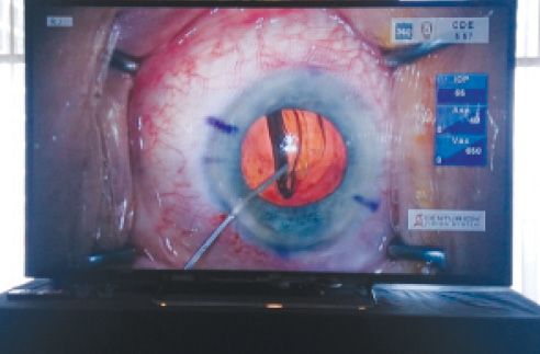
Figure. Dr. Mackool's surgery as seen using Sony's 3-D technology.
IMPROVING YOUR TECHNIQUE
The 3-D system not only helps observers, but it also helps the surgeon. During a case, the surgeon is focused on the task at hand. Once the case has ended, however, he or she can watch the 3-D video and gain an expanded perspective by critically analyzing the procedure and developing modifications or new approaches for the future. My colleagues and I routinely review complex and interesting cases on the 3-D system. With 2-D systems of the past, a true understanding of intraoperative events was often lost. With the 3-D system, we have been able to relive procedures, and this added perspective has helped us analyze techniques and innovate.
CONCLUSION
These easily implemented high-resolution 3-D systems provide an opportunity to revolutionize ophthalmic teaching. As these systems evolve, they will likely play a greater role in the surgical suite as well.n
Richard Mackool Jr, MD
• assistant director of The Mackool Eye Institute and Laser Center
in Astoria, New York
• (718) 728-3400
• acknowledged no relevant financial interest

