CASE PRESENTATION
A 70-year-old woman is referred for cataract surgery. The patient is an equestrian and was kicked in the face by her award-winning horse about 10 years ago. She experienced an orbital fracture of the left eye, which was not treated surgically. The injury left her with mild enophthalmos and progressive vision loss in that eye. Her BCVA is counting fingers at 3 feet OS with light projection in all quadrants. She has traumatic glaucoma in the left eye that is controlled with one medication and an angle recession for 30º. An iridoplegia with pupil sphincter ruptures; a dense subluxated cataract; and 5 clock hours of zonulolysis, vitreous herniation, and anterior vitreous membrane rupture into the anterior chamber are visible at the slit lamp.
The anterior and posterior segment examinations of the fellow eye are normal aside from 2+ to 3+ nuclear sclerosis that leaves the patient with 20/50 UCVA and BCVA OD, which she finds bothersome when driving.
How would you counsel her, and how would you proceed? Please describe your surgical technique and contingency plan.
— Case prepared by Lisa Brothers Arbisser, MD

CRISTOS IFANTIDES, MD, MBA
Assuming the B-scan ultrasound and Purkinje tree entoptic test are normal in the traumatized left eye, surgery would be scheduled for the end of the day. The goal would be to clear all vitreous from the anterior segment, normalize the IOP, place a secured IOL, and perform a pupilloplasty.
A retrobulbar block would be administered. After the creation of a peritomy, a 23-gauge trocar would be placed followed by an anterior chamber maintainer. Triamcinolone acetonide would be injected, and a pars plana anterior vitrectomy would be performed until all vitreous has been pulled back toward the posterior segment.
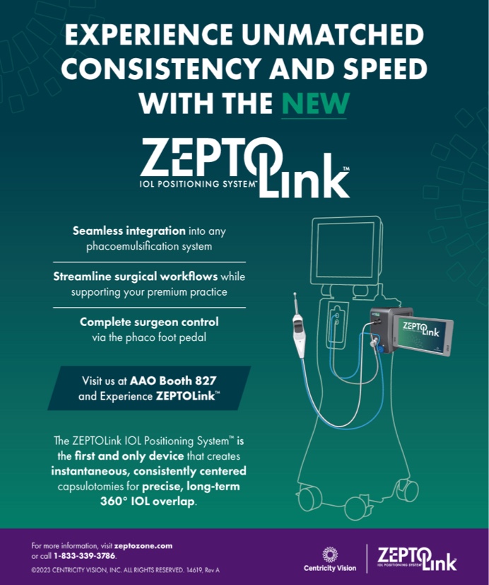
The Zepto IOL Positioning System (Centricity Vision) would be used to create a strong capsulotomy to support an Ahmed Capsular Tension Segment (CTS; Morcher). Capsular hooks would then be placed.
The normal steps of cataract surgery, including vertical and possibly rotary chop, would be performed on the dense lens. A capsular tension ring (CTR) would be inserted. Next, either a three-piece CT Lucia 602 (Carl Zeiss Meditec) or a one-piece enVista IOL (Bausch + Lomb) would be implanted. Both lenses are aspheric, and their power is the same from the center to the edge, minimizing the impact of decentration. An Ahmed CTS would be fixated with either a double-flanged 5-0 polypropylene (Prolene, Ethicon) or PTFE (Gore-Tex, W.L. Gore & Associates) suture in the area of capsular weakness. Finally, multiple single-pass four-throw 10-0 polypropylene sutures would be placed to repair the iridoplegia.

TOBIAS H. NEUHANN MD, FEBOS-CR
Relatively rapid rehabilitation with the option of improving the patient’s visual acuity and ability to drive could be achieved by correcting her better-seeing eye with laser cataract surgery and the implantation of an extended depth of focus IOL such as the RayOne EMV (Rayner). If the patient is dissatisfied with her postoperative result, surgery on the second eye to address the cataract and repair damage from the earlier trauma would be planned.
The capsulotomy and nuclear fragmentation would be performed with a femtosecond laser. The corneal incisions would be created manually. The ports would then be prepared for a pars plana vitrectomy (PPV). If a PPV is not feasible because the view into the vitreous cavity is obscured by the cataract, the vitreous prolapse would be removed as much as possible from the anterior chamber with a vitrector, and triamcinolone acetonide would be injected to facilitate the detection of vitreous striae.
If laser cataract surgery on the left eye is not possible because of the iridoplegia, the pupil would be dilated with an expansion device such as a Malyugin Ring (MicroSurgical Technology). The capsulotomy would be performed under OVD protection. The capsular bag would be secured in the region of the zonulolysis with an AssiAnchor (Hanita Lenses) and a 5-0 polypropylene suture. If a single anchor is insufficient, a second would be placed. Gentle hydrodissection, a cautious attempt at rotating the lens nucleus, and phaco chop would ensue.
When to insert a CTR depends on the intraoperative outcome. Inserting one before phacoemulsification could make aspiration of the lens cortex more difficult. Alternatively, the device could be inserted before IOL implantation.
The same IOL design selected for the right eye would be implanted in the left. Triamcinolone would be injected into the anterior chamber, and a PPV would be performed to remove vitreous elements and thereby eliminate vitreomacular traction. Once the anterior chamber is clear of vitreous striae and the IOL is centered, surgery may be completed.

ZEBA A. SYED, MD
The most notable complications from the equestrian injury are significant lens subluxation and frank vitreous prolapse into the anterior chamber. When counseling the patient, I would emphasize the complex nature of her surgery and the increased risk of postoperative retinal complications. Given her history of traumatic glaucoma, I would also carefully review prior nerve imaging and visual fields to assess the patient’s visual potential.
The enophthalmos may present ergonomic challenges. I would be careful if performing a retrobulbar block given the risk of injury to the globe due to the patient’s anatomy. Preoperatively, I would examine her while she is supine to confirm that the lens does not fall posteriorly and become inaccessible.
The first step of surgery would be to clear vitreous from the anterior chamber. I typically inject triamcinolone to facilitate visualization and perform a thorough anterior vitrectomy from a pars plana approach. This can maximize the removal of vitreous strands. Depending on the degree of zonulopathy and visualization of the lens, it might be possible to preserve the capsular bag with a capsule-stabilization technique. In eyes with significant zonular loss, the entire lens-capsule unit is removed anteriorly.
After the anterior vitrectomy, an OVD would be injected under the lens to prolapse it into the anterior chamber for safe phacoemulsification. If concerned that the lens might drop posteriorly, I would consider using a Sheets glide or performing manual lens extraction. A benefit of working at an academic hospital is that retina colleagues are available in the adjacent OR. I would ask the retina team to be on standby to assist in case lens material drops posteriorly.
After lens removal, another anterior vitrectomy would be performed followed by scleral fixation of a CT Lucia IOL with the Yamane technique.1
Postoperatively, a dilated fundus examination would be performed. The patient’s IOP would be monitored closely.

TANYA TRINH, MBBS, FRANZCO, FWCRS
The patient would be counseled that the goals of surgery are to provide her with the best quality of vision possible through long-term fixation of an IOL and to minimize future complications. Given the density of the cataract in the left eye, B-scan ultrasound of the posterior segment would be performed to rule out a subclinical retinal detachment. Ultrasound biomicroscopy would be performed to determine the degree of lens tilt and subluxation, and a supine examination would be conducted with a portable slit lamp. These would facilitate preoperative surgical planning. A vitreoretinal team should be on standby at the time of surgery in case the lens falls entirely posteriorly. Specular microscopy would be obtained preoperatively to ascertain the probability of postoperative endothelial dysfunction and the patient counseled accordingly.
A triamcinolone-assisted anterior vitrectomy from a pars plana approach would be performed to release traction on the retina before the anterior segment is cleared of vitreous.
The capsulorhexis would be initiated in the area of intact zonules for adequate countertraction. Capsular hooks would be placed as necessary to maintain a centered bag and facilitate countertraction. Nuclear disassembly would be performed with a gentle zonule-sparing chop technique. Gentle cortical viscodissection would be performed. A gentle two-piece I/A approach would be used for cortical removal with a CTR placed as early as necessary but as late as possible.
To achieve long-term fixation of the bag, one or more CTSs would be secured to the sclera with PTFE sutures given the degree of zonular dialysis. A three-piece IOL would be placed in the bag to allow future intrascleral haptic fixation if necessary.
If capsular bag support cannot be preserved, the cataract, capsular remnants, and zonular processes would be completely removed. Four-point IOL fixation with a quadruple-looped haptics approach would be performed via an intrascleral PTFE suture technique (two 3-mm intrascleral paths 2 mm posterior and parallel to the limbus and 180º apart). First, the needle is removed. Then, the first arm of the PTFE suture (proximal to the surgeon) is internalized into the anterior chamber, posterior to the iris, and brought out through the main incision with a handshake technique. Next, the free end of the PTFE suture is looped through two of the four haptics, brought back into the anterior chamber and behind the iris, and externalized via a second sclerostomy at the distal end of the intrascleral PTFE passage path. This allows adjustment and centralization of the IOL, avoids worry about tilt, and addresses the potential concern of PTFE suture exposure over time. The suture is tied off, the knot is pushed into the sclerostomy holes, and the ends are trimmed to prevent protrusion. (This technique was originally described by Jamin Brown, MD.)
A pupilloplasty would then be performed, and the diameter would be titrated to approximately 3.5 mm.

WHAT I DID: LISA BROTHERS ARBISSER, MD
The patient wore contact lenses on occasion and was willing to continue doing so if necessary after surgery. She had also worn safety glasses routinely since her accident. I hoped to save the capsular bag and implant an IOL within it but explained to the patient that additional surgery might be required. She was still driving, so attempted rehabilitation of the traumatized left eye was planned. MIGS devices to address the elevated IOP at the time of cataract surgery were not available when this case was performed.
On the day of surgery, capsular support devices and backup IOLs were available in the OR. A trocar was inserted through a scleral tunnel. A sutureless transconjunctival pars plana incision was made, followed by a corneal incision. Triamcinolone acetonide was instilled in the anterior chamber to identify prolapsed vitreous for removal. Under OVD control of the chamber, a continuous curvilinear capsulorhexis was performed, and Yaguchi capsule hooks were placed2 (watch the surgery below).
The anterior-posterior vitreous connections were amputated with a dry technique through the trocar cannula, and normotension was maintained with an OVD. Hydrodissection was difficult owing to greater zonulolysis than was initially evident. Additional capsular hooks were placed.
Vertical chop phacoemulsification could not be completed before part of the nucleus dropped. In retrospect, it is possible that the vitrector port faced up rather than sideways or down. I will never know whether a spot in the posterior capsule had been weakened by the earlier trauma or if an instrument violated the capsule during the anterior vitrectomy.
The iris was floppy from posttraumatic iridoplegia, which was a contraindication for iris fixation of an IOL. The distinct possibility of a low endothelial cell count from prior inflammation and the abnormal angle were contraindications for an anterior chamber IOL. The patient, moreover, would need a three-port vitrectomy and lens fragmentation in the future, making primary scleral IOL fixation redundant. For these reasons, all lens material was removed from the anterior segment, the iris was pulled out of the angle, and the eye was left aphakic.
After surgery, the patient was referred to a posterior segment colleague. The eye quieted, and the IOP was easily controlled following the vitrectomy and lensectomy. Subsequent secondary implant surgery resulted in an excellent outcome.
A few months later, I performed uneventful cataract surgery on the patient’s right eye.
1. Yamane S, Sato S, Maruyama-Inoue M, Kadonosono K. Flanged intrascleral intraocular lens fixation with double-needle technique. Ophthalmology. 2017;124(8):1136-1142.
2. Yaguchi S, Yaguchi S, Asano Y, et al. Repositioning and scleral fixation of subluxated lenses using a T-shaped capsule stabilization hook. J Cataract Refract Surg. 2011;37(8):1386-1393.


