CASE PRESENTATION
A 73-year-old man presents with a complaint of painless vision loss in both eyes. The patient reports experiencing difficulty viewing signs at a distance, especially with glare from oncoming traffic at night, during the past year. At age 14, he sustained a traumatic injury to his right eye from a BB gun, and he has had decreased vision and a decentered pupil in that eye ever since. The patient underwent laser peripheral iridotomy (LPI) in his left eye 2 years ago to address an anatomically narrow angle.
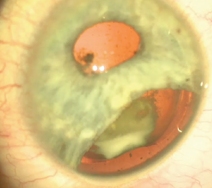
On examination, distance BCVA is 20/150 with a manifest refraction of +4.50 -1.25 x 040º OD and 20/25 with a manifest refraction of +3.00 -1.25 x 095º OS, decreasing to 20/50 OS with glare testing. The remainder of the examination is remarkable for an iridodialysis and zonular dialysis from 6 to 10 o’clock and posterior synechiae in the right eye and an LPI at 12 o’clock in the left eye (Figure 1). Each eye has 2+ nuclear sclerotic cataract. The right eye has 2+ to 3+ cortical cataract, and the left eye has 1+ cortical cataract.

Figure 1. Cataract, iridodialysis, posterior synechiae, and zonular dehiscence 59 years after a BB injury.
After discussion of the risks and benefits of cataract surgery and iridodialysis repair, the patient decides to proceed with cataract surgery on his right eye, but he is unsure whether to undergo surgery to repair the iris. What further discussion would you have with him? What would your surgical approach be?
—Case prepared by Cathleen M. McCabe, MD

GERD U. AUFFARTH, MD, PhD, FEBO
I would advise this patient to consider undergoing surgery on both eyes in an effort to achieve emmetropia. Cataract surgery on the right eye could be challenging. To repair the iris from 6 to 10 o’clock, I would place 9-0 polypropylene sutures (Prolene, Ethicon) via the iris root through the sclera and bury them in scleral tissue.
Adrenaline injection might or might not dilate the pupil. Cataract surgery might therefore involve some stabilization of the iris with hooks, an I-Ring Pupil Expander (BVI), or a Malyugin Ring (MicroSurgical Technology). I would inspect the anterior chamber for prolapsed vitreous. If cataract removal proved uneventful, I would insert a capsular tension ring (CTR). In addition, one could use a modified CTR with a black iris segment (aniridia implant, Morcher, not available in the United States) that could be placed at the site of the iridodialysis.
The choice of IOL is debatable. The safest option would be a monofocal IOL targeted for emmetropia (if both eyes are to undergo cataract surgery). In Europe, an increasingly popular choice for a case like this would be the IC-8 IOL (AcuFocus, not available in the United States), which has a small-aperture inlay in the optic. This implant would overcome problems related to the IOL power calculation, astigmatism, and surface irregularities from corneal scarring. In addition, the IC-8 offers some depth of focus, which would allow consideration of an extended depth of focus or low-add multifocal IOL for the left eye to achieve spectacle independence. Of greatest importance, however, is performing complication-free surgery on the right eye.

KENDALL E. DONALDSON, MD, MS
Traumatic cataracts carry a risk of unpredictability, necessitating a thorough discussion with this patient about the potential risks and complications of surgical intervention, including the potential need for vitrectomy or further surgery as well as alternative IOL positioning. Given his history of narrow angles and the need for anterior segment reconstruction, I would recommend preoperative specular microscopy. I always use a peribulbar or retrobulbar block in cases like this one to ensure the best patient experience, and I prepare for any potential needs that may arise during the case.
I would begin by repairing the iridodialysis. After making a peritomy, I would pass a double-armed 10-0 polypropylene suture on a CIF-4 needle (Ethicon) from a paracentesis or through the primary wound and then through the iris twice over to reapproximate the appropriate iris position. This maneuver can be executed under a Hoffman pocket or under a scleral flap. Once the iris was properly repositioned, I would turn my attention to the cataract.
Because of the synechiae, I would use iris hooks or a Malyugin Ring to enlarge the pupil sufficiently for safe phacoemulsification. If I encountered phacodonesis (common in such cases), I would consider capsular hooks to ensure stability during cataract removal. After removing the cataract, assuming no vitreous prolapse, I would place a CTR. It appears that the area of zonular weakness is limited to about 4 clock hours, so placing a nonsutured CTR would be appropriate before implantation of the IOL. In an eye with more diffuse zonular loss, I would consider using an Ahmed Capsular Tension Segment (Morcher, US distributor FCI Ophthalmics), which could be fixated with an 8-0 PTFE suture (Gore-Tex, W.L. Gore & Associates, off-label use).
I prefer three-piece monofocal IOLs in these cases because I find they help support the capsular bag and offer reliability as far as effective lens position. That said, I have listened to many impressive presentations of similar cases in which multifocal one-piece IOLs were used. Based on my experience, however, whenever zonular loss is present, the effective lens position may be altered, and unpredictable IOL positioning can yield substandard visual results.
In general, traumatic cataracts with associated iris defects are extremely rewarding cases because they give surgeons an opportunity to improve both the vision and the aesthetics of the eye.

WHAT I DID: CATHLEEN M. MCCABE, MD
After a discussion of the surgical options for cataract removal and iridodialysis repair, the patient was initially reluctant to undergo the latter procedure. I had outlined the additional steps and risks associated with iridodialysis repair and zonular weakness. He was very concerned about the additional risk but, after further conversation, agreed to proceed.
I created a conjunctival peritomy and performed three stab incision paracenteses at the limbus in the area of the iridodialysis. I used iris hooks to stabilize the iris and to expand and center the pupil. The capsulorhexis was created without complication, despite significant zonular instability in the area of the iris defect. Careful hydrodissection was performed, and the nucleus was removed using a vertical chopping technique and slow-motion phaco settings. I performed cortical cleanup with additional dispersive OVD to stabilize the capsular bag and then placed a CTR. In eyes with zonular instability, I prefer to implant the hydrophilic acrylic Akreos AO60 IOL (Bausch + Lomb) because its four open-loop haptics provide additional support and redistribute stress on the weak or absent zonules.
Watch It Now
Next, I began iridodialysis repair. A long cannula was used to facilitate placement of double-armed 10-0 polypropylene suture needles through a paracentesis and then through the peripheral iris in the area of the dialysis. This technique prevents accidental passage of the needle through the corneal fibers when entering the eye through the paracentesis. Intraocular forceps were used to facilitate passage of the needle through the iris tissue and then through the sclera posterior to the limbus. The two arms of the suture were passed several millimeters apart so that a loop of suture remained on the surface of the sclera. The knot was rotated into the sclera. When tying the suture, it is important to maintain a gap between the iris tissue and the sclera so that the angle structures are not covered. Two sets of double-armed 10-0 polypropylene sutures were placed and the knots buried.

Figure 2. Cosmetic appearance after iridodialysis repair and cataract surgery (left), with 10-0 polypropylene sutures visible under the conjunctiva (middle). Retroillumination outlines the gap between the iris and sclera in the area of the dialysis repair (right).
The patient was delighted with the eye’s postoperative cosmetic appearance (Figure 2), his 20/20 BCVA, and the resolution of his glare symptoms.


