
Like most steps of the phaco procedure, successful completion of hydrodissection and hydrodelineation is crucial to facilitate subsequent surgical events. If these steps do not achieve their desired outcomes, the rest of the surgery may be made longer and more difficult, with a higher risk for complications.
Other steps in the cataract procedure may be more technically challenging, but I often see beginning cataract surgeons struggle to achieve a good hydrodissection. Admittedly, even after more than 10,000 cataract operations, I am still occasionally somewhat dissatisfied with my hydro performance.
I am indebted to Khiun F. Tjia, MD, for a pedagogic demonstration a few years ago during an ESCRS instructional course. He showed the course attendees a most efficient, effort-saving, simple, and atraumatic way to perform hydrodissection, a technique that has helped me to improve my surgical efficacy and precision.
Before going further through the hydro steps, here is a recommendation regarding instrumentation: For the injection of balanced saline solution, use a 2- to 3-mL syringe armed with a blunt hydrodissection cannula with angulation close to the tip, 1 to 2 mm maximum, as seen in the surgical video images referenced later in this article. Cannulas with this design are available from several manufacturers. Straight cannulas or cannulas that are bent 4 to 8 mm from the tip do not support effective hydrodissection, mobilization, and hydrodelineation. Use those cannulas for something else: for example, for injecting intracameral antibiotics at the end of operation … or not at all (Figure 1).
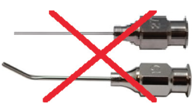
Figure 1. Straight cannulas or cannulas that are bent 4 to 8 mm from the tip do not support effective hydrodissection, mobilization, and hydrodelineation. Do not use them for these steps. Use one with angulation close to the tip, 1 to 2 mm maximum, as seen in Figures 2 and 3.
HYDRODISSECTION
The aim of the hydrodissection step is to break adhesion between the inside of the lens capsule and the outermost cortical layer of the lens. The technique I describe here, including figures, is partially based on my previous article, “Mobilizing a Nucleus From the Capsule.”2
For hydrodissection, the cannula is positioned with the bent tip horizontal, just beneath the anterior edge of the capsulorhexis (Figure 2). The point of the tip is first lifted to tent the anterior capsule slightly upward, and at the same time, it is gently advanced under the capsule (and, depending on pupil size, under the iris as well) toward the lens equator. Balanced saline solution is then injected in small portions to create a visible fluid wave that traverses the pupil across the posterior surface of the lens. Alternatively, with dense cataract, this wave is seen as a visible elevation of the lens.
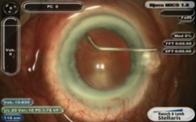
Figure 2. For hydrodissection, the cannula is positioned with the bent tip horizontal, just beneath the anterior edge of the capsulorhexis.
Keeping the syringe at the same angle, the hydrodissection cannula tip is then rotated clockwise, up to approximately 90º, so that the tip is pointing downward, and the tip is used to gently press the lens pole downward (Figure 3). Note that the difference between Figures 2 and 3 is quite small. This technique ensures that the lens is not moved excessively posteriorly, and the stress on the zonules is thus limited.
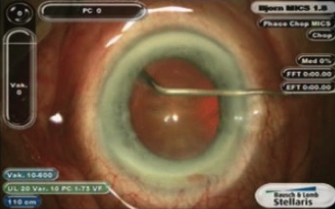
Figure 3. The tip is then rotated clockwise, up to approximately 90º, so that the tip is pointing downward, and is used to gently press the lens pole downward.
This movement loosens adhesions between the cortical fibers and the inside of the capsule. If needed, the balanced saline solution injection can be repeated on the opposite side from the first injection, followed by the same rotating of the tip downward to depress the nucleus slightly. Added injections can be made similarly in other meridians as necessary. Usually, though, one or two such maneuvers will suffice to break corticocapsular adhesions enough to continue with the next step.
HYDRODELINEATION
The primary aim of this step is to separate the inner, harder nucleus from the softer epinuclear bowl and thereby be able to preserve the epinucleus as protection for the capsule during phacoemulsification. A secondary aim is to gather useful information about the size and hardness of the nucleus.
The technique starts with the syringe and the downward-pointing tip in exactly the same position as at the end of previous step. The cannula tip, which is now farther into the lens, is sunk deeper until it reaches the harder nucleus proper. At this point you will feel that it is not possible to advance the tip deeper without pushing the whole lens backward, which is not what you want to do! Stop there.
You now have some information on the lens size and hardness. In young patients with soft nuclei (not always easy cases), you may be able to advance the tip to the middle of the soft lens. Voilá, you now know that you are dealing with a lens that can be aspirated without ultrasound, maybe even with the I/A handpiece, but other measures are needed for nuclei of greater hardness and different sizes.
Remember the angle and position of the tip: pointing virtually vertically downward, resting on the surface of the harder nucleus. Now, inject balanced saline solution. Be advised that resistance may now be much greater in comparison with the hydrodissection step, and some force may be needed to start the solution flow. Therefore, be very careful and be prepared to decrease pressure on the syringe plunger as soon as the resistance gives way. When this happens, you should be able to see a curved line appear in the sector where the cannula tip is placed—or in the best case, a golden ring—provided the pupil is large enough.
MOBILIZATION OF THE LENS
The primary aim of this step is to ensure that the lens can be rotated inside the capsule without risking zonular integrity. Secondary aims are to get further information on the hardness and size of nucleus and to separate the central harder nucleus from the softer epinuclear bowl.
After the previous step, your cannula tip should now be resting on the anterior surface of the harder nucleus, slightly peripheral to the capsulorhexis edge. Now use the cannula tip as a crank and make a rotating movement in the direction of your choice. The movement should be parallel to the rhexis margin and/or the normal dilated pupil.
If there are remaining cortical adhesions to the inside of the capsule making rotation difficult, go back to the previous steps to perform more hydrodissection or hydrodelineation. If only a small amount of rotation is achieved, you may want to try rotating in the opposite direction. This sometimes loosens the remaining adhesions. Avoid using too much force, which can damage zonules.
In eyes in which the zonules seem to be weak or even absent, the hydro steps may still be possible once the capsulorhexis margin has been stabilized with iris hooks.
When the nucleus can be rotated 45º to 90º, inject saline again to extend the curved line. With repeated injections, it will form a circle. This circle, which may be dark or a shining golden ring, shows the outline of the harder central nucleus, which will soon be attacked in subsequent steps with the phaco handpiece.
For further insights into the hydro steps, I recommend the instructive chapter by Fine, Hoffman, and Packer in the book Minimizing Incisions and Maximizing Outcomes in Cataract Surgery.1
1. Fine IH, Hoffman RS, Packer M. Hydrodissection and hydrodelineation. In: Alió J, Fine IH, eds. Minimizing Incisions and Maximizing Outcomes in Cataract Surgery. Berlin: Springer; 2010.
2. Johansson B. Mobilizing a nucleus from the capsule. Cataract & Refractive Surgery Today Europe. November/December 2013.


