Small-aperture implants have recently drawn increased attention. However, the idea of using a small aperture in an IOL is not new. The first attempt to incorporate a pinhole aperture into an intraocular implant dates back to 1964, when renowned British ophthalmologist D. Peter Choyce, FRCS, FRCOphth, introduced the Mark V implant (Rayner), an anterior chamber PMMA IOL with an embedded pinhole mask.
Since that time, the concept of using a pinhole aperture to provide increased depth of field has been employed in intrastromal corneal implants and IOLs. In 2014, a proof-of-concept study of the XtraFocus Pinhole Implant (Morcher) was published.1 A fourth version of the XtraFocus Pinhole Implant received the CE Mark in 2016. Since then, the device has been commercially available in Europe, Australia, New Zealand, and some countries in Latin America and the Middle East. It is not yet available in the United States.
DEVICE DETAILS
The device is made of a foldable black hydrophobic acrylic material. The optic has an occlusive portion with a diameter of 6 mm that contains a 1.3-mm clear central opening with no refractive power. The implant was designed to be placed in the ciliary sulcus of a pseudophakic eye in a piggyback fashion (Figure 1).
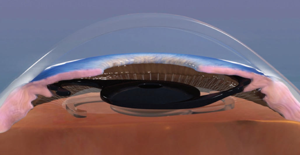
Figure 1. The implant was designed to be placed in the ciliary sulcus of a pseudophakic eye in a piggyback fashion.
The primary indication for the XtraFocus is for the treatment of irregular corneal astigmatism, for example in eyes after radial keratotomy, penetrating keratoplasty, and keratoconus (Figure 2), or in eyes with post-LASIK ectasia. Depth of focus extension, to provide near vision enhancement, is also a good indication, with the added benefit of reversibility.
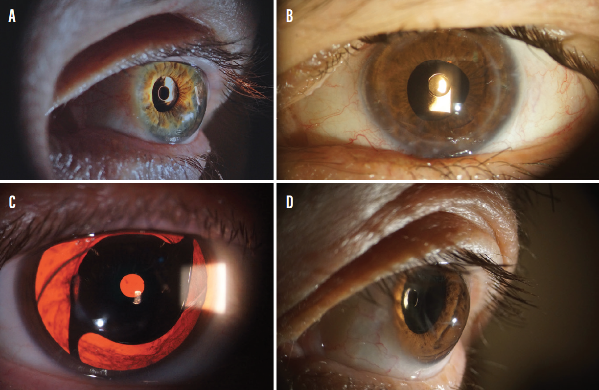
Figure 2. The XtraFocus in eyes after radial keratotomy (A), penetrating keratoplasty (B), and keratoconus (C, D).
The implant can be used with any other IOL, including toric versions. This is especially important in postpenetrating keratoplasty eyes and in patients with keratoconus. Although the pinhole effect can neutralize some regular astigmatism, in patients with a high magnitude of astigmatism the combination of the XtraFocus with a high-cylinder toric IOL can have a strong synergistic effect. Confirmation of topographic stability is mandatory in this scenario.
PATIENT SELECTION
Ideal patients are those with significant corneal aberrations with a transparent central cornea and a large pupillary diameter. A good example would be a cataract patient with keratoconus, mean keratometry of 58.00 D, central corneal thickness of approximately 380 µm, and a 4-mm pupil in mesopic conditions. Visual acuity in such a patient can achieve unexpectedly high levels, such as 20/30 UCVA.
To demonstrate postoperative acuity to the patient preoperatively, placing pinhole occlusion on top of the best refraction can provide a fair simulation. However, a favorable pinhole acuity test does not guarantee success. Corneal opacities (corneal dystrophies, scars, severe haze) represent a relative contraindication for this treatment.
Another important point to determine is the ideal refractive target. For an eye with irregular corneal astigmatism, a refractive target of -2.00 D is advisable. This provides solid uncorrected near visual acuity, while the extended depth of focus from the pinhole provides satisfactory uncorrected distance vision that can be further improved with glasses.
OTHER USES, PLUSES, AND MINUSES
The XtraFocus can also be used for the treatment of iris defects (Figure 3). Reduction of glare, ghost images, and light sensitivity can be expected with this approach.
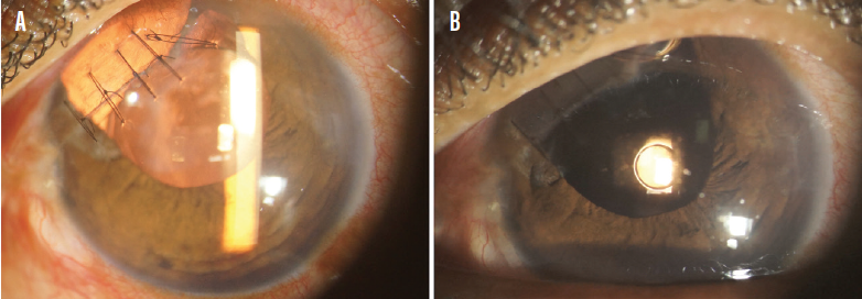
Figure 3. A patient who experienced penetrating ocular injury with iris loss before (A) and after (B) the XtraFocus was implanted.
Another interesting application for this implant is for the treatment of dysphotopsia after multifocal IOL implantation (Figure 4). The piggyback design provides a versatile solution when an IOL exchange might present significant risks, such as when a previous posterior capsulotomy is present.
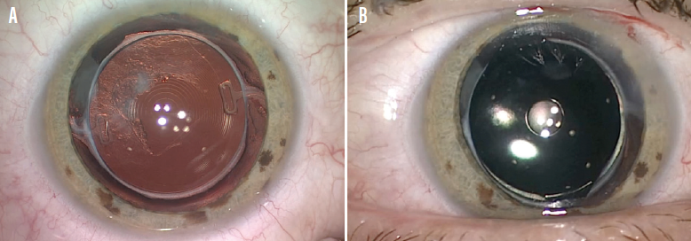
Figure 4. In an eye with dysphotopsia after multifocal IOL implantation (A), the XtraFocus implant can mitigate symptoms and preserve an extended range of functional vision (B).
The downside of pinhole implants is their inherent reduction of light entry. In some patients, this may translate into a sensation of dark vision under low-light conditions. This perception is usually well tolerated, and it is often outweighed by the remarkable improvement in visual acuity that good candidates experience. However, this is an important point to be discussed with patients preoperatively.
The black acrylic material has the unique feature of being transparent to infrared light, therefore allowing examination of structures located behind the implant with the use of infrared-based equipment (Figure 5). The occlusive body has a very slim profile (180 µm around the pinhole), with a concave-convex shape to avoid contact with the primary IOL. Because of the reduced thickness, the device can be implanted through a 2-mm corneal incision. It can also be safely implanted in the bag together with the primary IOL.
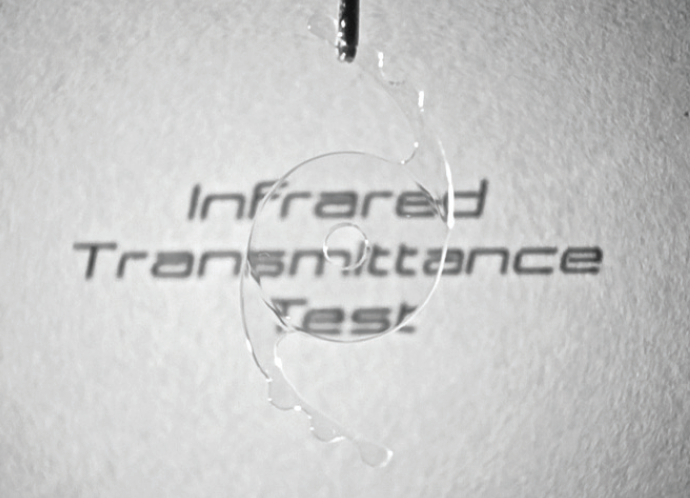
Figure 5. The black acrylic material is transparent to infrared light, allowing examination of structures located behind the implant using infrared-based equipment.
CONCLUSION
Although follow-up data are limited, the XtraFocus seems to be a promising new type of intraocular implant. Future studies will provide additional information.
1. Trindade CLC, Trindade BLC. Novel pinhole intraocular implant for the treatment of irregular corneal astigmatism and severe light sensitivity after penetrating keratoplasty. J Cataract Refract Surg Online Case Rep. 2015;3(1):4-7.
Suggested Reading:
• Trindade BLC, Trindade FC, Trindade CLC. Intraocular pinhole implantation for irregular astigmatism after planned and unplanned posterior capsule opening during cataract surgery. J Cataract Refract Surg. 2019;45(3):372-377.
• Trindade BLC, Trindade FC, Trindade CLC, Santhiago MR. Phacoemulsification with intraocular pinhole implantation associated with Descemet membrane endothelial keratoplasty to treat failed full-thickness graft with dense cataract. J Cataract Refract Surg. 2018;44(10):1280-1283.
• Trindade CC, Trindade BC, Trindade FC, Werner L, Osher R, Santhiago MR. New pinhole sulcus implant for the correction of irregular corneal astigmatism. J Cataract Refract Surg. 2017;43(10):1297-1306.
• Tsaousis KT, Werner L, Trindade CLC, Guan J, Li J, Reiter N. Assessment of a novel pinhole supplementary implant for sulcus fixation in pseudophakic cadaver eyes. Eye (Lond). 2018;32(3):637-645.