INTERESTING AND ARTISTIC
SERGEI LUZHETSKIY, MD
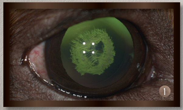
A Dog’s Cataract
1. This is an image of an inherited cataract in a dog.
DANIEL DE SOUZA
COSTA, MD
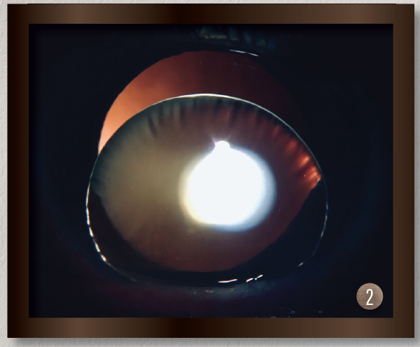
A Spontaneous Displacement of the Lens
2. This photo is of the eye of a 46-year-old woman who presented with displacement of the lens into the anterior chamber with no history of ocular trauma.
GRAND PRIZE WINNER
PATRIK RAJS
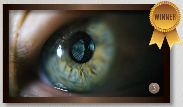
Gramophone Record in the Eye
3. This photo is of the eye of a patient with presbyopia who underwent laser cataract surgery. A multifocal IOL was captured with a combination of two vintage Zeiss and Pentax lenses.
RARE AND UNUSUAL
YONG
KAM, MD
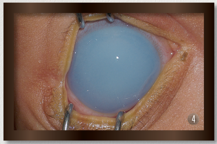
Anterior
Segment
Dysgenesis
4. This photo shows the eye of a newborn patient with increased IOP; a diffusely edematous, hazy, and enlarged cornea; and an absent Schlemm canal and trabecular meshwork on surgical exploration, consistent with severe anterior segment dysgenesis.
GRAND PRIZE WINNER
DANIEL
DE SOUZA
COSTA, MD
A Prominent
Symblepharon
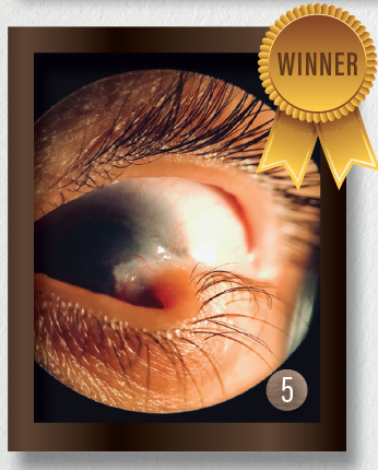
5. The eye of this 5-year-old girl has epidermolysis bullosa and serious ocular manifestations of this condition. Biomicroscopy examination revealed the presence of symblepharon in both eyes. The ocular complication was so severe that it deformed the palpebral anatomy.
AARON S. WANG, MD
Fish Tank
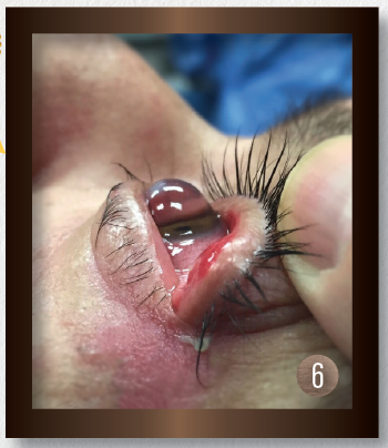
6. Significant keratoglobus in the eye of this patient with Down Syndrome, who frequently rubs his eyes. This picture was taken just prior to corneal transplantation.
SLIT LAMP
KYLE WENZEL AND ROBERT J. WEINSTOCK, MD
Silicone Oil in the Anterior Chamber
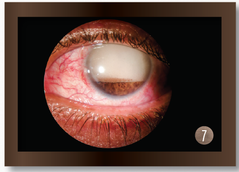
7. This photo depicts silicone oil leaking into anterior chamber.
MALLIKARJUN M.
HERALGI, MD
Morning Glory IOL
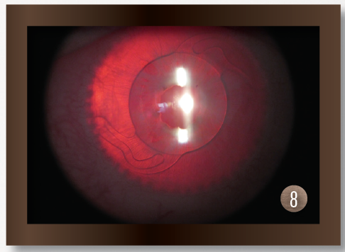
8. The patient in this photo has albinism and has undergone phacoemulsification with IOL implantation in the bag. The entire lens can be observed with retroillumination because of very little iris pigmentation.
GRAND PRIZE WINNER
CHAN-JAN BOND, MBBS,
MMed(Ophth)
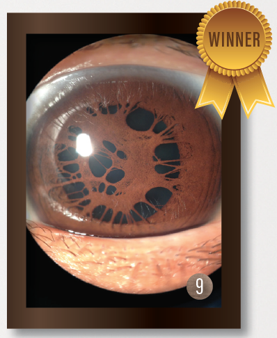
Spider Web-Like Persistent
Pupillary Membrane
9. This eye has a large, thick, persistent pupillary membrane.
SURGICAL COMPLICATIONS
GRAND PRIZE WINNER
MATTHEW FENG, MD
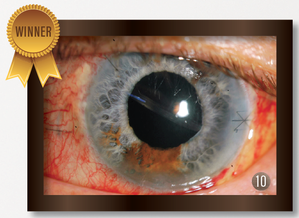
Living on the Edge
10. A scleral-fixated glued IOL is found to be tilted 90° on postoperative day 1.
BRUNO MIOLO, MD
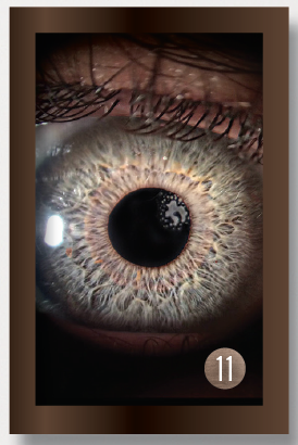
Epithelial Growth After SMILE Surgery
11. A 27-year-old man who underwent small-incision lenticule extraction (SMILE) developed an epithelial ingrowth 7 days postoperative.
MARIANA
CASTAÑEDA, MD
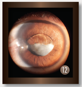
IOL Pocket
12. This image shows a subluxated IOL with posterior capsular opacification.
TRAUMA
HARSH V. SINGH, MD
Blunt Ocular Trauma
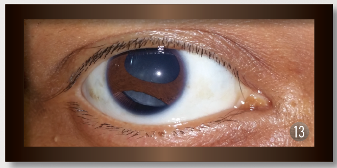
13. A 27-year-old woman with a history of blunt trauma experienced decreased visual acuity. The photograph of her right eye shows iridodialysis from the 5 to 8 clock positions with a superonasally subluxated lens.
SÉBASTIEN GAGNÉ, MD, FRCSC
4-Year Postop Capsular Tension
Segment
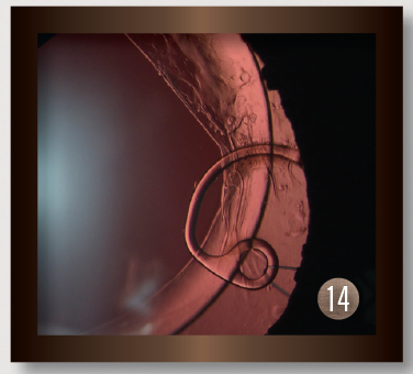
14. This image shows a capsular tension segment sutured to the sclera 4 years after surgery to remove a traumatic cataract.
GRAND PRIZE WINNER
GABRIEL CAMARGO
CORRÉA, MD
Gunpowder
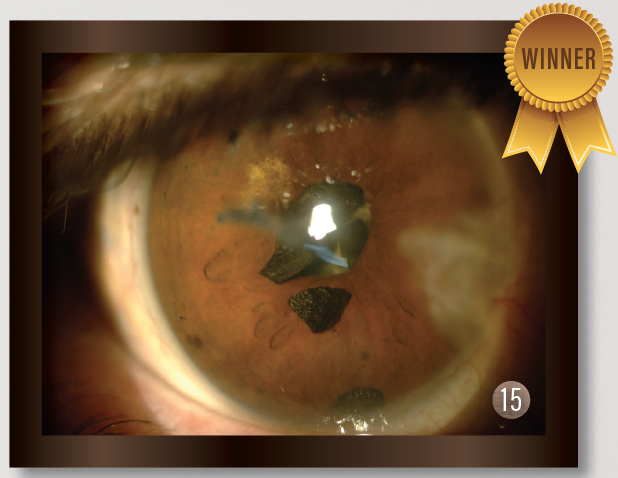
15. This patient was referred to the office after a bomb explosion. The eye was stable despite a gunpowder foreign body in the anterior chamber.

