
Influences of SMILE and FS-LASIK on Corneal Sub-basal Nerves: A Systematic Review and Network Meta-analysis
Jiang X, Wang Y, Yuan H, et al1
Industry support: None
ABSTRACT SUMMARY
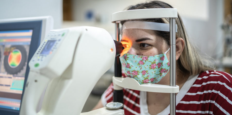
A systematic review and network meta-analysis compared postoperative corneal nerve injury and repair after SMILE versus femtosecond LASIK (FS-LASIK). Twelve prospective comparative studies (n = 755) met the criteria, including corneal nerve injury and recovery measurements such as corneal nerve density and the number of corneal nerve trunks and branches as determined by in vivo confocal microscopy.
Study in Brief
Corneal nerve injury and repair may be assessed by measuring corneal nerve density and the number of corneal nerve trunks and branches with in vivo confocal microscopy. A meta-analysis showed greater corneal nerve injury with femtosecond LASIK compared to SMILE. Six months after surgery, however, there was no significant difference in corneal nerve density and number between the two groups.
WHY IT MATTERS
Few studies have directly compared the outcomes of femtosecond LASIK and SMILE, particularly corneal nerve injury and repair. The meta-analysis showed that confocal microscopy can be used to assess corneal nerve morphology and provide insight into corneal changes after refractive surgery.
Corneal nerve density decreased postoperatively in both study groups. It was lower at 1 month in the FS-LASIK group, but the difference was negligible between the groups after 3 months. A separate analysis of studies directly comparing the two groups detected differences at 1 and 3 months but not at 6 months postoperatively.
DISCUSSION
The technology for refractive surgery has improved significantly during the past 25 years, but the procedures still often injure corneal nerves, contributing to decreased corneal function and dry eye symptoms. In FS-LASIK, the creation of a flap in the anterior corneal stroma damages the subbasal nerve, and photoablation of the stroma damages the stromal nerve.2 In SMILE, the lateral incision to remove the lenticule is 30º to 40º instead of the nearly 300º corneal incision created during FS-LASIK. The difference explains why the meta-analysis found that SMILE resulted in better corneal nerve density during the first few postoperative months. By 6 months, however, the difference between the two groups resolved.
The number of corneal nerve trunks and branches is an indicator of postoperative corneal nerve regeneration and reinnervation, respectively. SMILE appeared to be associated with less nerve damage and faster nerve regeneration than FS-LASIK in the observed time frame. Postoperative corneal nerve reinnervation, as indicated by corneal nerve branch number, did not differ significantly between groups. The primary repair method in FS-LASIK, known as sprouting,3 may account for the difference in corneal nerve branch number, but FS-LASIK is known to cause greater nerve damage in the early postoperative period.
Correlation of Hair Cortisol and Interleukin 6 With Structural Change in the Active Progression of Keratoconus
Stival LR, Avila LP, Araujo DC, et al4
Industry support: None
ABSTRACT SUMMARY
A prospective study of 133 eyes of 74 patients compared the concentrations of interleukin (IL) and hair cortisol in eyes with progressive keratoconus (n = 53) versus in those with stable keratoconus (n = 28) and healthy controls (n = 52). Patients with active allergic conditions were excluded to prevent confounding with inflammatory cytokine levels.
Study in Brief
A prospective study demonstrated that interleukin-6 and hair cortisol concentrations were higher in eyes with progressive keratoconus than in eyes with stable keratoconus. Further, interleukin-6 and hair cortisol concentrations showed a significant correlation with corneal structural damage.
WHY IT MATTERS
Monitoring patients at the molecular level may improve the evaluation of keratoconus progression by allowing earlier detection and intervention.
Overall, eyes with stable and progressive keratoconus had higher mean inflammatory mediator concentrations (IL-6, IL-8, tumor necrosis factor [TNF] alpha) than did healthy eyes. IL-6 levels were significantly higher in eyes with progressive versus stable keratoconus. When stratified by keratoconus grade, IL-6 levels were significantly elevated in grade 1 versus grade 4 keratoconus, but significant differences were not detected between other stages (ie, 1 vs 2, 2 vs 3). Hair cortisol concentration was higher in eyes with progressive keratoconus than in eyes with stable keratoconus and healthy controls. When stratified by keratoconus grade, hair cortisol concentration was significantly higher in eyes with grade 1 versus 4 disease. Significant differences were not detected between other keratoconus stages.
DISCUSSION
Keratoconus is a progressive corneal ectasia that results in the steepening and thinning of the cornea. Prior studies have hypothesized that inflammatory mediators such as IL, TNF, and transforming growth factor influence disease progression.5,6 IL-6, for example, has been shown to induce the expression of corneal collagenases (matrix metalloproteinase [MMP]-9), which could contribute to tissue degradation and disease progression.
The study by Stival et al improved upon previous methodology by separating eyes with progressive keratoconus (> 1.00 D increase in maximum keratometry per year), which allowed the investigators to assess the relationship between inflammatory mediators and disease progression.4 Based on this methodology, IL-6 was the only inflammatory cytokine found to be significantly elevated in eyes with progressive keratoconus.
Inflammatory mediator levels were further correlated with disease severity and structural changes in the cornea. Specifically, IL-6 was significantly more elevated in eyes with grade 4 versus grade 1 keratoconus and had a positive correlation with maximum keratometry readings. IL-6 is known to regulate MMP-9, which degrades collagen fiber types 1 and 4 in the corneal epithelium. When this is taken into consideration along with the study’s results, it suggests that monitoring IL-6 levels could assist with the detection of keratoconus progression. Cyclosporine A, an antiinflammatory medication, has been documented to inhibit MMP-9 and inflammatory cytokines (TNF-alpha, IL-6). The drug’s use in keratoconus management could therefore be beneficial.7 Further study is warranted.
1. Jiang X, Wang Y, Yuan H, et al. Influences of SMILE and FS-LASIK on corneal sub-basal nerves: a systematic review and network meta-analysis. J Refract Surg. 2022;38(4):277-284.
2. Yesilirmak Davis Z, Yoo SH. Refractive surgery (SMILE vs. LASIK vs. phakic IOL). Int Ophthalmol Clin. 2016; 56(3):137-147.
3. Bech F, González-González O, Artime E, et al. Functional and morphologic alterations in mechanical, polymodal, and cold sensory nerve fibers of the cornea following photorefractive keratectomy. Invest Ophthalmol Vis Sci. 2018;59(6):2281-2292.
4. Stival LR, Avila LP, Araujo DC, et al. Correlation of hair cortisol and interleukin 6 with structural change in the active progression of keratoconus. J Cataract Refract Surg. 2022;48(5):591-598.
5. Lema I, Durán JA. Inflammatory molecules in the tears of patients with keratoconus. Ophthalmology. 2005;112(4):654-659.
6. Jun AS, Cope L, Speck C, et al. Subnormal cytokine profile in the tear fluid of keratoconus patients. PLoS One. 2011;6(1):e16437.
7. Shetty R, Ghosh A, Lim RR, et al. Elevated expression of matrix metalloproteinase-9 and inflammatory cytokines in keratoconus patients is inhibited by cyclosporine A. Invest Ophthalmol Vis Sci. 2015;56(2):738-750.


