
Interesting and Artistic Images
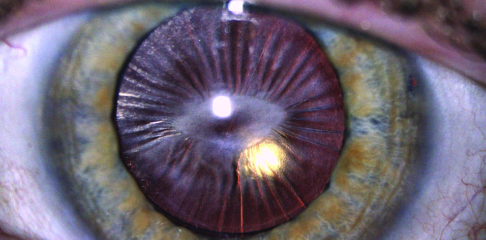
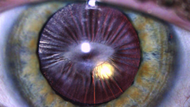
The Apple of My Eye
By Taj Nasser, MD, and Anthony Vanrachack, OD
tajnassermd@gmail.com
The slit-lamp photograph shows posterior synechiae from chronic uveitis.
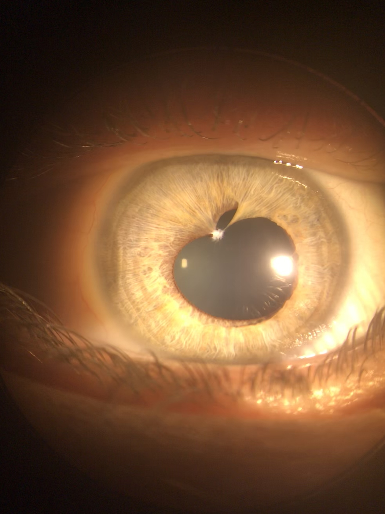
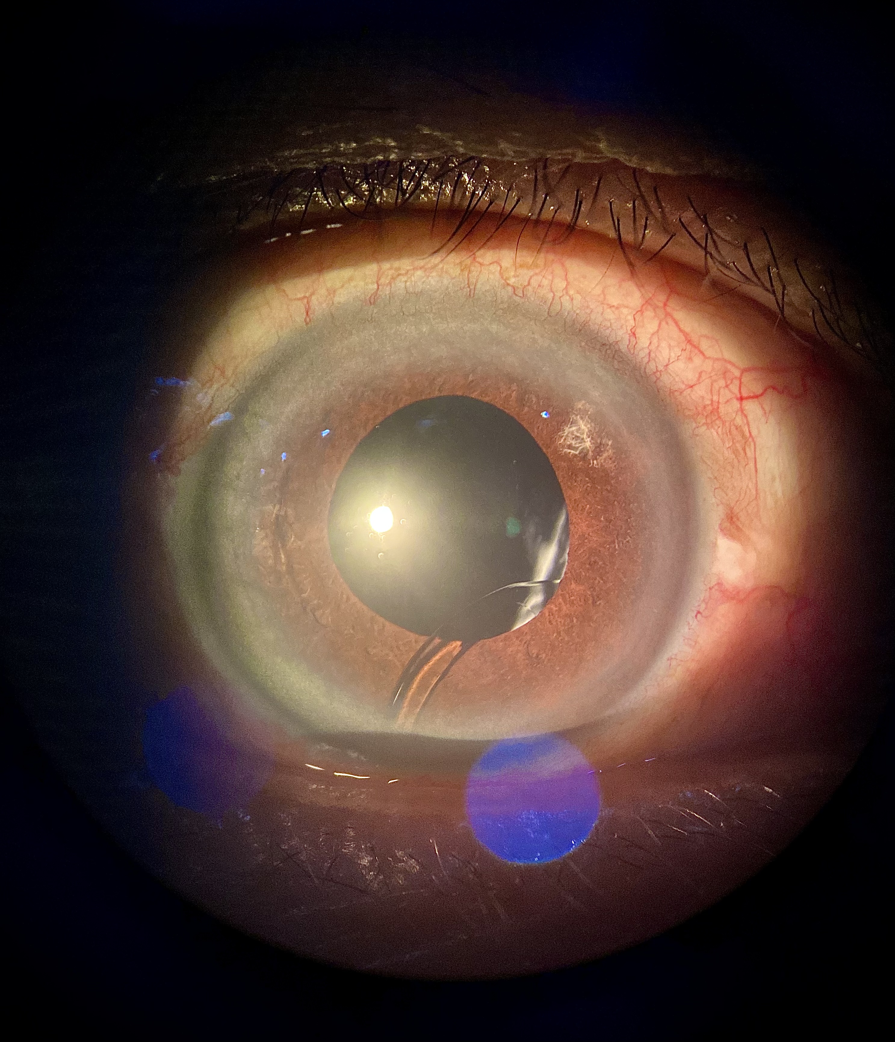
Pupillary Bridge
By Rodolfo Bonatti, MD
rodolfobonatti@gmail.com
A persistent pupillary membrane connects the embryological to the present.
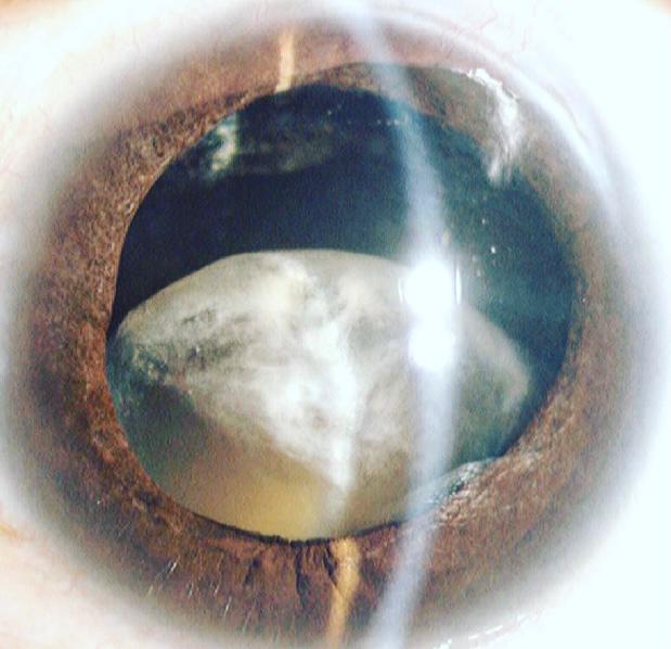
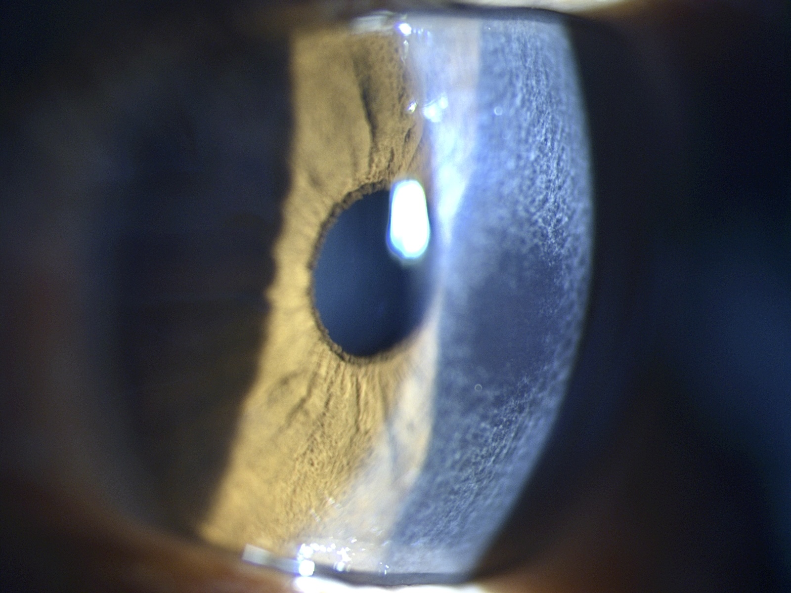
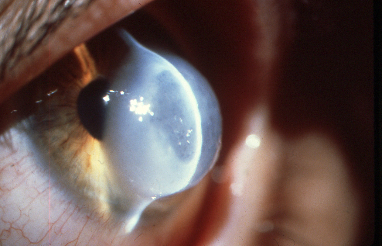
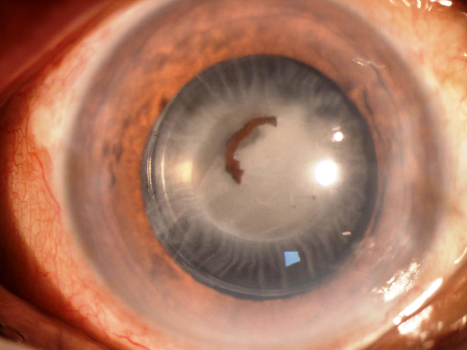
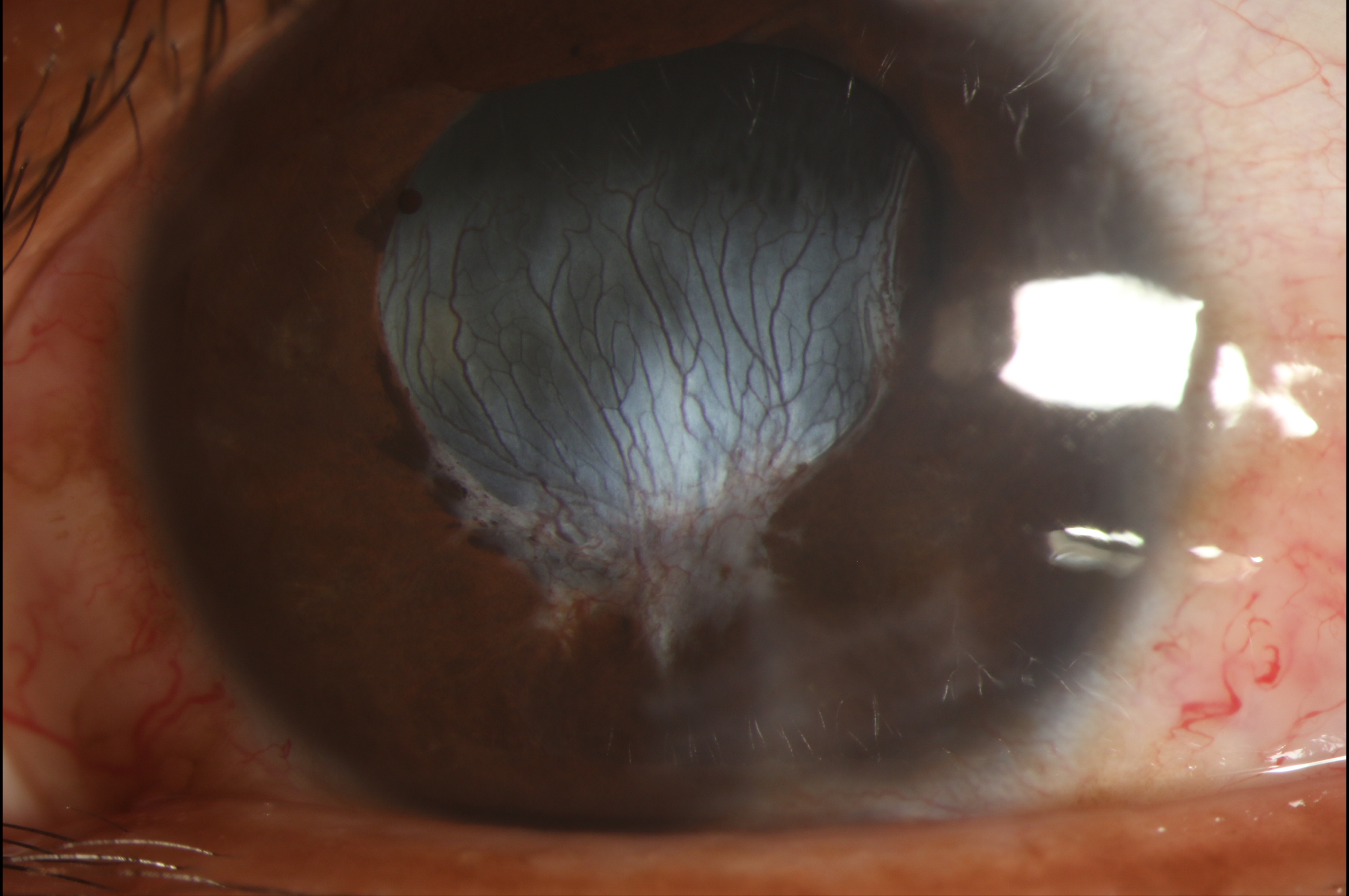
Moonrise
By Edgar Rafael Carazo Vargas, MD
carazoed@hotmail.com
The image shows a subluxated cataract in the right eye of a patient with pseudoexfoliative glaucoma.
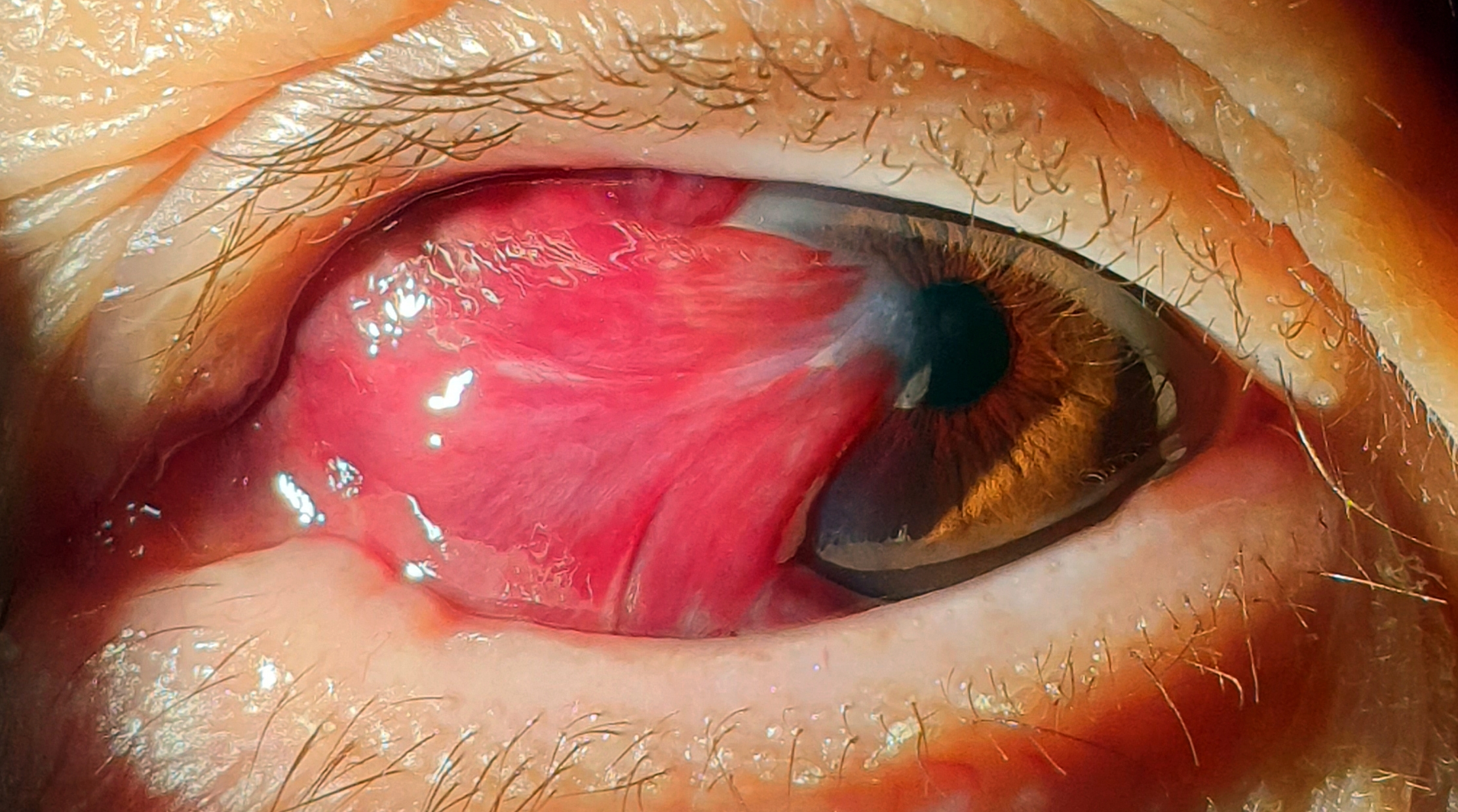
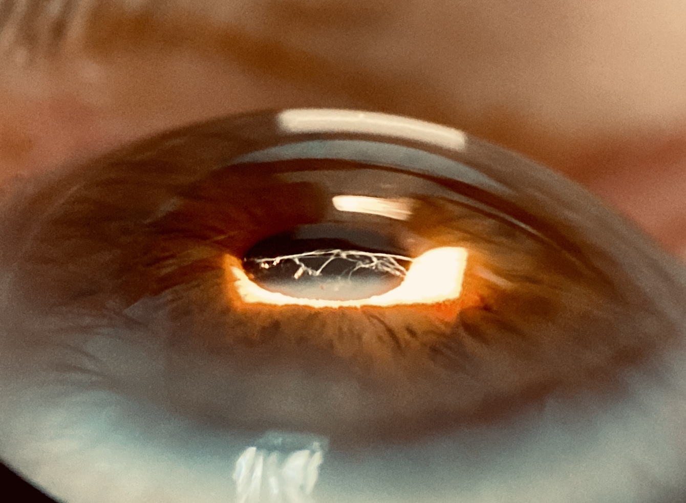
Pterygosphagma
By Luis Alcalde Blanco, MD, PhD
luisalcaldeblanco93@gmail.com
The eye has a hyposphagma underlying a preexisting pterygium.
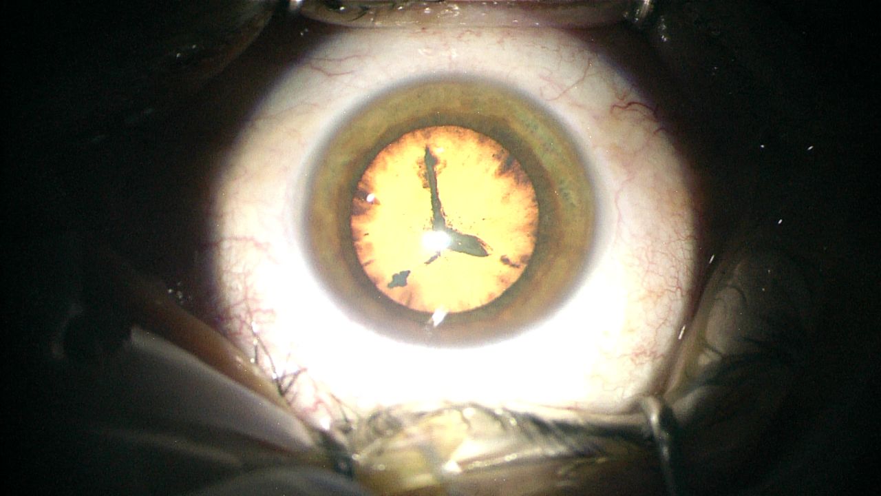
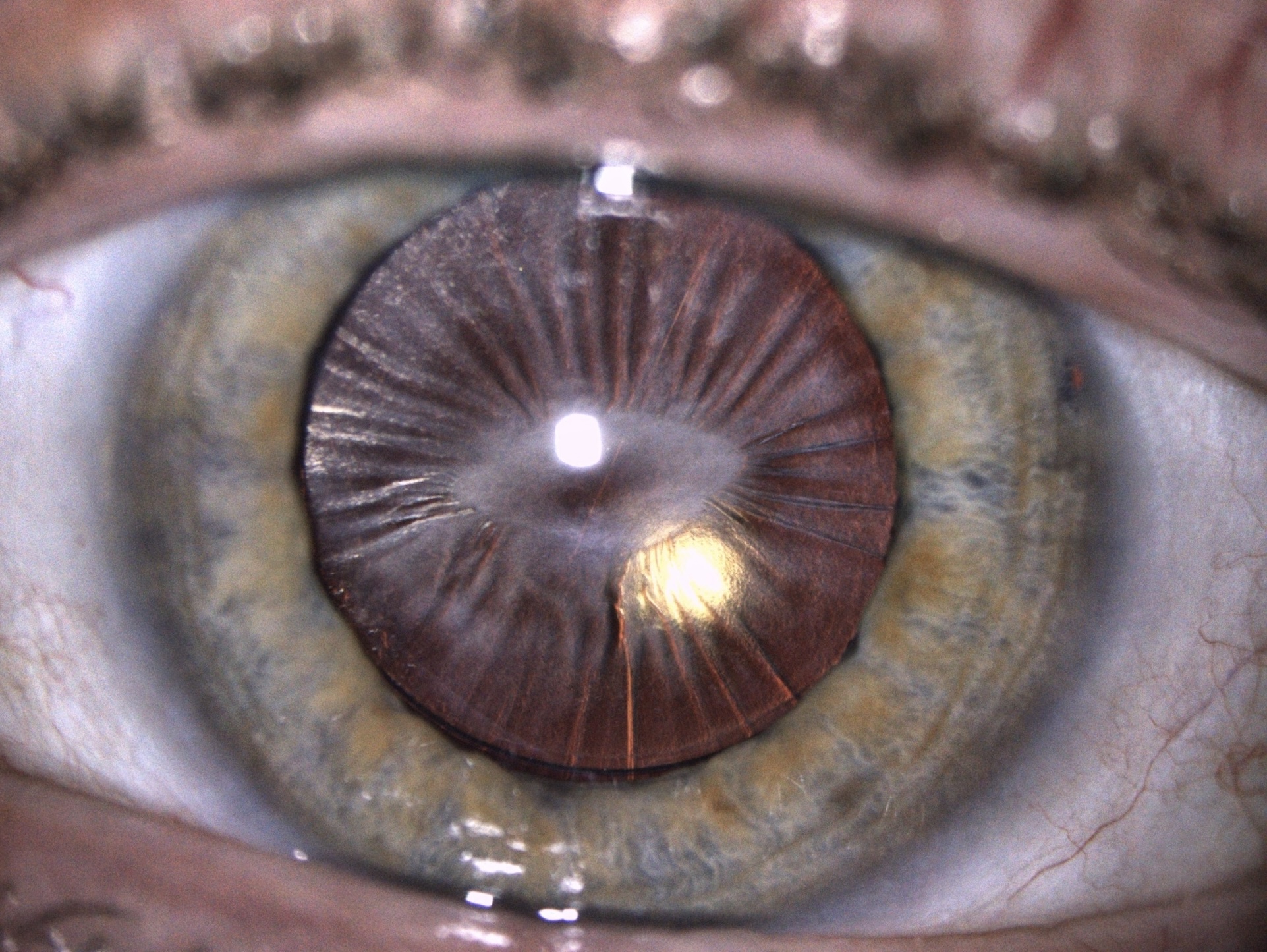
Time for Cataract (Eyeclock)
By Konstantinos Moschou, MD
moschouk@otenet.gr
The cataract in this eye resembles a clock.
Rare and Unusual Diseases
Anterior Spiderweb
By Zaira Lizette Jiménez Díaz, MD
zairajim@gmail.com
The image shows late spontaneous dislocation of the IOL–capsular bag complex in the eye of a 44-year-old patient with Usher syndrome who underwent cataract surgery 20 years ago.
Thiel-Behnke Corneal Dystrophy
By Pablo Larco Jr, MD, and Luis Izquierdo Jr, MD, PhD
drpablolarco@gmail.com
The eye has an unusual subepithelial dystrophy known as honeycomb dystrophy. It is transmitted as an autosomal dominant trait and is classified as a variant of Reis-Bücklers corneal dystrophy.
Conjunctivalization on LASIK Flap
By Suphi Taneri, MD, FEBOS-CR
taneri@refraktives-zentrum.de
Conjunctiva grows over the cornea in an eye that underwent myopic LASIK 13 years ago.
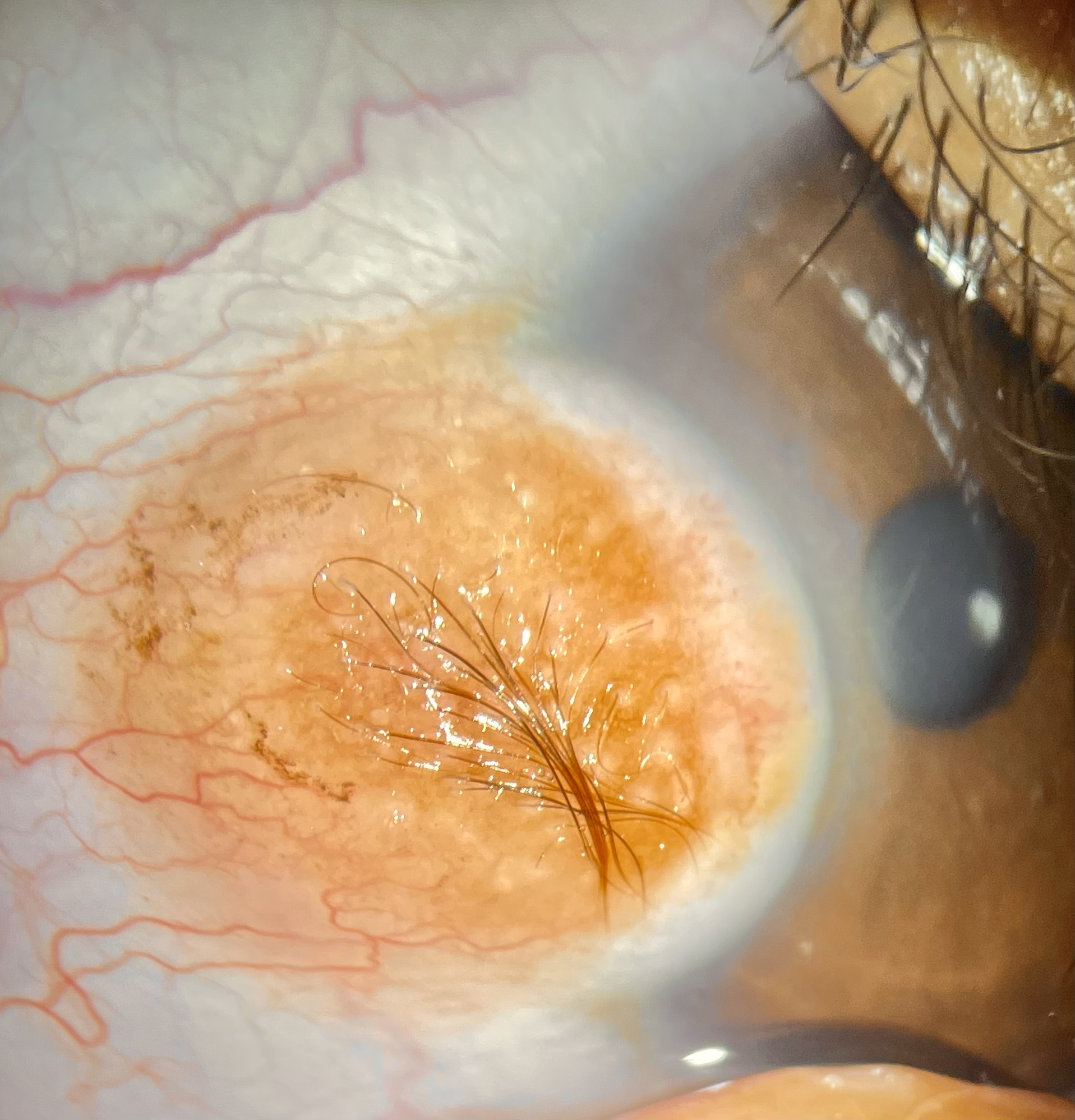
Limbal Dermoid
By Amit Mishra, MS
amit.mishra@hotmail.com
This photograph shows an eye that has a limbal dermoid with hairs.
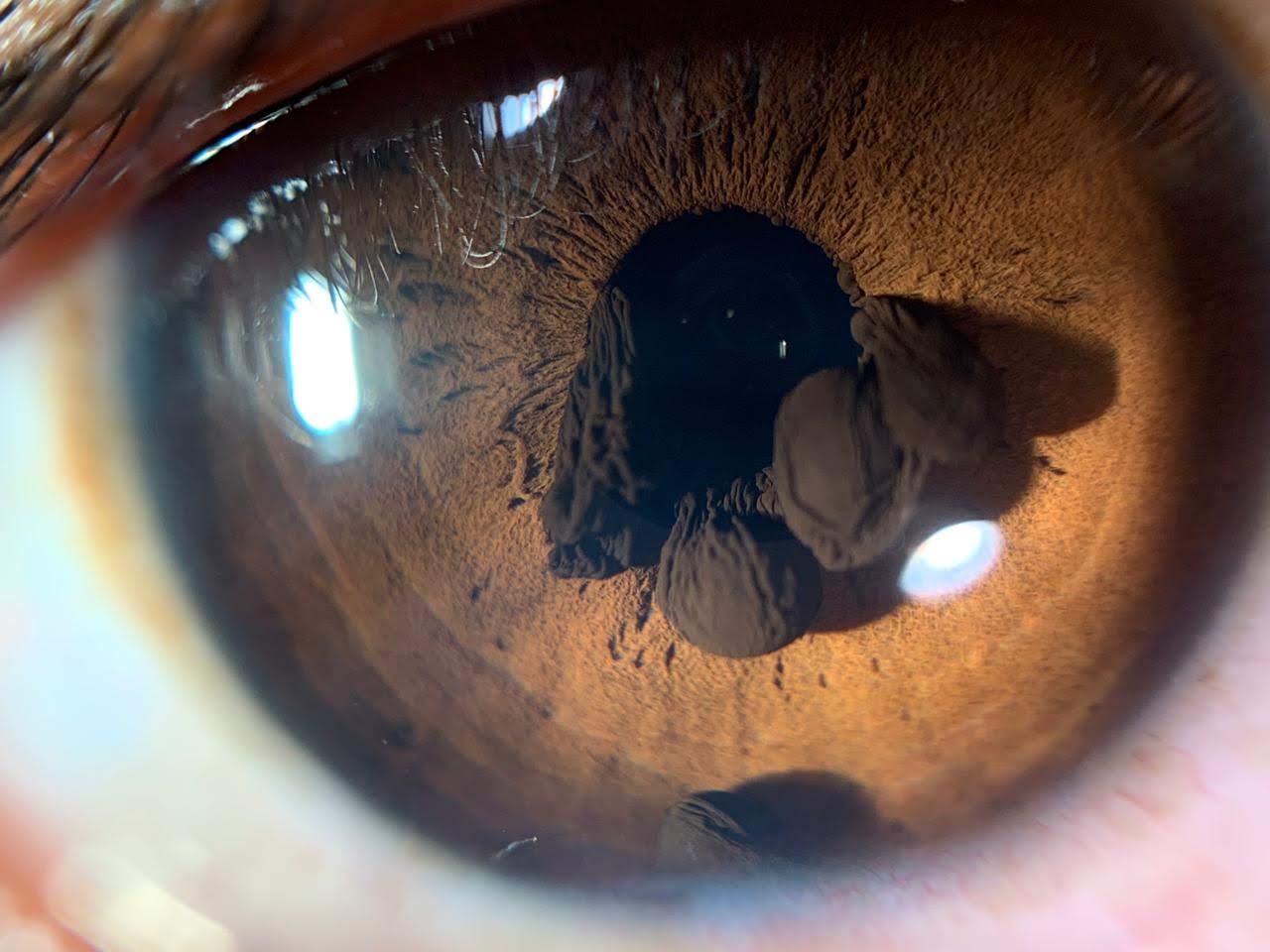
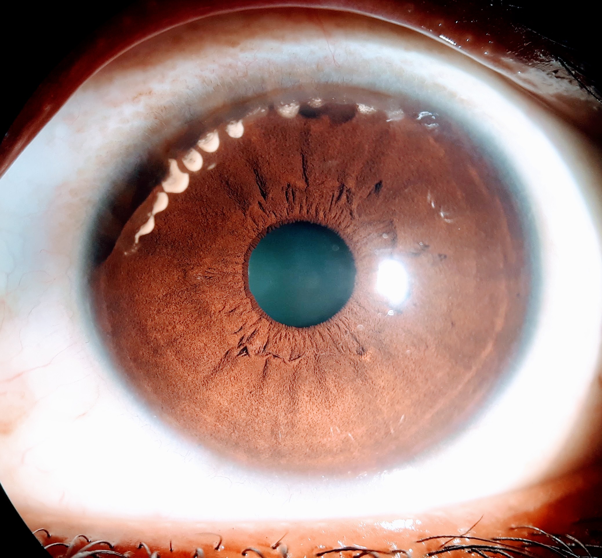
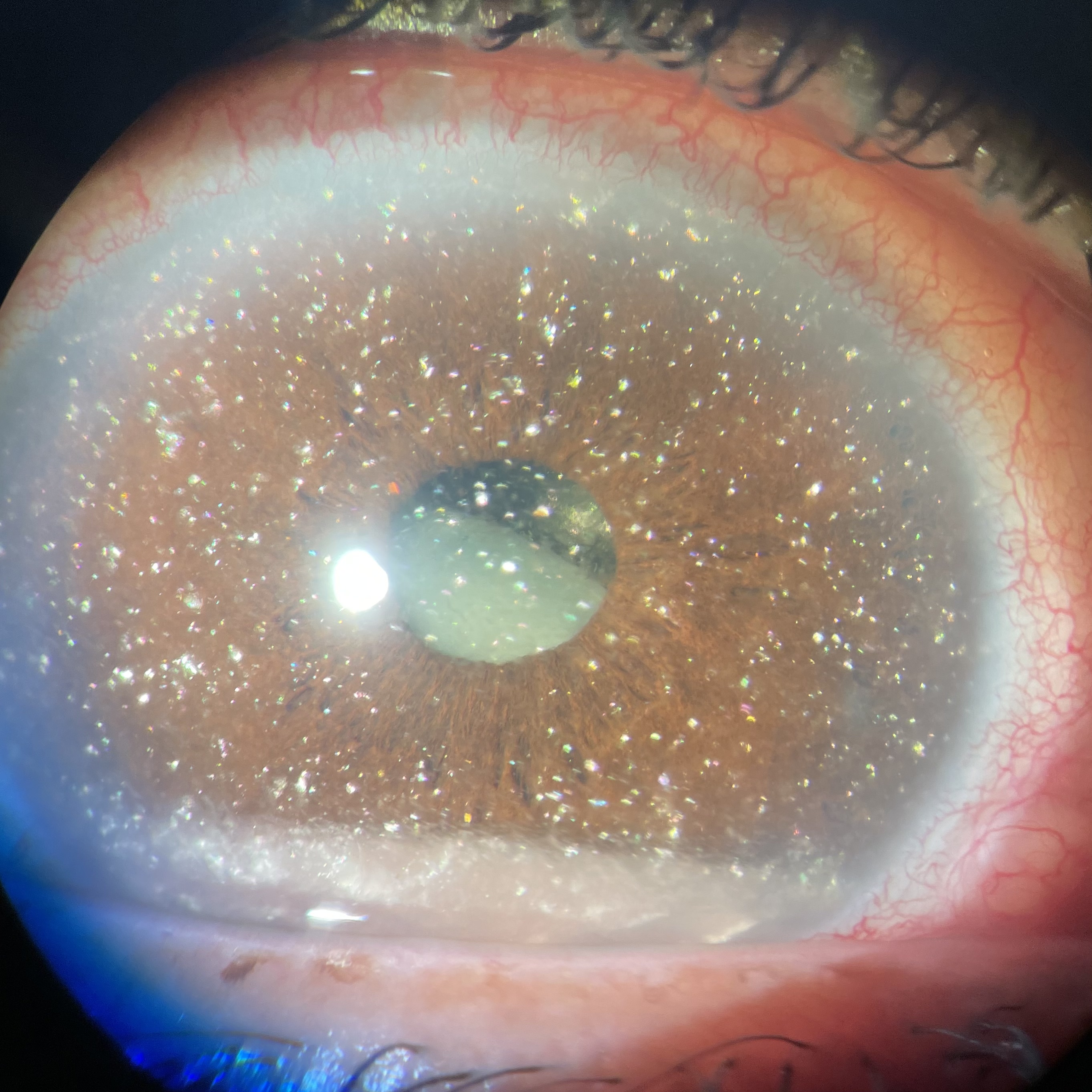
Iris Pigment Epithelial Cysts
By Joel Antonio Morales López, MD
joelaml@yahoo.com
This is a photograph of an eye with a pupillary iris cyst and angle dislodged iris pigment epithelial cysts.
Slit Lamp
Blood Stream in the Anterior Chamber
By Jason Calhoun, COA
gbjay692002@gmail.com
A thin line of blood has leaked into the anterior chamber following glaucoma surgery.
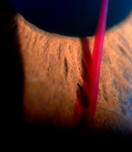
Terrien Marginal Degeneration With Lipid Deposition
By Neha Pathak, MBBS, MS
pthknh@gmail.com
A yellow line of lipid deposits has formed at the central edge of the furrow in an eye with Terrien marginal degeneration.
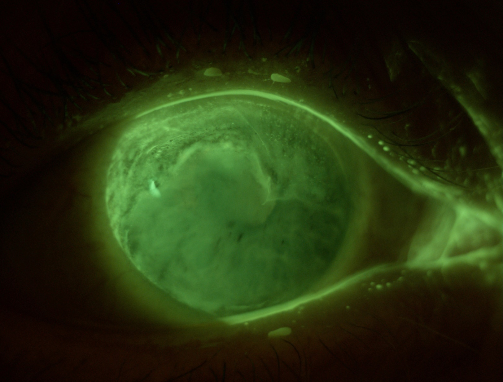
Acute Corneal Hydrops
By Thomas Tien, MD, and Irving M. Raber, MD
The slit-lamp photograph shows severe corneal hydrops in a patient with keratoconus. The thickness of the edematous cornea was greater than the anterior chamber depth.
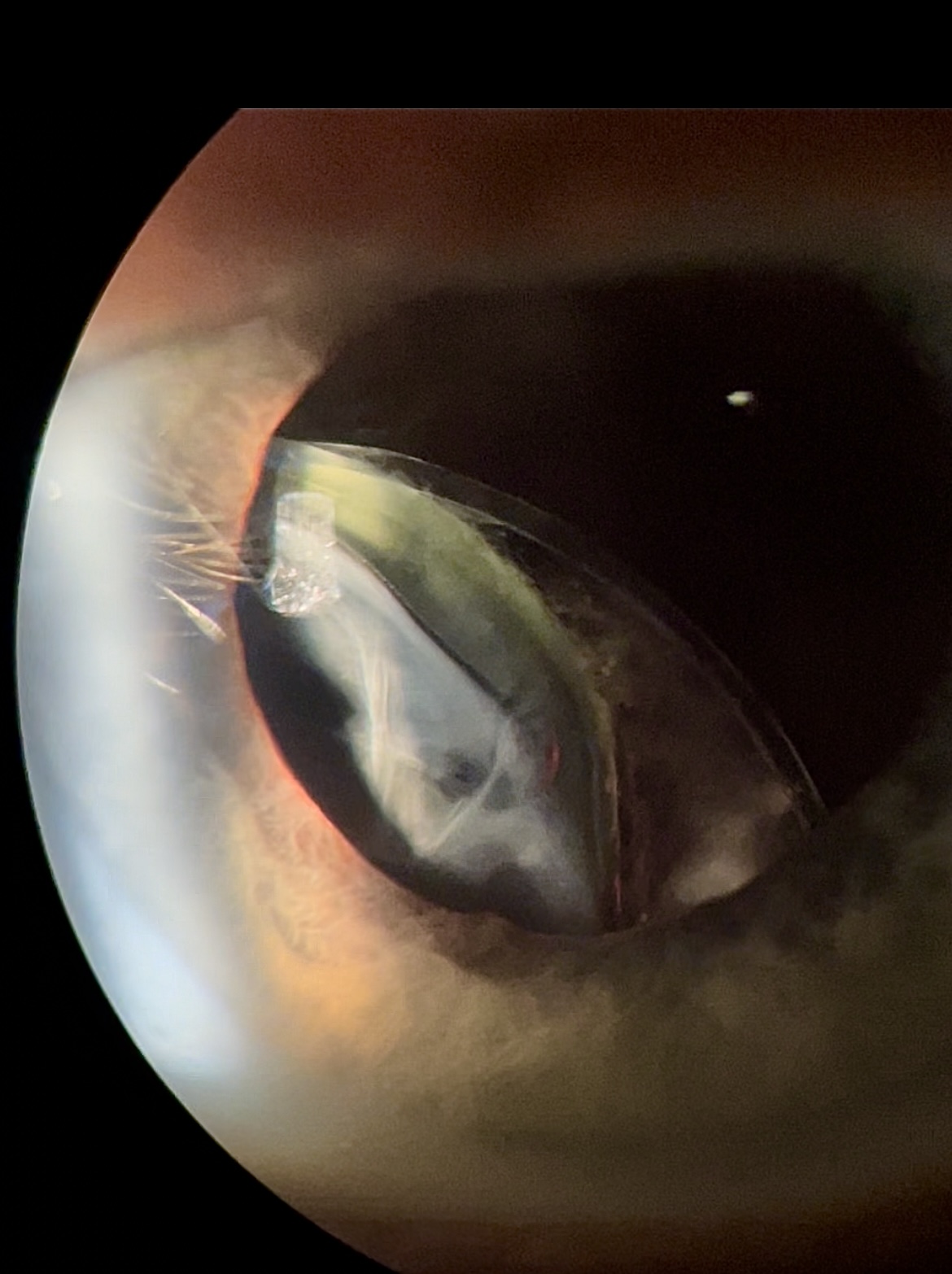
Hanging by a Haptic
By Jay Lim, MD
jtecsonlim@yahoo.com
The IOL has dislocated in the right eye of a 50-year-old patient who presented to the clinic with a complaint of blurry vision after accidentally hitting his eye against a bike handle.
GRAND PRIZE WINNER
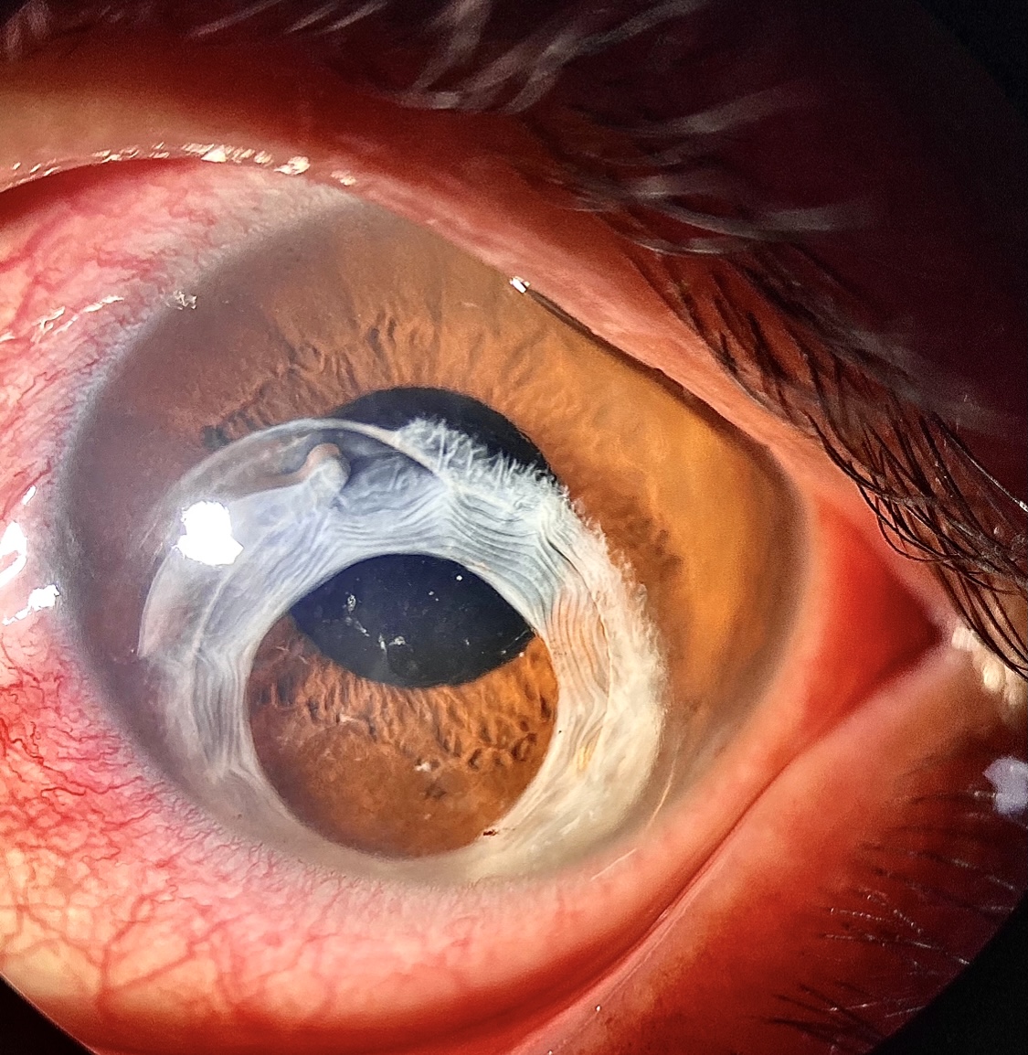
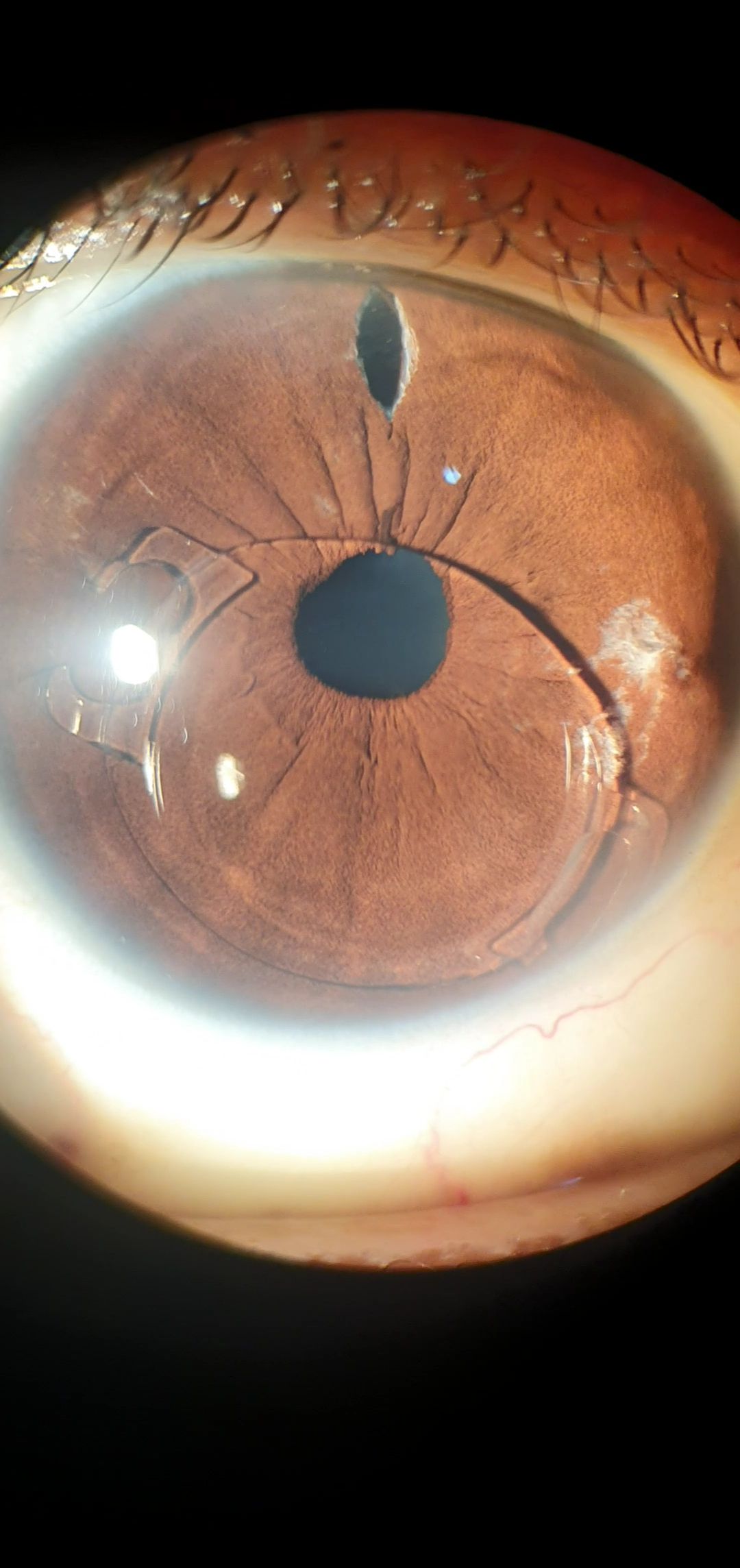
Total Anterior Capsular Phimosis
By Radhika Rampat, MBBS, BSc(Hons), FRCOphth, and Mark Wilkins, MD, FRCOphth
radhikarampat@me.com
The photograph provides a rare illustration of a central island of scarred tissue with radial lines and 360º of fibrosis of the anterior capsule in the eye of a patient who experienced a rapid loss of vision in the months following uneventful cataract surgery.
Surgical Complications
Vitreous Prolapse
By Jorge Neaves, MD
jorgeneaves93@gmail.com
This is a photograph of a vitreous block in the eye of an aphakic patient whose posterior capsule tore during phacoemulsification. The hyaloid membrane is intact.
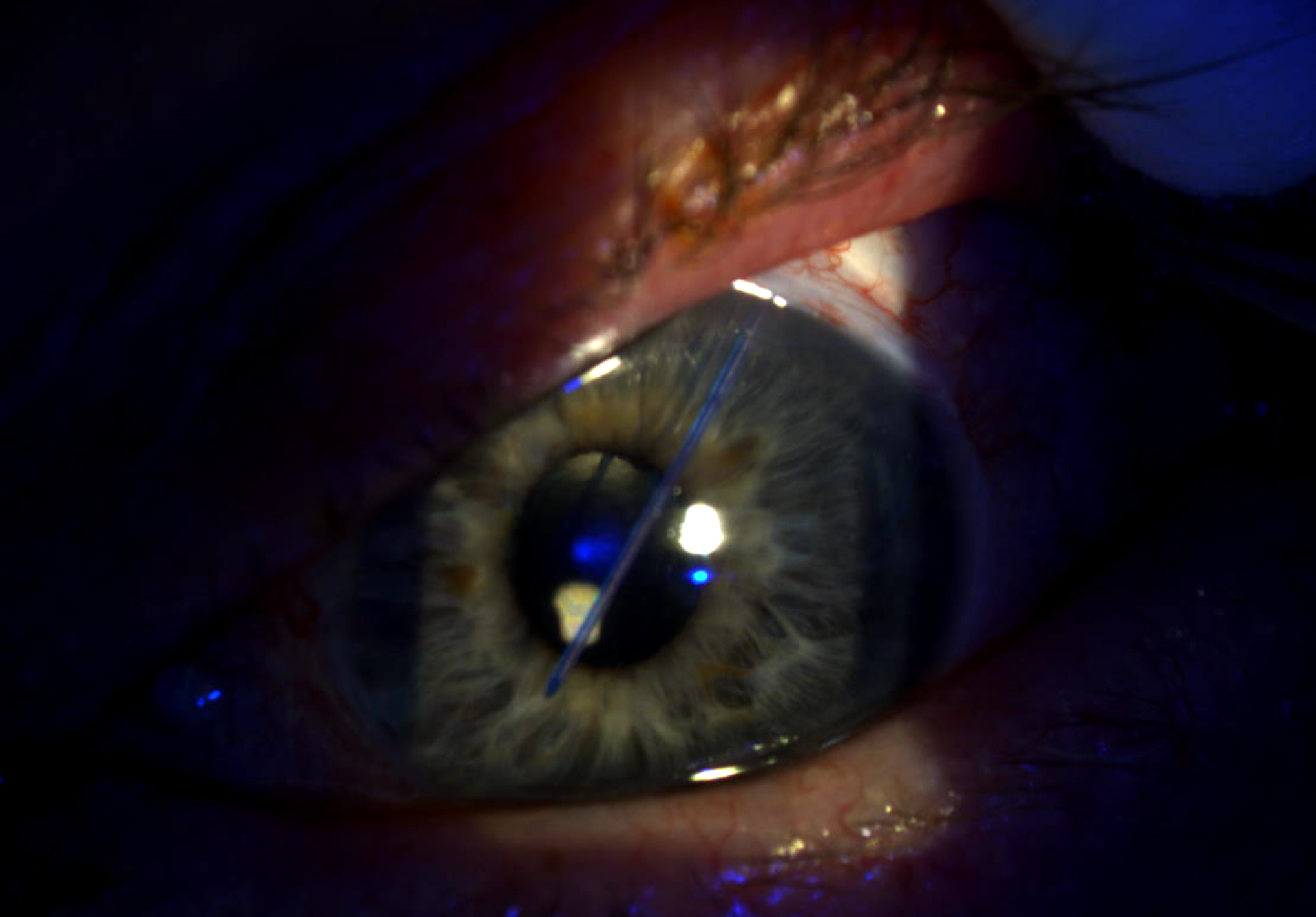
Falling From the Iris
By Luciano Rabello Netto Cirillo, MD
lucianocirillo22@gmail.com
An Artiflex IOL (Ophtec) has dislocated in the eye of a patient who experienced diminished visual acuity beginning the morning after a party.
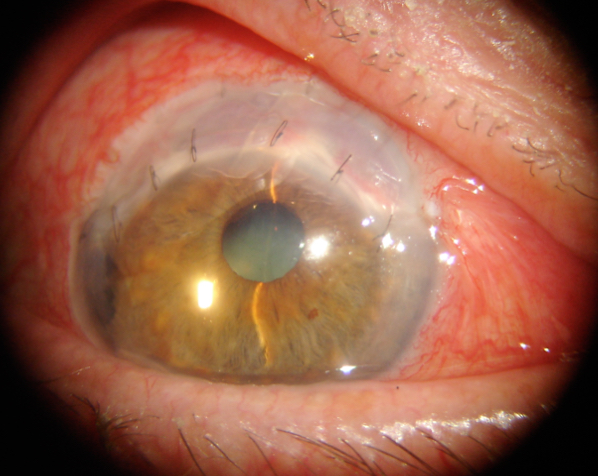
Graft Rejection in Terrien Marginal Degeneration
By C. Manuel Nicoli, MD
manuelnicoli@hotmail.com
Stromal graft rejection occurred following a C-shaped lamellar keratoplasty in the eye of a patient with Terrien marginal degeneration.
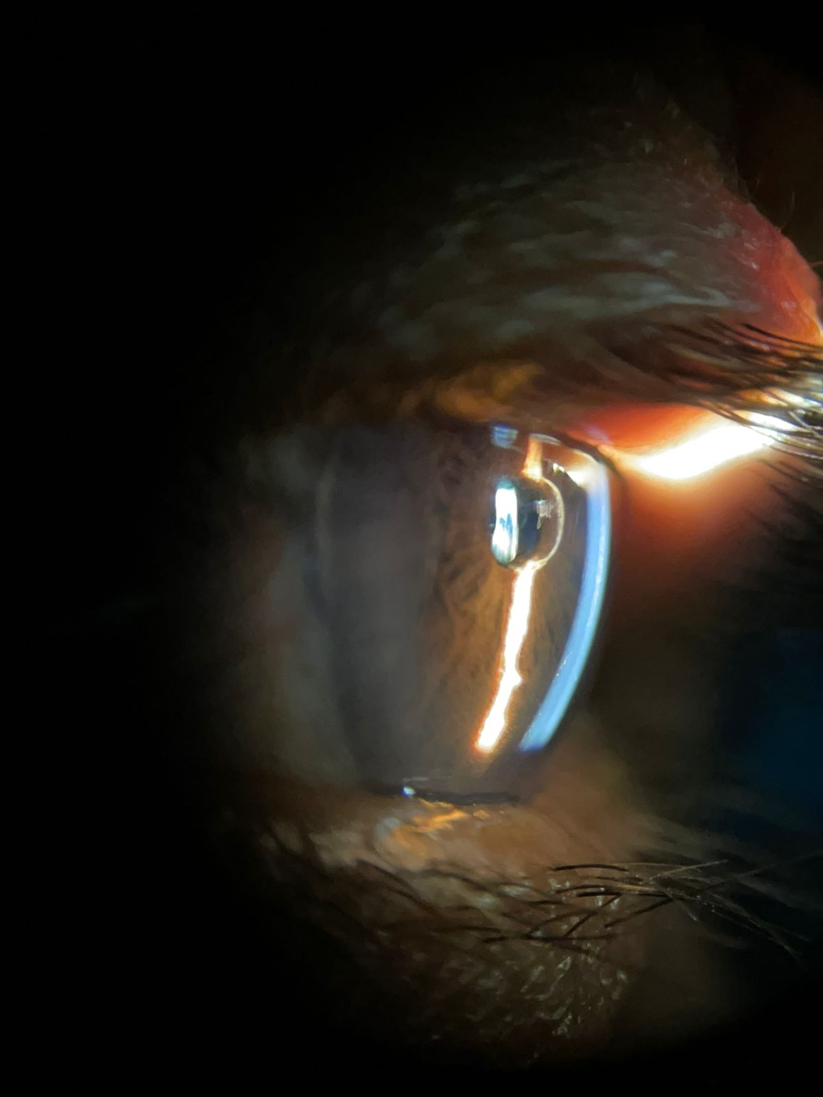
Migration of Glaucoma Drainage Device Stent Suture
By Andrew Tatham, MBChB, FRCOphth, FRCS(Ed), FEBO
andrewjtatham@gmail.com
An intraluminal 3-0 polypropylene stent suture migrated into the anterior chamber 2 weeks following the implantation of a 350-mm2 Baerveldt glaucoma implant (Johnson & Johnson Vision). The stent was withdrawn at the slit lamp through a conjunctival incision.
Fibrotic Membrane Over IOL
By Gerson López-Moreno, MD
gerlopezm@gmail.com
A fibrotic membrane has formed on the IOL in the eye of a 61-year-old patient who underwent cataract surgery 3 days earlier.
Trauma
Hex Marks the Spot
By Hasenin Al-khersan, MD
hxa358@miami.edu
The image shows a ruptured globe in a patient with a history of a hexagonal keratotomy.
The Christmas Eye
By Alonso Meza-Anguiano, MD
dralonsomeza@gmail.com
The image shows subluxated hypermature cataract crystals secondary to old ocular trauma.
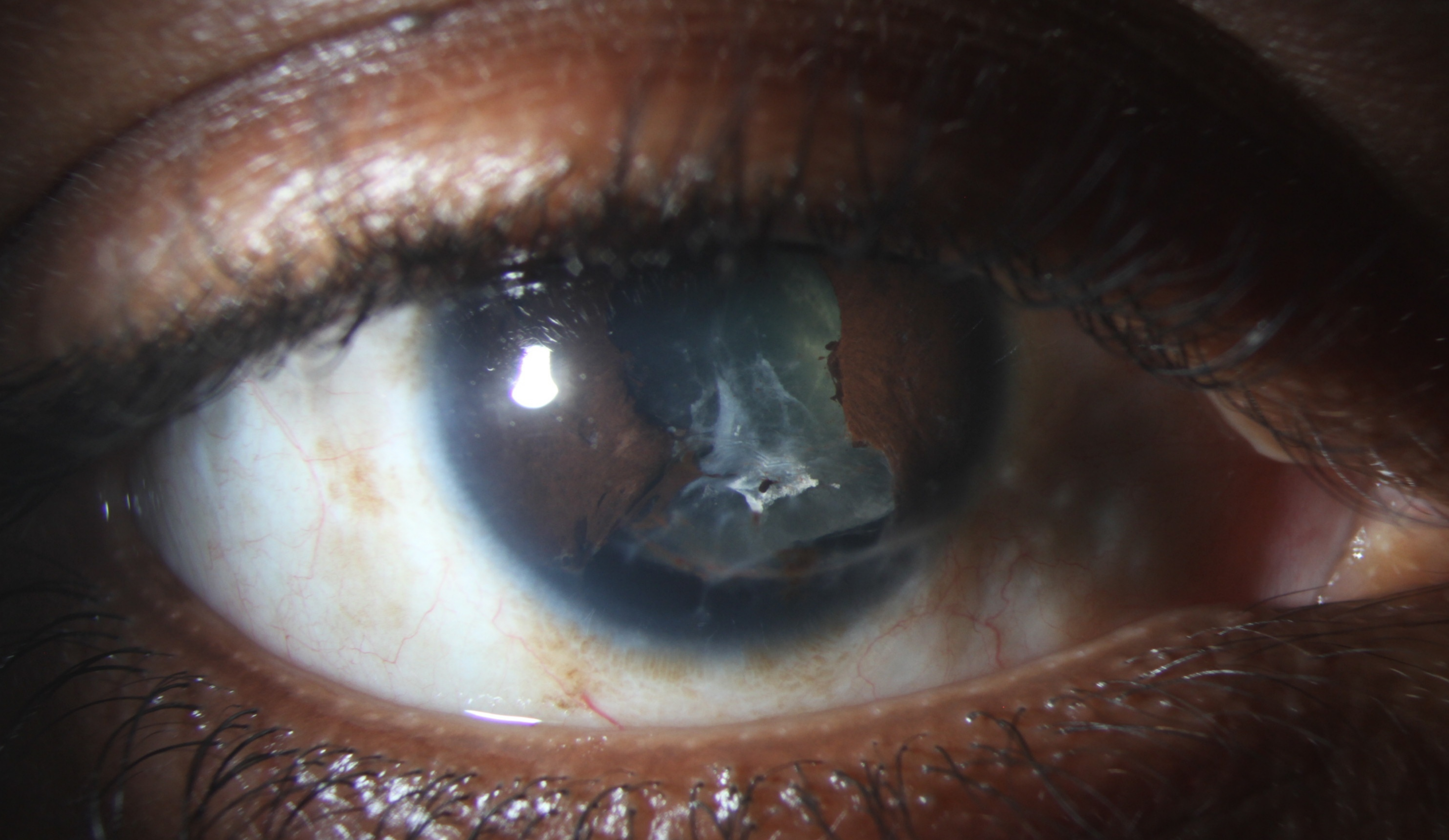
Fibrous Downgrowth After Ruptured Globe Repair
By Jason C. Fan, MD
A 22-year-old man experienced a traumatic corneoscleral laceration.
The photograph was taken 7 months after the initial repair. A
vascularized pupillary membrane appears to extend from the iris
to the anterior lens capsule. A biopsy of the membrane revealed
stratified mucosal epithelium consistent with fibrous downgrowth.
A Traumatic Experience
By Jonathan Tijerina, MD, MA
jdt105@miami.edu
This is a photograph of the right
eye of a 58-year-old patient who
experienced traumatic subluxation
of the IOL–capsular bag complex
during a basketball game.
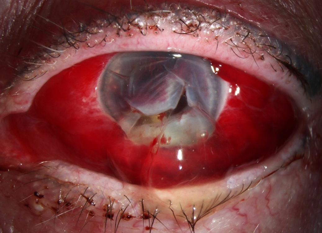
A Lock With No Key
By Francesco Pozzo Giuffrida, MD, and Florence Cabot, MD
francescopozzo@yahoo.com
This photograph was taken
months after a ruptured globe
injury and shows a sectorial
inferior iris defect from the 4 to
8 clock positions with complete
zonular loss resembling a lock.

