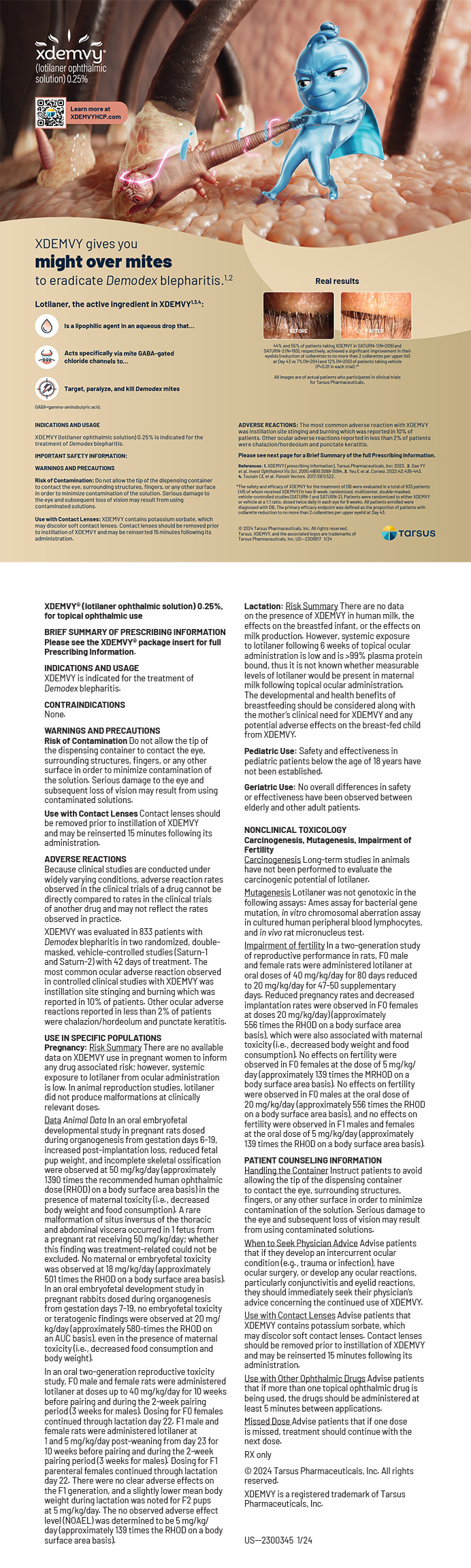
Those of us who have been practicing ophthalmology for the past 3 decades have been fortunate to witness the birth and evolution of modern refractive surgery. A wide array of procedural options are currently available to us, and many once-promising procedures have been abandoned in favor of safer, more effective techniques. In the late 1990s, there were limited options for patients seeking less dependence on spectacles. By then, incisional techniques such as radial keratotomy (RK) had, by and large, been relegated to the history books, and the age of excimer laser–based refractive surgery had dawned.
Before LASIK entered the lexicon, PRK was the primary method for correcting myopia. A significant percentage of patients who underwent PRK—particularly those with high myopia—developed late-onset corneal haze. It often became visually significant and was a major source of postoperative dissatisfaction.
Theories on the etiology of the haze ranged from environmental exposure and demographic factors to a virus. What was clear was that higher refractive ablations were associated with a greater risk of corneal haze, which occurred frequently, with a reported incidence of more than 20% of cases in one series.1 Various treatments were proposed for addressing postoperative corneal haze, including cooling the cornea, the administration of topical steroids, and the use of adjunctive agents such as diclofenac, thiotepa, and interferon alpha.
MMC FOR EXISTING CORNEAL HAZE
In 1991, investigators evaluated mitomycin C (MMC) for the modulation of wound healing after excimer laser ablation.2 This alkylating agent inhibits DNA synthesis and prevents cell proliferation. It wasn’t until a study3 on the use of MMC for the treatment of corneal intraepithelial neoplasia/dysplasia was published in 1997 that my colleague Randy J. Epstein, MD, seriously began to consider using MMC in refractive surgery.
At the time, several of his patients had developed dense, central fibrotic scars after RK that recurred after surgical removal. Dr. Epstein observed that the morphologic appearance of the dysplastic change was similar to that seen in preneoplastic proliferation. MMC had been, for many years, used successfully to prevent recurrent pterygium and fibrosis of the filtering bleb after trabeculectomy. Use of the agent was met with trepidation, however, because of numerous reports of scleral melt after its application.4
The first patient of Dr. Epstein’s to receive MMC directly on the corneal stromal surface had undergone multiple superficial keratectomies for central fibrosis after RK, but unfortunately, the subepithelial fibrosis recurred every time. Dr. Epstein prescribed topical MMC drops, which did not really do much. Because the patient was on the verge of needing a corneal transplant, MMC was applied directly to the corneal stromal surface after superficial keratectomy. Amazingly, the fibrosis did not recur.
MMC FOR CORNEAL HAZE AFTER PRK
Soon after that first case, I encountered a woman who had undergone multiple PRK procedures at another practice in 1994 and 1995 to correct a refractive error of -11.00 D. Repeated regression and treatment had left her with grade 4 corneal subepithelial fibrosis bilaterally. The patient had also developed a cataract from months of topical steroid therapy to treat her corneal haze. When she presented to my clinic, her BCVA was 20/400 OU, and she was ready for a penetrating keratoplasty.
I decided to explore MMC as a potential treatment option. Informed consent was obtained, and MMC 0.02% (0.2 mg/mL) was applied for 2 minutes. This was the same dose Dr. Epstein had used on his patient, but it was considerably lower than that used in glaucoma and pterygium surgery. I was not aggressive about removing the haze, so the scar remained. This makes sense knowing what we know now about the mechanism of action of MMC in that it does not remove haze. Instead, the agent prevents the reactivation of stromal keratocytes and their differentiation into myofibroblasts. We realized that near-complete removal of haze followed by MMC application would be necessary. Reepithelialization was timely, and the patient’s cornea remained clear. Her BCVA was 20/20 after cataract surgery. I performed a similar procedure on the fellow eye, and the patient did well for many years until she was lost to follow-up. This case marked the first MMC application (as far as I am aware) in humans for post-PRK haze, and it proved to be a successful treatment option.
My colleagues and I subsequently published the first series describing the use of topical MMC for corneal haze in post-RK and post-PRK patients.5 It is among the top 100 most cited articles in the history of refractive surgery.6 Our article was followed by the publication of a large, double-blind series by Vigo et al, which helped solidify MMC as a treatment option.7
PROPHYLACTIC USE OF MMC IN PRK
Once we had a model for treating existing corneal haze, our focus shifted to developing guidelines for the prophylactic use of MMC in PRK.8-10 Through collaborations with surgeon-investigators around the world, we determined how and when to administer MMC to prevent corneal haze in high-risk patients undergoing PRK.8-10 We proposed using the agent for excimer laser ablation depths exceeding 75 µm, which is the criterion I still use today. Additionally, we established that shorter application times (12 seconds vs 2 minutes) are appropriate for prophylaxis11 but a full 2-minute application is often required after haze has formed.
Multiple studies, including a large meta-analysis from the AAO, have shown that MMC is a safe and effective adjunctive treatment in high-risk PRK.12 Advances in laser technology have reduced the incidence of post-PRK haze. The complication, however, can still occur. It is gratifying to know that more than 93% of refractive surgeons worldwide administer MMC to modulate wound healing following PRK.13
CONCLUSION
As with most advances in medicine, the success of MMC as a treatment option and prophylactic measure in high-risk PRK patients was not created in a vacuum. Rather, it required a bit of luck, outside-the-box thinking, and standing on the shoulders of giants who laid the foundation upon which we could build.
1. Sia RK, Ryan DS, Edwards JD, Stutzman RD, Bower KS. The U.S. Army Surface Ablation Study: comparison of PRK, MMC-PRK, and LASEK in moderate to high myopia. J Refract Surg. 2014;30(4):256-264.
2. Talamo JH, Gollamudi S, Green WR, De La Cruz Z, Filatov V, Stark WJ. Modulation of corneal wound healing after excimer laser keratomileusis using topical mitomycin C and steroids. Arch Ophthalmol. 1991;109(8):1141-1146.
3. Frucht-Pery J, Sugar J, Baum J, et al. Mitomycin C treatment for conjunctival-corneal intraepithelial neoplasia: a multicenter experience. Ophthalmology. 1997;104(12):2085-2093.
4. Rubinfeld RS, Pfister RR, Stein RM, Foster CS, Martin NF, Stoleru S, Talley AR, Speaker MG. Serious complications of topical mitomycin-C after pterygium surgery. Ophthalmology. 1992;99(11):1647-1654.
5. Majmudar PA, Forstot SL, Dennis RF, et al. Topical mitomycin-C for subepithelial fibrosis after refractive corneal surgery. Ophthalmology. 2000;107(1):89-94.
6. Randleman JB, Asroui L, Tarib I, Scarcelli G. The most cited articles and authors in refractive surgery. J Refract Surg. 2023;39(2):78-88.
7. Vigo L, Scandola E, Carones F. Scraping and mitomycin C to treat haze and regression after photorefractive keratectomy for myopia. J Refract Surg. 2003;19(4):449-454.
8. Carones F, Vigo L, Scandola E, Vacchini L. Evaluation of the prophylactic use of mitomycin-C to inhibit haze formation after photorefractive keratectomy. J Cataract Refract Surg. 2002;28(12):2088-2095.
9. Lee DH, Chung HS, Jeon YC, Boo SD, Yoon YD, Kim JG. Photorefractive keratectomy with intraoperative mitomycin-C application. J Cataract Refract Surg. 2005;31(12):2293-2298.
10. Netto MV, Mohan RR, Ambrósio R Jr, Hutcheon AE, Zieske JD, Wilson SE. Wound healing in the cornea: a review of refractive surgery complications and new prospects for therapy. Cornea. 2005;24(5):509-522.
11. Virasch VV, Majmudar PA, Epstein RJ, et al. Reduced application time for prophylactic mitomycin-C in photorefractive keratectomy. Ophthalmology. 2010;117:885-889.
12. Majmudar PA, Schallhorn SC, Cason J, et al. Mitomycin-C in corneal surface excimer laser ablation techniques: a report by the American Academy of Ophthalmology. Ophthalmology. 2015;122(6):1085-1095.
13. Hasson M. Survey shows slight increase in laser refractive surgery volume. Healio. April 11, 2012. Accessed March 13, 2023. https://www.healio.com/news/ophthalmology/20120412/survey-shows-slight-increase-in-laser-refractive-surgery-volume