Dry eye disease (DED) and allergic conjunctivitis are two separate but overlapping conditions. Ocular allergy can cause exacerbation of DED, which can increase inflammation, hyperosmolarity, and tear film instability, which in turn exacerbates ocular allergy. Inflammation is both the cause and effect of dry eye, resulting in a vicious cycle.
The Dry Eye Workshop II (DEWS II) authors included allergic conjunctivitis as one of the most common risk factors for DED.1 Further, Galor et al demonstrated a direct correlation between seasonal allergens and dry eyes, with both pollen and DED reaching a yearly peak in the month of April.2
Managing patients with one or both conditions can mean double trouble. This article presents five things you need to know about the relationships between allergy and DED, the treatments available, and the best steps for managing your patients affected by these entities.
No. 1: Differentiate between allergic conjunctivitis and DED. Red, watery eyes are the hallmark of both allergy and dry eye. Differentiating between allergic conjunctivitis and DED can be a challenging task (see Symptoms of Dry Eye Disease and Seasonal Allergy). Pollen and other seasonal particles may trigger DED or make existing symptoms worse.
Symptoms of Dry Eye Disease (DED) and Seasonal Allergy
DED Symptoms
- Watering and irritation
- Burning or stinging
- Foreign body sensation
- Eye fatigue
- Fluctuation in vision
- Redness
Allergy Symptoms
- Itchy eyes
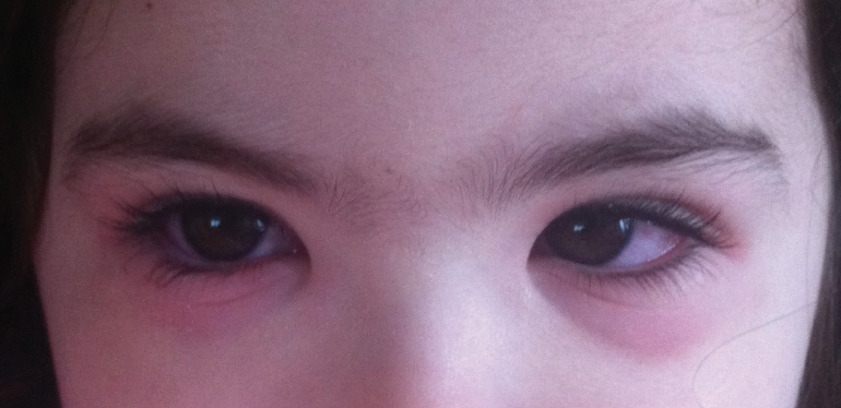
- Red eyes (Figure)
- Burning or stinging
- Swollen eyelids
- A runny nose, nasal congestion and a watery discharge from the eyes
- Light sensitivity
- White, ropey discharge

Figure. A child with seasonal ocular allergies.
Photo courtesy of Christopher E. Starr, MD
Patients with either condition may complain of nonspecific symptoms such as eye redness, irritation, and tearing, but the symptom of itching is the key differentiator. We can generally exclude allergic conjunctivitis if there is no itching. Itchiness produces a strong desire in patients to rub their eyes, creating an itch-rub cycle that leads to releasing of more cytokines and worsening of signs and symptoms.
By contrast, symptoms of DED may include foreign body sensation, scratching, burning, and fluctuation in vision, especially when one is concentrating on visual tasks such as reading or computer work. Diagnosis can be more challenging if patients are unable to recall which situations exacerbate their symptoms. To make matters more challenging, DED and ocular allergy often occur simultaneously.2,3
For a patient with suspected allergy, examination by an eye care professional is crucial to rule out underlying conditions such as blepharitis, DED, meibomian gland dysfunction (MGD), and preservative toxicity. Fluorescein staining of the cornea or conjunctiva, reduced tear breakup time, MGD, and reduced tear meniscus are indicative of DED. The diagnosis of ocular allergy is traditionally made by history and clinical examination. Common hallmark signs and symptoms include redness, itching, lid swelling, chemosis, and a papillary reaction involving the palpebral conjunctiva.
It may be difficult to distinguish whether ocular itchiness is due to allergic conjunctivitis or blepharitis associated with Demodex infestation. Asking patients to describe the location of the itch can help to pinpoint the etiology. Ocular allergy involves itching of the conjunctiva, whereas Demodex-related itching involves the lid along with pathognomonic cylindrical collarettes that can be seen at the slit lamp.
Allergy skin testing (Doctor’s Rx Allergy Formula, Bausch + Lomb) can be performed to help the eye care practitioner differentiate between OSD and ocular allergy. This test can also help pinpoint which allergens the patient should avoid. The tests are designed specifically for each area of the country and its allergic triggers. Skin testing, however, may fail to reveal the allergen causing the allergic conjunctivitis. Thus, a combination of testing and questioning is necessary.
An ocular allergy is caused by sensitivity to a substance. When the allergen interacts with mast cells, histamine is released, causing itching, redness, and swelling. Allergic disorders of the ocular surface include seasonal and perennial allergic conjunctivitis, vernal keratoconjunctivitis (VKC), atopic keratoconjunctivitis (AKC), and drug-induced blepharoconjunctivitis.
If a patient experiences intense itching and redness while outdoors, suspect seasonal ocular allergies. Perennial ocular allergens such as dust, pet hair or dander, and mold can present a constant aggravation all year around. Vernal and atopic allergies tend to be more severe, often with corneal involvement. Drug-induced allergies usually affect the lower lids and usually resolve once the patient discontinues the offending agent.4
MGD, the most common cause of DED,5 occurs due to blockage of the meibomian glands that preclude the glands from producing the oils that lubricate the eyes. Because symptoms (eye irritation, redness, a scratchy sensation, and watery eyes) often overlap with allergy, the key to successful treatment is a correct diagnosis.
There is an association between ocular allergy and an unstable tear film,6-9 and patients affected by ocular allergy can develop changes in the structure of the meibomian glands that may be attributed to damage from rubbing, ocular inflammation, or a combination of these factors.10
The DEWS II definition of DED includes hyperosmolarity of the tear film.11 Studies have demonstrated an increase in tear osmolarity and the inflammatory marker matrix metalloproteinase-9 (MMP-9) in patients with allergic and vernal keratoconjunctivitis. These factors can also be associated with dry eye symptoms and increased conjunctival and corneal staining.12-14
Neurosensory abnormalities may also play a significant role in OSD, which is also incorporated in the DEWS II definition of DED.11 Inflammation and hyperosmolarity of the tear film, both of which can result from ocular allergies, can alter corneal sensory receptors and result in subsequent nerve damage.
It is imperative that eye care practitioners examine the entire ocular surface and treat the underlying condition or conditions.
No. 2: Treat allergic conjunctivitis without exacerbating DED. Many people use over-the-counter products such as oral antihistamines to self-treat ocular allergy and DED. However, these medications may not be safe or effective and they may exacerbate preexisting DED.
Avoidance of the causative agent is an essential first step in managing allergies. For mild allergy, consider the combination of preservative-free artificial tears, cool compresses, and avoidance of eye-rubbing. When this isn’t enough, pharmacologic intervention such as a topical antihistamine and/or mast cell stabilizer may help alleviate the symptoms of allergic conjunctivitis. NSAIDs, oral antihistamines, steroids, and some off-label treatments can also be used.
Remember, treatment options for one condition can worsen other coexisting conditions. Studies have demonstrated that oral antihistamines, including recent-generation antihistamines, cause a significant reduction in tear volume.15-17 This is counterproductive, as the reduced tear volume leads to inadequate flushing of allergens and an increased concentration of inflammatory mediators.
Topical treatment of ocular allergies is superior to systemic treatment because it ensures direct delivery to the tissue of concern. In fact, systemic treatment is rarely needed when ocular allergy is the main manifestation. Additionally, topical treatment prevents the drying effects that result from the use of systemic antihistamines.
Therapeutic options to optimize the health of the tear film include treating the meibomian glands and the underlying inflammation and using preservative-free artificial tears. Instilling chilled drops helps to soothe the eye and reduce vascular permeability of the conjunctival vessels and chemosis.
Topical drops such as cetirizine ophthalmic solution 0.24% (Zerviate, Eyevance Pharmaceuticals) and bepotastine besilate ophthalmic solution 1.5% (Bepreve, Bausch + Lomb) are excellent drops for patients with concomitant DED and allergy. They also can be used to treat nonocular symptoms such as rhinitis, allowing many patients to use fewer oral systemic medications and nasal sprays. These products are unique because they are highly specific to H1-receptor antagonists. They have minimal adverse events, comfortable formulations, rapid onset, and long duration of action with b.i.d. dosing.
Steroid drops such as fluorometholone acetate ophthalmic suspension 0.1% (Flarex, Eyevance Pharmaceuticals), loteprednol etabonate 0.2% (Alrex, Bausch + Lomb) or loteprednol etabonate ophthalmic gel 0.5% (Lotemax Gel, Bausch + Lomb) can be used for acute, severe symptoms of allergic conjunctivitis and exacerbations of DED.
Prescribing an immunomodulator to reduce inflammation and increase tear production is also recommended for patients who experience dry eye symptoms. Cyclosporine ophthalmic emulsion 0.05% (Restasis, Allergan) has shown benefits when used off-label in patients with allergic keratoconjunctivitis.18,19
Punctal occlusion works well in patients with aqueous deficient DED; however, if there is inflammation in the tear film or related allergies, plugs should be avoided.
No. 3: Cosmetics and beauty products can worsen allergic and dry eye conditions. Improper makeup application or removal may aggravate or result in the clogging of meibomian gland orifices. The resulting deficiency in the oil layer of the tears can allow premature evaporation of the tear film, causing aqueous deficiency that leads to ocular dryness.
The use of eyelash extensions, dyes, or curlers can result in allergic blepharitis and dermatitis. Top allergens found in cosmetics include preservatives, antioxidants, resins, pearlescent additives, fragrances, nickel, and surfactants in waterproof makeup removers. Patients should avoid ingredients such as arsenic, beryllium, cadmium, carmine, lead, selenium, and thallium, which can cause toxicity to the ocular surface. Hypoallergenic brands usually have a lower likelihood of containing these irritants. A patient who is allergic to a component of a cosmetic may exhibit dermatitis or an allergic reaction along the lid or eyelash line.
Educate patients that makeup should be removed before bed to avoid clogging the meibomian glands and to prevent infection. It is best to use gel-based makeup removal products that are oil- and paraben-free and to avoid irritants such as mineral oil, sodium lauryl sulfate, and diazolidinyl urea. A gentle eyelid scrub applied after makeup is removed can help prevent clogging of the meibomian glands. Tell patients to replace makeup every 3 to 6 months, avoid sharing products, and avoid using saliva with makeup application. (For a more in-depth look at eye cosmetics, see the article by Laura M. Periman, MD.)
No. 4: Lifestyle changes for allergy and DED. Whether a patient’s problem is due to allergies or DED, simple lifestyle changes can help to improve comfort and possibly vision (see Lifestyle Changes: Tips for Patients).
Lifestyle Changes: Tips for Patients
Outdoors
- Avoid exposure to allergens by trying to stay inside when the pollen count is high
- When outdoors, wear glasses or goggles to keep the pollen out of the eyes and the moisture in
Indoors
- Use air filters, especially at the height of allergy season when pollen counts are high
- Add a humidifier in the winter months
- Decrease excessive air movement from ceiling fans or air blowing from a car heater or air conditioner
- Keep the home free of dust to reduce exposure to irritants and allergens
- If contact lenses exacerbate dry eye disease and/or allergies, changing the type of contacts or cleaning solution may be necessary; sometimes patients need to discontinue wearing them all together
- Take frequent breaks from screen time and implement the 20/20/20 rule (ie, every 20 minutes look at something 20 feet away for 20 seconds)
Other considerations
- Consumption of reesterified omega-3 fatty acids can improve tear osmolarity, omega-3 index levels, tear breakup time, metalloproteinase-9 levels, and symptom scores1
- Incorporating foods rich in fatty acids such as fish, walnuts, and olive oil can help promote a healthy ocular surface
- Consider that some systemic medications (eg, diuretics, antihypertensives, antihistamines, anticholinergics, hormones, antidepressants, and pain relievers) can exacerbate dry eye symptoms
1. Epitropoulos AT, Donnenfeld ED, Shah ZA, et al. Effect of oral re-esterified omega-3 nutritional supplementation on dry eyes. Cornea. 2016;35:1185-1191.
No. 5: Be aggressive about treating sight-threatening atopic and vernal keratoconjunctivitis. Prompt treatment should be initiated in patients with atopic and vernal keratoconjunctivitis, and they should be monitored closely for corneal complications. Aggressive use of topical and/or oral steroids should be initiated for acute, severe symptoms, with tapering as symptoms abate. Patients can be weaned onto a drop that can be safely used long-term, such as a combination mast cell stabilizer/antihistamine. I may prescribe tacrolimus ointment off-label for vision-threatening atopic or vernal keratoconjunctivitis. Further, a topical immunomodulator can be effective in treating these challenging and sight-threatening forms of allergic conjunctivitis.18,19
CONCLUSION
DED and allergic conjunctivitis often coexist, and diagnosis and treatment can be challenging, despite recent innovations in diagnostic testing. Keeping in mind how lid disease, ocular allergies, and dry eyes are interconnected and executing careful examinations, diagnosis, and patient education can lead to effective tailored and preventive management.
1. Gomes JAP, Azar DT, Baudouin C, et al. TFOS DEWS II iatrogenic report. Ocul Surf. 2017;15(3):511-538.
2. Kumar N, Feuer W, Lanza NL, Galor A. Seasonal Variation in Dry Eye. Ophthalmology. 2015;122(8):1727-1729.
3. Hom MM, Nguyen AL, Bielory L. Allergic conjunctivitis and dry eye syndrome. Allergy Asthma Immunol. 2012;108(3):163-166.
4. Abelson MB, Chapin MJ. Current and future topical treatments for ocular allergy. Comp Ophthalmol Update. 2000;1:303-317.
5. Lemp MA, Crews LA, Bron AJ, et al. Distribution of aqueous-deficient and evaporative dry eye in a clinic-based patient cohort: a retrospective study. Cornea. 2012;31:472-478.
6. Villani E, Strologo M, Pichi F, et al. Dry eye in vernal keratoconjunctivitis: A cross-sectional comparative study. Medicine (Baltimore). 2015;94(42):e1648.
7. Chen L, Pi L, Fang J, et al. High incidence of dry eye in young children with allergic conjunctivitis in Southwest China. Acta Ophthalmol. 2016;94(8):e727-e730.
8. Akil H, Celik F, Ulas F, Kara IS. Dry eye syndrome and allergic conjunctivitis in the pediatric population. Middle East Afr J Ophthalmol. 2015;22(4):467-471.
9. Kim T, Moon N. Clinical correlations of dry eye syndrome and allergic conjunctivitis in Korean children. J Pediatr Ophthalmol Strabismus. 2013;50(2):124-127.
10. Mizoguchi S, Iwanishi H, Arita R, et al. Ocular surface inflammation impairs structure and function of meibomian gland. Exp Eye Res. 2017;163:78-84.
11. Craig JP, Nichols KK, Akpek EK, et al. TFOS DEWS II definition and classification report. Ocul Surf. 2017;15:276-283.
12. Leonardi A, Brun P, Abatangelo G, et al. Tear levels and activity of matrix metalloproteinase (MMP)-1 and MMP-9 in vernal keratoconjunctivitis. Invest Ophthalmol Vis Sci. 2003;44(7):3052-3058.
13. Kumagai N, Yamamoto K, Fukuda K, et al. Active matrix metalloproteinases in the tear fluid of individuals with vernal keratoconjunctivitis. J Allergy Clin Immunol. 2002;110(3):489-491.
14. Villani E, Rabbiolo G, Nucci P. Ocular allergy as a risk factor for dry eye in adults and children. Curr Opin Allergy Clin Immunol. 2018;18(5):398-403.
15. Foutch BK, Sandberg KA, Bennett ES, Naeger LL. Effects of oral antihistamines on tear volume, tear stability, and intraocular pressure. Vision (Basel). 2020;4(2):E32. Published 2020 Jun 20.
16. Nally L, Emory TB, Welch DL. Ocular drying associated with oral antihistamines (loratadine) in the normal population – effect on tear flow and tear volume as measured by fluorophotometry. Invest Ophthalmol Vis Sci. 2002;43(13):92.
17. Ousler GW 3rd, Workman DA, Torkildsen GL. An open-label, investigator-masked, crossover study of the ocular drying effects of two antihistamines, topical epinastine and systemic loratadine, in adult volunteers with seasonal allergic conjunctivitis. Clin Ther. 2007;29(4):611-616.
18. Akpek EK, Dart JK, Watson S, et al. A randomized trial of topical cyclosporin 0.05% in topical steroid-resistant atopic keratoconjunctivitis. Ophthalmology. 2004;111(3):476-482.
19. Velasco P, Baca O, Velasco R, Viggiano D, Cardona C. Topic cyclosporine A in the management of allergic conjunctivitis. Invest Ophthalmol Vis Sci. 2003;44(13):3798.


