We cataract surgeons are constantly seeking advances that enhance both the safety and precision of our procedures. The shift from conventional optical microscopes to 3D heads-up display (HUD) systems is one such innovation. These systems allow us to operate in a more natural, heads-up posture, potentially reducing physical strain during long procedures. Despite these ergonomic benefits, concerns persist regarding potential operational delays and inferior image resolution compared to traditional optics.

This article reviews the latest clinical data and technical analyses, which demonstrate that modern 3D HUDs not only match but, in some instances, surpass the optical quality of conventional microscopes.
LATENCY CONCERNS
Three-dimensional heads-up visualization systems were designed to overcome the limitations of traditional optical microscopes, which include poor surgeon ergonomics, eye strain, and monocular depth perception. Stereoscopic images are captured with high-definition cameras and displayed in real time on a 3D monitor. This allows us to operate while viewing the monitor and eliminating the need to look directly through the microscope’s oculars.
Modern systems such as the Ngenuity 3D Visualization System (Alcon), SeeLuma 3D Surgical System (Bausch + Lomb),1 and Beyeonics One (Beyeonics)2 integrate advanced image processing technologies that enhance visualization, contrast, and depth perception. Ultrafast processors minimize latency, ensuring that the image on the monitor is perfectly synchronized with our hand movements. Additionally, high-resolution cameras and sophisticated image processing algorithms provide visual clarity that meets or exceeds that of traditional optical microscopes.
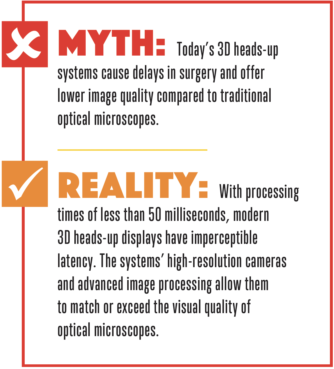
Concerns about latency with 3D heads-up systems originated with early models, whose slower processing and data transmission speeds led to noticeable delays. The high-speed cameras, graphics processing units, and data transmission technologies of contemporary systems have reduced latency to imperceptible levels. For instance, the Ngenuity system operates with a latency of less than 50 milliseconds,3 which is below the threshold detectable by the human eye and ensures seamless synchronization with hand movements.
Diakonis et al4 found no statistically significant difference in the completion time of critical surgical steps, such as the capsulorhexis and phacoemulsification, between procedures performed using traditional optical microscopes versus 3D HUDs. The study findings suggest that latency in modern systems does not compromise surgical performance.
IMAGE QUALITY
Superior Visual Quality With Modern 3D HUDs
A common misconception is that image quality—particularly resolution, contrast, and depth of field—is worse with 3D heads-up systems than with optical microscopes. This belief likely stems from early models that had lower-resolution cameras and less advanced image processing. Modern systems use 4k cameras to capture high-resolution images with a wide dynamic range. Advanced algorithms enhance contrast, adjust brightness, and sharpen details in real time to provide a crisp, clear view of the surgical field.
Depth of field is often a point of comparison between optical and digital systems. It is effectively managed by 3D HUDs through real-time focus adjustments and image stacking techniques. Studies have demonstrated that 3D HUDs provide comparable or superior image quality across various parameters, with enhanced visualization of ocular structures, especially in complex cases involving small pupils or dense cataracts.5
ERGONOMICS
Traditional optical microscopes require us to maintain a fixed posture, which can lead to neck and back strain during long procedures. In contrast, 3D HUDs allow us to sit in a more natural, upright position, reducing physical strain and fatigue. In a study by Weinstock et al,6 surgeons using 3D HUDs reported lower levels of musculoskeletal discomfort and were able to maintain focus and precision for longer periods. This can improve surgical performance, particularly during lengthy or complex procedures.
WHY 3D HUDS ARE THE FUTURE OF CATARACT SURGERY
The belief that 3D heads-up visualization introduces delays and offers inferior image quality compared with optical microscopes is rooted in outdated perceptions. Advances in camera technology, image processing, and data transmission have effectively eliminated latency concerns. Modern 3D HUDs deliver image quality that meets or exceeds that of traditional optical systems. Moreover, the ergonomic benefits of 3D heads-up systems and their digital enhancement capabilities make them an attractive choice for cataract surgery.
As this technology continues to evolve, 3D heads-up visualization may become the standard of care for ophthalmic surgery. Surgeons who have been hesitant to adopt this technology owing to concerns about delays or image quality can find reassurance in the evidence supporting the efficacy and benefits of modern 3D HUDs in cataract surgery.
1. Bausch + Lomb. Bausch + Lomb and Heidelberg Engineering announce the introduction of SeeLuma fully digital surgical visualization platform. April 13, 2023. Accessed September 23, 2023. https://ir.bausch.com/press-releases/bausch-lomb-and-heidelberg-engineering-announce-introduction-seelumatm-fully-digital
2. Beyeonics. Beyeonics One: revolutionizing visualization in cataract surgery. Beyeonics Technical Overview. 2024.
3. Alcon. NGENUITY 3D Visualization System: technical specifications and clinical applications. Alcon Product Literature. 2024.
4. Diakonis VF, Tsaousis KT, Watson C, Castellano K, Weinstock RJ. Cataract surgery using two 3D visualization systems: complication rates, surgical duration & comparison with traditional microscopes. Eur J Ophthalmol. Published online February 28, 2024. doi:10.1177/11206721241237298
5. Del Turco C, D’Amico Ricci G, Dal Vecchio M, et al. Heads-up 3D eye surgery: safety outcomes and technological review after 2 years of day-to-day use. Eur J Ophthalmol. Published online April 30, 2021. doi:10.1177/11206721211012856
6. Weinstock RJ, Ainslie-Garcia MH, Ferko NC, et al. Comparative assessment of ergonomic experience with heads-up display and conventional surgical microscope in the operating room. Clin Ophthalmol. 2021;15:347-356.


