What intrigues me the most about small-incision lenticule extraction (SMILE) are its refractive precision, lack of corneal flap, and patient’s comfort. Because the technique allows an accurate and precalculated lenticule and incision, it has proven to be a dependable procedure that enhances the surgical process for patients as well as surgeons. Currently in the United States, the approved treatment range is 1.00 to 10.00 D of spherical myopia.1 This article provides tips for performing SMILE.
AT A GLANCE
- Although small-incision lenticule extraction looks easy, the dissection component can be tricky in the first few cases, as the surgeon learns to identify the anterior and posterior aspects of the lenticule and obtain optimal centration prior to beginning photodisruption.
- Patients typically need to be more relaxed for SMILE than they do for PRK or LASIK, because eye movement or eyelid squeezing during the refractive photodisruption phase of the procedure can cause a break in suction and force the surgeon to abort the case.
PREPARATION
The Room
As with any refractive surgical procedure, there is preparation involved. Prior to performing SMILE, my setup is much as it is for LASIK. The necessary instruments include a locking speculum and a corneal dissector with two parts: (1) a pick end (similar to a Sinskey IOL rotator) to enter the interface, identify the edge of the lenticule, and delineate the anterior and posterior aspects of the lenticule and (2) a flat, spoon-like end to perform the dissection of the anterior and posterior aspects of the lenticule. I also keep a lenticule rhexis forceps (Rupal Shah, MD) on hand to free the lenticule from its bed if necessary and a toothless forceps (Pearse-Hoskins) for globe stabilization if needed.
The Patient
To physically prepare patients for SMILE, I instruct them to apply a topical antibiotic for several days prior to surgery. On the day of surgery, the eyelid skin and lashes are prepared with Betadine skin solution (Purdue Frederick). I then irrigate the conjunctival fornix for meibomian gland secretions. After entering the laser suite, the patient receives three doses of generic 0.5% proparacaine hydrochloride ophthalmic anesthetic drops in each eye. The first dose is administered no more than 2 minutes before I proceed to operate on the first eye.
There is another side of patients’ readiness for SMILE—emotional preparedness. Patients typically need to be more relaxed for SMILE than for PRK or LASIK, because eye movement or eyelid squeezing during the refractive photodisruption phase of a SMILE procedure, which lasts 14 seconds, can cause a break in suction and force the surgeon to abort the case.
I look for nonverbal cues that a patient may have an elevated propensity for eyelid squeezing or Bell phenomenon and supraduction leading to a break in vacuum. The first sign is if I find it difficult to instill eye drops or to place the speculum. Second, if a partner or technician is holding the patient’s hand and he or she is markedly squeezing the other person’s hand in return, that patient is still “too energized” for SMILE. To calm anxious patients, I increase the sedative and employ what I call verbal anesthesia, which entails relaxing the patient with oral coaching. These patients need to be constantly reminded to avoid a hard squeeze of their eyelids as well as looking up or over their head.

THE PROCEDURE
The Four Phases of Treatment
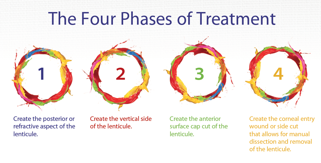
When the patient is under the laser, the green fixation light should be in the geometric center of the pupil before suction is initiated. Once the surgeon activates the vacuum, there are essentially four phases to the treatment (see above). The first phase is a photodisruptive pass to create the posterior or refractive aspect of the lenticule. The second phase is to create the vertical side of the lenticule. The third phase is to create the anterior surface or what is termed the cap cut of the lenticule. The fourth phase is the corneal entry wound or side cut that allows for manual dissection and removal of the lenticule.
The refractive lenticule will typically be 6.5 mm in diameter. The cap cut is typically 1 mm larger than the refractive lenticule and 0.5 mm on either side of the lenticule’s peripheral diameter. Additionally, the cap cut is parallel to the corneal surface and has zero refractive power. After manually dissecting the lenticule, the surgeon removes it through the small incision, leaving the remainder of the superficial cornea intact. The subsequent change in the cornea’s shape achieves the refractive correction.
Making Contact With the Cornea
The femtosecond laser designed to perform SMILE (VisuMax; Carl Zeiss Meditec) only contacts the cornea at the limbus. Without vacuum applied to the sclera, suction is weaker than that created by femtosecond lasers. The curved interface of the VisuMax is essential, because it avoids flattening or applanation of the cornea, which would have a significantly negative effect on refractive precision. A typical corneal flattening applanation will create stromal folds and result in irregular astigmatism if two surfaces such as a lenticule are created. This is not an issue with flap creation, but it is a significant issue when the goal is lenticule creation.
SURGICAL PEARLS
When Suction Breaks
As mentioned, if the Bell phenomenon occurs during the first pass or refractive cut, the case must be aborted. In such instances, the patient can no longer undergo SMILE but can undergo LASIK or PRK instead at a later date. If the surgeon gets past the refractive photodisruption phase and loses vacuum during any of the later three phases of the SMILE procedure, the surgeon may redock the laser, continue with the procedure, and then complete the manual dissection.
Bennett Walton, MD, demonstrates his small-incision lenticule extraction technique.
Identification of the Lenticule’s Anterior and Posterior Aspects
The second and most important part of SMILE is the manual dissection. The goal is to definitively identify the anterior and posterior aspects of the lenticule; otherwise, the case becomes challenging. The surgeon must identify the anterior aspect of the anterior and posterior lenticule prior to beginning the manual dissection with the corneal dissector. It is easiest to complete the anterior interface dissection followed by the posterior dissection. After removing the lenticule and directly visualizing the unfolded tissue, the surgeon withdraws the speculum and administers antibiotic and corticosteroid drops. There is no need for an eye shield. The patient will continue using the eye drops for a total of 5 days and will be seen for follow-up at 1 day, 2 weeks, and 3 months.
Visual disparities between LASIK and SMILE have been reported with earlier versions of SMILE. With laser energy optimization via adjusting individual spot energy and assessing ideal spot and track spacing, the return of visual acuity quantity and quality of vision can be identical for SMILE and LASIK.
Early in a surgeon’s experience, a disparity compared with LASIK may be noted, but with relatively quick femtosecond laser optimization, this difference either completely collapses or becomes virtually clinically insignificant. Greater biomechanical stability and less dry eye have been reported repeatedly with SMILE as compared with LASIK.
As with LASIK, maintaining epithelial integrity is important with SMILE. If an abrasion occurs on the endothelial cap, a prophylactic diffuse lamellar keratitis topical steroid regimen should begin on the day of the surgery. This complication will likely be rare.
CONCLUSION
SMILE is a new procedure. Surgeons completing their due diligence prior to performing the surgery will help ensure that it is not an anxiety-ridden experience. Although SMILE looks easy, the dissection component can be tricky in the first few cases. If the surgeon learns how to accurately identify the anterior and posterior aspects of the lenticule and then proficiently completes the manual dissection, he or she will find the procedure rewarding, and the patients will be most appreciative of their new improved unaided visual abilities.
1. FDA approves VisuMax Femtosecond Laser to surgically treat nearsightedness [news release]. September 13, 2016. http://www.fda.gov/NewsEvents/Newsroom/PressAnnouncements/ucm520560.htm. Accessed May 26, 2016.


