CASE PRESENTATION
An 85-year-old man with a 9-year history of severe neck contracture secondary to ankylosing spondylitis was referred for cataract evaluation. The patient had initially presented to the practice 2 years earlier with a complaint of decreased vision and difficulty reading and watching television. Cataract surgery was not performed at that time because of challenges associated with performing the surgery.
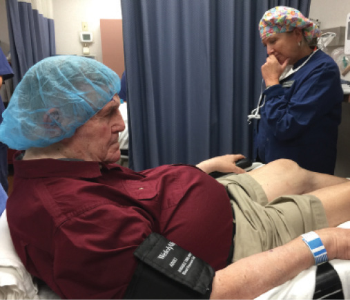
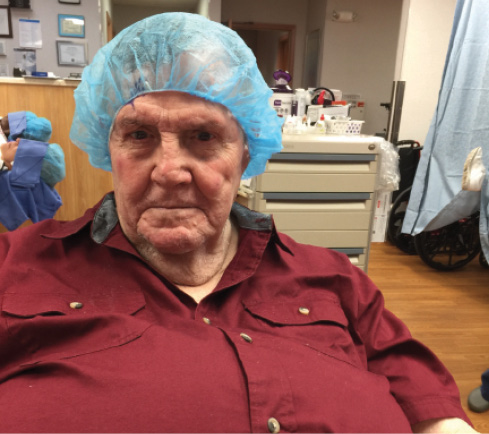
An examination of the patient is remarkable for his fixed position, with his neck immobile and fixed in a contracted position such that his face is parallel to the floor, 90º to normal (Figure). In addition, he has kyphosis and is unable to move his head to the right at all and only minimally to the left. Distance BCVA is 20/70 OU, with a manifest refraction of +1.00 -0.50 × 095 OD and +2.00 -1.25 × 095 OS. A handheld slit-lamp examination and indirect biomicroscopy are remarkable for 3+ nuclear sclerotic cataracts and mild retinal pigment epithelial changes in the macula.
What considerations are there for this patient with regard to preoperative measurements, intraoperative positioning, and the mechanics of surgery? What additional risks need to be discussed with him? What would your plan be for postoperative care? What is the refractive goal?
—Case prepared by Cathleen M. McCabe, MD.


Figure. The patient’s neck is immobile and fixed in a contracted position such that his face is parallel to the floor, 90º to normal.

ASHA BALAKRISHNAN, MD
Standard optical biometry and topography measurements are not feasible owing to patient positioning. Portable keratometry and A-scan devices can be used for preoperative measurements. Prior to surgery, I would discuss the risks of posterior pressure from positioning and prolonged postoperative inflammation and pressure spikes, along with regular operative risks. Given the intraoperative challenges, one could consider operating on one eye only to improve vision and minimize risk. Under those circumstances, I would choose the left eye and aim for a refractive target of plano or slight myopia. Considering the retinal pigment epithelial changes, I would highlight the possibility that visual potential may not be 20/20, but cataract surgery would optimize the vision notwithstanding retinal issues.
In the OR, pillows would have to be placed under the patient’s legs and a foam support under the head to recline the bed and tilt the head into a more supine, parallel position to the floor. It will be challenging to recline the head into usual operative positioning, so the head will be at an angle to the floor. It will be important to be mindful of posterior pressure, because the patient’s legs will be above the level of his head. If the angle of the head does not allow the ophthalmologist to sit for surgery, he or she may have to consider an assistant surgeon to help operate the microscope and phaco machine pedals. I would consider administering acetazolamide (Diamox; Wyeth Pharmaceuticals) preoperatively to decompress the vitreous in the event of excessive posterior pressure with the positioning. Intraoperative aberrometry could be performed to provide additional lens measurements.

JAMES T. BANTA, MD
I have dealt with a number of patients with severe kyphosis over the years, but this extreme example is daunting to say the least. To place this patient in a normal surgical position, he would need to be strapped to a bed capable of a nearly vertical incline, something that is neither available nor safe. Trendelenburg positioning can usually create approximately 50º of tilt, and the bed is elevated to bring the horizontal plane high enough to use the operating microscope. Both the phaco and microscope pedals are controlled by the outside foot, because the surgeon’s knees will not fit under the head of the bed, allowing the use of only one foot.
The patient may very well need to be strapped in to maintain a safe, stable position, and shoulder wedges may need to be used to prevent slipping. Although his brow would still create a difficult view, the surgery could likely be undertaken safely. I always counsel these patients preoperatively that I will abort the surgery if I do not feel comfortable under the microscope once the best positioning has been accomplished. Needless to say, these cases should be booked with extra time, and anesthesia staff needs to be involved early on to make certain that extreme Trendelenburg positioning can be used safely.

LISA M. NIJM, MD, JD
Detailed preoperative planning is the key to success in challenging cases involving patients with limited mobility. I would confirm preoperative measurements with a manual A-scan and keratometry, because automated, rigid devices are prone to error with these patients. To decrease stress at the time of surgery, I would have the patient visit the OR first for a “trial run” of proper positioning. Several surgical techniques have been described to address kyphosis, from operating vertically1 to using an orthopedic table2 to maximal reverse Trendelenburg with multiple pillows behind the head and legs.3 One could also place a single 7–0 Vicryl corneal traction suture (Ethicon) inferiorly to better manipulate the eye into position.4
These cases often require creativity on the part of the surgeon, and my technique of choice varies depending on the results of my trial run. I would counsel the patient on the type of anesthesia (general vs heavy sedation), risks of capsular rupture from increased posterior pressure owing to positioning (trypan blue, mannitol, iris expanders available), and increased risk of intraocular inflammation secondary to ankylosing spondylitis.
In conjunction with the patient’s rheumatologist, I would use any additional pre- or postoperative anti-inflammatory agents needed to decrease the risk of complications. The target refraction will depend on the patient’s goals. He has difficulty seeing at distance and near but also has retinal pigment epithelial changes, which may limit the use of a multifocal lens. A toric lens (if indicated) with monovision may be an option, depending on the patient’s preference.

WHAT I DID: CATHLEEN M. McCABE, MD
Preoperatively, I counseled the patient that it might be impossible to achieve a position that would enable safe and effective cataract surgery. Additionally, we discussed the increased risks of (1) an intraocular complication related to elevated vitreous pressure and (2) posterior capsular rupture or corneal endothelial damage owing to unusual biomechanics and fluidics. I also reviewed with him the difficulties that would be present in managing an intraoperative complication.
Biometry was performed preoperatively with immersion A-scan and a handheld keratometer. It was difficult to obtain reproducible keratometry readings, so an average of the obtained measurements was used for the right eye. I chose an IOL power for a myopic result of -1.25 D, given the patient’s fixed head position and near focal point for most tasks.
He was brought into the preoperative area for a trial run with regard to positioning. It became apparent that maximal reverse Trendelenburg of the stretcher would only result in orienting the patient’s head perpendicular to the floor and facing forward. A second stretcher was placed underneath the primary operating table to provide a failsafe, in case the increased load on the head of the bed resulted in unexpected movement of the patient. On the day of surgery, the operating microscope was positioned parallel to the floor facing the patient’s left eye. As a result, it was necessary for me to operate with arms extended to reach the operative eye. I placed a dispersive viscoelastic on the cornea, and surgery proceeded uneventfully despite altered fluidics and the patient’s coughing during the capsulorhexis (see video below).
Another consideration in this case was the patient’s difficulty instilling eye drops. At the conclusion of surgery, I used a 27-gauge cannula for transzonular administration of an antibiotic and anti-inflammatory medication (triamcinolone/moxifloxacin; ImprimisRx Compounding Pharmacy). I used ReSure Sealant (Ocular Therapeutix) to ensure watertight wound closure. Postoperatively, the patient instilled topical drops (besifloxacin ophthalmic suspension 0.6% [Besivance; Bausch + Lomb], bromfenac ophthalmic solution [Prolensa; Bausch + Lomb], and difluprednate ophthalmic emulsion 0.05% [Durezol; Alcon]) with a unique silicone wand (SimplyTouch; SimplyTouch) that does not require the head to be tilted back.
The patient was extremely grateful for the effort that was made to perform cataract surgery and improve his vision. The postoperative period was uncomplicated, and he was happy to be watching television and reading again. Positioning of the right eye proved to be a much more complicated endeavor. Watch the video at bit.ly/mccabe1217.
1. Ang GS, Ong JM, Eke T. Face-to-face seated positioning for phacoemulsification in patients unable to lie flat for cataract surgery. Am J Ophthalmol. 2006;141(6):1151-1152.
2. Prasad S, Kamath GG, Phillips RP. Phacoemulsification in a patient with marked cervical kyphosis. J Cataract Refract Surg. 2000;26(8):1258-1260.
3. Gordon MI, Rodríguez AA, Olson MD, Miller KM. Pillow case. J Cataract Refract Surg. 2005;31(9):1824-1825.
4. Kooner KS, Barte FM. Intraocular surgery in kyphosis: an easier approach. Case Rep Ophthalmol. 2013;4(2):34-38.


