Primary headaches are a set of complex pain disorders that can have a heterogeneous presentation. Some of these presentations can often involve symptoms that suggest otolaryngological and ophthalmological diagnoses. In this second installment of a two-part series, the symptoms and diagnoses that overlap between ophthalmology and neurology will be addressed (please see the March 2014 issue of Cataract & Refractive Surgery Today for the otolaryngology installment of this series). The two main complaints that patients present with for ophthalmology consultations are eye pain and visual disturbances. When ophthalmological evaluations including slit-lamp examinations and tonometry are unremarkable, other diagnostic possibilities should be considered.
MIGRAINE AND CLUSTER HEADACHE
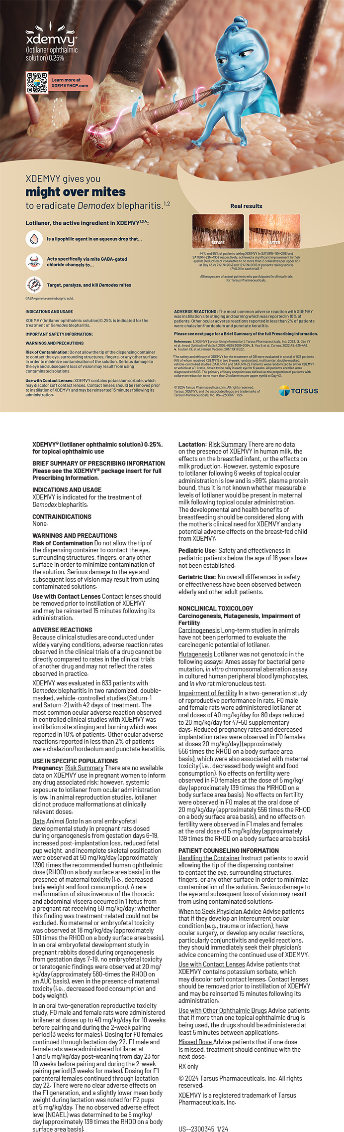
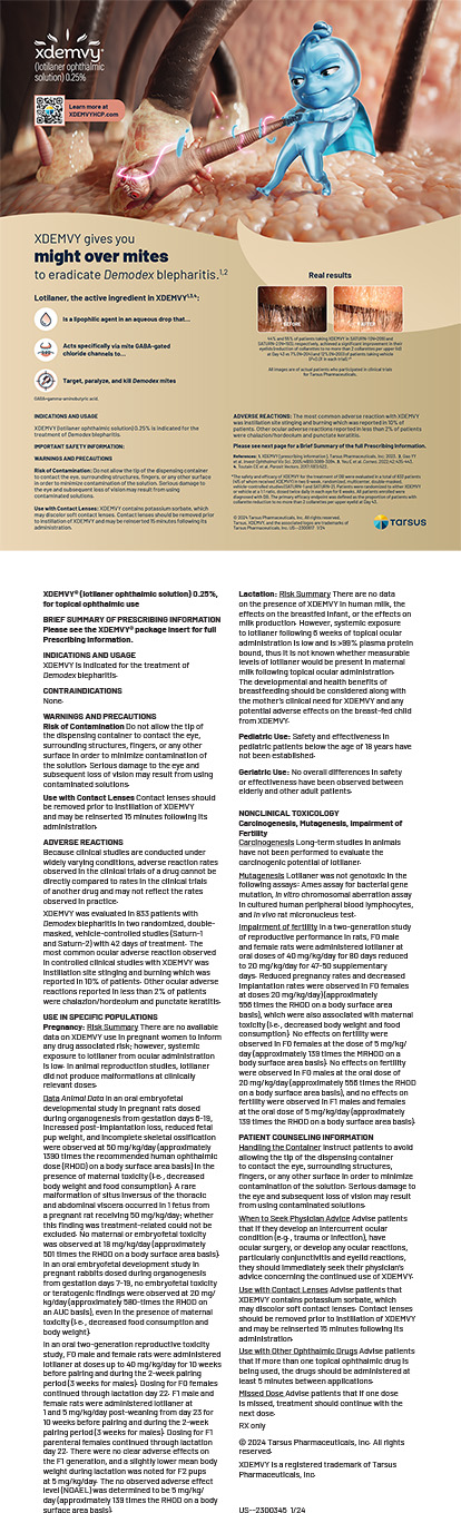
The numerous causes of eye pain include blepharitis, iritis, keratitis, conjunctivitis, glaucoma, and corneal abrasions. Patients with both migraine and cluster headache can also present with pain that is focused around the orbit. Patients with cluster headache often describe the pain as stabbing in quality with a location in or behind the eye. The pain of cluster headache can also be accompanied by an orbital foreign body sensation. Cluster headache is a trigeminal autonomic cephalgia (cluster headache, short-lasting unilateral neuralgiform headache with conjunctival injection and tearing or SUNCT, short-lasting unilateral neuralgiform headache attacks with cranial autonomic symptoms or SUNA, paroxysmal hemicrania, hemicrania continua), which consists of hemicranial headaches that can involve ipsilateral ptosis, conjunctival injection, and lacrimation. These features, along with eye pain, can be very suggestive of an ophthalmological disorder to the unknowing patient or provider, rather than a primary headache disorder.1 Although autonomic features are not part of the diagnostic criteria for migraine, in a study of 786 migraine patients (625 women, 61 men), 56% of the subjects experienced autonomic symptoms, but these symptoms tended to be bilateral.2
In addition to pain around the orbit, migraines can also commonly involve blurry vision,3 which tends to occur during the headache phase of the migraine and increases as the intensity of the pain peaks. Blurry vision should not be confused with migraine visual aura. Visual auras by definition are fully reversible, homonymous visual symptoms that can involve positive features (scintillating scotomas, fortification spectrum) and/or negative features (areas of visual loss). Visual auras typically last from 5 to 60 minutes and can be accompanied by other aura phenomena in succession such as sensory or speech disturbances.1 A major misconception about migraine aura is that it must occur prior to the onset of the headache. This is often not the case in clinical practice. A multicenter study that included 267 patients demonstrated that the majority of patients experienced aura during the early headache phase of their migraine.4
A migraine subtype known as basilar migraine can involve visual aura symptoms simultaneously in both temporal and nasal fields of both eyes as well as diplopia. The term basilar migraine was coined by Bickerstaff in 1961, who suggested that basilar artery vasoconstriction is part of the pathophysiology of this migraine subtype.5 To date, there has not been any significant evidence suggesting that posterior circulation oligemia or ischemia occur as part of the pathophysiology of basilar migraine. As such, migraine with brainstem aura is the new terminology that is used for this disorder in the The International Classification of Headache Disorders 3rd edition beta version, a test version of the classification that was released prior to the pending release of the finalized version.6 Monocular positive and/or negative visual disturbances associated with migraines are categorized as retinal migraines. Making this diagnosis can be difficult, as many patients will often consider the hemianopsia associated with classical visual aura to be a problem of a single eye rather than a binocular defect. Examining a patient during an attack would be an optimal but impractical solution. As such, having the patient evaluate the visual disturbance with one eye closed can help to confirm this diagnosis.1 As with any migrainerelated visual disturbance, retinal migraine is a diagnosis of exclusion, and other causes of monocular visual loss should be considered.
ARTERIAL DISSECTIONS
For patients with eye pain in the setting of trauma, arterial dissections should be considered. In one study of 135 subjects with internal carotid artery dissection, 60% presented with anterior headache that was typically ipsilateral to the side of the dissection, and 10% had eye, facial, or ear pain without headache.7 Even after radiographic resolution of the dissection, up to 25% of patients can experience chronic persistent headaches and/or pain.8
It is estimated that about 25% of patients with carotid artery dissections experience Horner syndrome due to damage of the oculosympathetic pathway. Horner syndrome due to damage to the internal carotid artery can result in ptosis and miosis but typically not anhydrosis. The sympathetic fibers responsible for facial sweating have a more proximal branching point and travel along the external carotid artery. Up to 56% of patients with carotid dissections can experience cerebral ischemia in the form of a transient ischemic attack or stroke.9 In another study, 28% of subjects suffered from transient monocular visual loss due to ischemic optic neuropathy, and 16% experienced scintillations that were thought to be due to acute choroidal hypoperfusion. Of these patients who experienced monocular visual symptoms due to carotid artery dissection, 36% subsequently experienced a nonreversible ocular or hemispheric stroke.10 As such, a history of even mild neck trauma in a patient with ipsilateral pain, monocular visual symptoms, and/ or Horner syndrome should trigger further evaluation, because the early diagnosis and treatment of carotid dissection can potentially prevent permanent deficits.
TEMPORAL ARTERITIS
In the setting of a patient over the age of 50 years with eye pain with or without visual changes, temporal arteritis (TA) should certainly enter the differential diagnosis. Other symptoms suggestive of this diagnosis are temporal artery prominence, temporal regional tenderness, jaw claudication, tongue claudication, and fever. Erythrocyte sedimentation rate and C-reactive protein are both reasonable first-line tests; however, up to 1.2% of patients with TA can have normal values.11 TA also involves branches of the aorta with frequent involvement of the extracranial branches of the carotid artery. TA involves areas of segmental mononuclear cell inflammatory infiltrates, often with giant cells, which can lead to normal temporal artery biopsy results in up to 15% of the cases.12 Even in cases with negative test results, if there is a high index of suspicion based on clinical presentation, treatment with steroids should be pursued, as permanent vision loss or blindness can result from untreated TA.
OPTIC NEURITIS
Consider optic neuritis in younger patients that present with monocular vision loss and eye pain. Some of the hallmark features of optic neuritis include pain with eye movement and color desaturation. Although optic neuritis can occur in isolation, it can be a presenting symptom of multiple sclerosis or neuromyelitis optica. With both of these disorders, patients will often have a remote history of transient focal weakness, numbness, and vision changes that resolved spontaneously. Based on clinical suspicion, magnetic resonance imaging of the brain with contrast, spinal imaging, lumbar puncture, and/or aquaporin-4 antibody testing should be considered. Treatment with intravenous methylprednisolone for 3 days followed by an oral prednisone taper has demonstrated a decrease in optic neuritis recovery time, and patients tended to have better outcomes at 6 months.13
IDIOPATHIC INTRACRANIAL HYPERTENSION
Visual deficits in an obese patient with headaches can be suggestive of idiopathic intracranial hypertension (IIH; pseudotumor cerebri). Although normal-weight individuals can also have IIH, it is a diagnosis that mostly affects obese women. A lumbar puncture demonstrating an opening pressure of more than 200 mm H2 O in normalweight individuals or more than 250 mm H2 O in obese patients can confirm the diagnosis. In addition to being diagnostic, lumbar punctures can be therapeutic, as the withdrawal of cerebrospinal fluid to reduce pressure to 120 to 170 mm H2 O can improve headaches.1
Patients with IIH can suffer from transient visual obscurations, which, in untreated cases, can progress to optic neuropathy and even blindness. In such cases, emergency optic nerve fenestration should be pursued. Headaches in the setting of IIH tend to improve when the individual is upright and worsen with lying flat. As such, many patients can experience their worst pain upon awakening in the morning. Patients with IIH can also present with papilledema, sixth-nerve palsy, nausea, and vomiting. In clinical practice, vomiting can lead to rapid improvement of headache pain in patients with IIH. This contrasts with the vomiting associated with migraine, which does not typically involve an associated improvement of pain.
Weight loss has proven to be one of the most effective treatments of IIH, although the exact mechanism of improvement is unclear. As such, gastric bypass surgery has demonstrated some efficacy in improving IIH.14 This option may be particularly useful in patients with comorbid sleep apnea, diabetes, hypertension, and osteoarthritis.15 In refractory cases, ventriculoperitoneal or lumboperitoneal shunts can be considered.
CONCLUSION
There are numerous symptoms that can suggest both ophthalmological and neurological disorders. A careful history and physical examination can often lead to the correct diagnosis, eliminating the need for unnecessary testing. In refractory or atypical cases, further evaluation should be pursued, including imaging studies and formal ophthalmological evaluations.
Paul G. Mathew, MD, FAHS is a member of the Harvard Medical School faculty. He is the director of continuing medical education at Brigham & Women’s Hospital, Department of Neurology, John R. Graham Headache Center, and the director of headache medicine at the Cambridge Health Alliance. He is board certified in neurology and headache medicine. Dr. Mathew may be reached at pmathew@partners.org.
- Headache Classification Committee of the International Headache Society: The International Classification of Headache Disorders. 2nd ed. Cephalalgia. 2004;24(suppl 1):9-160.
- Lai TH, Fuh JL, Wang SJ. Cranial autonomic symptoms in migraine: characteristics and comparison with cluster headache. J Neurol Neurosurg Psychiatry. 2009;80(10):1116-1119.
- Silberstein SD. Migraine symptoms: results of a survey of self-reported migraineurs. Headache. 1995;35(7):387-396.
- Hansen JM, Lipton RB, Dodick DW, Silberstein SD, et al. Migraine headache is present in the aura phase: a prospective study. Neurology. 2012;79(20):2044-2049.
- Bickerstaff ER. Basilar artery migraine. Lancet. 1961;1:15-17.
- Headache Classification Committee of the International Headache Society (IHS). The International Classification of Headache Disorders. 3rd edition (beta version). Cephalalgia. 2013;33(9):629-808.
- Silbert PL, Mokri B, Schievink WI. Headache and neck pain in spontaneous internal carotid and vertebral artery dissections.Neurology. 1995;45(8):1517-1522.
- Schytz HW, Ashina M, Magyari M, at al. Acute headache and persistent headache attributed to cervical artery dissection: field testing of ICHD-III beta [posted online ahead of print February 5, 2014]. Cephalalgia. doi: 10.1177/0333102414520767.
- Lee VH, Brown RD Jr, Mandrekar JN, Mokri B. Incidence and outcome of cervical artery dissection: a populationbased study. Neurology. 2006;67(10):1809.
- Biousse V, Touboul PJ, D’Anglejan-Chatillon J, et al. Ophthalmologic manifestations of internal carotid artery dissection. Am J Ophthalmol. 1998;126(4):565.
- Myklebust G, Gran JT. A prospective study of 287 patients with polymyalgia rheumatica and temporal arteritis: clinical and laboratory manifestations at onset of disease and at the time of diagnosis. Br J Rheumatol. 1996;35:1161.
- Nesher G. The diagnosis and classification of giant cell arteritis. J Autoimmun. 2014;48-49:73-75.
- Beck RW, Cleary PA, Anderson MM Jr, et al. A randomized, controlled trial of corticosteroids in the treatment of acute optic neuritis. The Optic Neuritis Study Group. N Engl J Med. 1992;326(9):581-588.
- Fridley J, Foroozan R, Sherman V, et al. Bariatric surgery for the treatment of idiopathic intracranial hypertension. J Neurosurg. 2011;114(1):34-39.
- Li JF, Lai DD, Lin ZH, et al. Comparison of the long-term results of roux-en-y gastric bypass and sleeve gastrectomy for morbid obesity: a systematic review and meta-analysis of randomized and nonrandomized trials. Surg Laparosc Endosc Percutan Tech. 2014;24(1):1-11.