Maintaining a physiologic IOP during cataract surgery can provide significant benefits in terms of safety and surgical efficiency. Recent advances in phaco technology help in this pursuit and ensure a stable and controlled surgical environment (see A Closer Look at Advanced Fluidics).
A CLOSER LOOK AT ADVANCED FLUIDICS
Advanced fluidics systems combine intelligent pressure sensing and real-time feedback with advanced algorithms to adjust fluid flow and vacuum levels automatically during surgery. Optimizing fluid dynamics during cataract surgery can improve surgeon control of the anterior chamber.
Intelligent Pressure Sensing and Real-Time Feedback
Advanced fluidics systems use pressure sensors to monitor and measure IOP continuously during surgery. The system provides real-time feedback on IOP levels, ensuring that the fluid flow and vacuum levels are adjusted to maintain a stable anterior chamber and prevent complications.
Automated Fluid Flow and Vacuum Level Adjustments
Based on the real-time feedback, the technology automatically adjusts fluid flow and vacuum levels to maintain the desired IOP.
Integration with Advanced Phaco Systems
Advanced fluidics technology can be found in some of the latest phaco systems, such as the Centurion Vision System (Alcon) and Stellaris Elite (Bausch + Lomb).
The benefits of a low IOP during cataract surgery are numerous and include improved safety and surgical efficiency.1-4 Emerging evidence also suggests that equivalent or improved clinical outcomes can be achieved when operating at lower IOPs with minimal changes in anterior chamber stability and working space between the corneal endothelium and lens. Additionally, I have found that operating at IOPs in the lower 40s mm Hg contributes to an enhanced overall surgical experience for patients.
AT A GLANCE
- Maintaining a low IOP during cataract surgery can improve patient safety and surgical efficiency.
- Technological advances in phaco machines can help maintain stable and low IOP during surgery.
- Sustaining a physiologic IOP during surgery can create better working space between the corneal endothelium and lens, maintain the posterior capsule’s position and anterior chamber stability, reduce fluid usage and loss, lessen the impact on the iris in eyes with intraoperative floppy iris syndrome, and decrease corneal edema and intraocular inflammation.
EMERGING EVIDENCE
I studied the anterior and posterior physiologic changes and vascular alterations of phacoemulsification performed at a traditionally high IOP versus a low, more physiologic IOP.1,2 A total of 25 patients with Lens Opacities Classification System grade III cataracts, no history of intraocular surgery, and unremarkable systemic and ocular health were enrolled.
Delayed sequential, uncomplicated bilateral phacoemulsification was performed with the Centurion Vision System with Active Sentry (Alcon). During surgery, IOP was maintained at 28 mm Hg or lower in one eye and 60 mm Hg or higher in the other.
Anterior chamber. Low IOP settings resulted in reduced fluid consumption compared with high IOP settings.1 Moreover, the cumulative dissipated energy was lower in the low IOP group than in the high IOP group. Alterations in corneal thickness were less pronounced in the low IOP group, both at the pupillary center and at the thinnest point at the 1-day and 1-week marks. By 1 month postoperatively, however, corneal thickness changes were equivalent between groups. Further, the low IOP group experienced a smaller reduction in endothelial cell density at 1 month postoperatively compared with the high IOP group. Visual acuity was consistent between the two groups at all measured time points.
Posterior chamber. Changes to the macular thickness; subfoveal choroidal thickness; foveal avascular zone (FAZ); and macular vasculature by OCT angiography were measured at baseline, postoperative day 1, and postoperative weeks 1, 4, and 12.2 A masked, expert third party read and provided quantified assessments of retinal and choroidal images.
Surgery with low IOP settings used less fluid than surgery with high IOP settings. Foveal and macular central subfield thicknesses showed a trend toward increasing in both groups, but statistical significance was not reached in either. The FAZ area did not change in any patients. In the low IOP group compared to the high IOP group, however, the FAZ 1-week change differed. At 1 month, FAZ changes from baseline were comparable between groups. Vessel density in the four quadrants was stable in all patients and across surgical IOP settings. Visual acuity was similar between the two groups at all time points.
ADVANTAGES of Low IOP Surgery
Working space and posterior capsule position. Protecting adjacent intraocular structures during cataract surgery and maintaining normal anatomic relationships are crucial. It is particularly important to preserve the working distance between the corneal endothelium and lens. Furthermore, reliable, safe surgery requires maintaining the posterior capsule’s position. Surge, which occurs when fluid outflow exceeds fluid inflow, can destabilize the anterior chamber depth and posterior capsule’s position. My experience with a phaco platform that maintains a more physiologic IOP is characterized by undetectable surge. Not only can more physiologic IOP settings maintain a good working space in the anterior chamber, but they can also reduce surge-type responses and resultant movement of the posterior capsule.
Surgical efficiency. This is largely driven by machine fluidics. In my experience, cataract surgery at a more physiologic IOP is just as efficient as surgery at a high IOP. I have noticed no difference in case times, cumulative dissipated energy values, and material delivery operating in both settings. During phaco chop, early fragment removal is facilitated by nuclear material appearing to float within the capsular bag. Low IOP surgery allows a roomier anterior segment and excellent surgical efficiency without the need for high-pressure fluidics.
Less fluid usage and loss. The amount of fluid that moves through the eye during cataract surgery is directly proportional to the average intraoperative IOP. At a high IOP, extensive fluid leakage can occur at the main wound and paracentesis incisions. Sleeve optimization at the phaco needle, quality wound construction, and surgeon technique can play a role in minimizing fluid loss. A phaco platform that maintains lower, more physiologic IOP settings, however, results in less fluid usage, even with dense cataracts. In my opinion, operating at a lower IOP plays a major role in decreasing fluid loss at the incisions.
Reduced impact on the iris. Successful and reproducible cataract surgery depends heavily on obtaining and maintaining adequate pupillary dilation throughout the case. Intraoperative floppy iris syndrome is defined by progressive miosis and iris billowing during cataract surgery and can be problematic for the surgeon. I have observed that the use of systemic alpha-blockers can be associated with greater maintenance of pupil size and stability of the iris when operating in a low IOP environment. This is thought to be related to less fluid usage and less fluid turbulence.5,6
Minimized corneal edema and endothelial cell loss. In a paired eye study comparing surgery on identically graded cataracts, procedures performed at low IOP settings caused less corneal edema in eyes on both days 1 and 7 when compared to procedures performed at high IOP. Additionally, low IOP surgery resulted in less endothelial cell loss at 1 and 3 months.2
Decreased intraocular inflammation. Intraocular inflammation can be associated with ocular pain and endothelial cell dysfunction. My analysis found less inflammation on day 1 postoperatively in low compared to high IOP surgery.
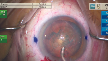
Reduced risk of reverse pupillary block. One of the insights I gained while transitioning from a high IOP environment to a low one is the notable reduction in reverse pupillary block. Reverse pupillary block was initially described in high myopes, but it can occur more often when operating at a high IOP.6 Introducing the phaco handpiece for nucleus removal at a high IOP can lead to a consistent backbowing of the lens-iris diaphragm. This phenomenon is significantly reduced with low IOP settings (Figure).

Figure. Iris configuration at high IOP resulting in more intense pupillary dilation and backbowing of the iris (A). Iris configuration at a physiologic IOP resulting in a less dilated pupil and backbowing of the iris (B).
THE FUTURE OF CATARACT SURGERY
Advances in phaco technology can support excellent cataract surgery performance at a lower, more physiologic IOP, maintain safety, and increase efficiency.
1. Rauen MP. Phacoemulsification at high IOP and physiologic IOP: Impact on anterior segment physiology. Paper presented at: ASCRS Annual Meeting; May 5-8, 2023; San Diego.
2. Rauen MP. Phacoemulsification at high IOP and physiologic IOP: Impact on posterior segment physiology. Paper presented at: ASCRS Annual Meeting; May 5-8, 2023; San Diego.
3. Beres H, de Ortueta D, Buehner B, Bernd Scharioth G. Does low infusion pressure microincision cataract surgery (LIPMICS) reduce frequency of post-occlusion breaks? Rom J Ophthalmol. 2022;66(2):135-139.
4. Zhao Z, Yu x, Yang X, et al. Elevated intraocular pressure causes cellular and molecular retinal injuries, advocating a more moderate intraocular pressure setting during phacoemulsification surgery. Int Ophthalmol. 2020;40(12):3323-3336.
5. Chang DF, Campbell JR. Intraoperative floppy iris syndrome associated with tamsulosin. J Cataract Refract Surg. 2005;31:664-673.
6. Cionni RJ, Barros MG, Osher RH. Management of lens-iris diaphragm retropulsion syndrome during phacoemulsification. J Cataract Refract Surg. 2004;30:953-955.


