
When a Book’s Cover Tells Opposing Stories
Four surgeons' readings of the ocular surface evaluation.
Key Takeaways
- Thicker corneal epithelium in an area of steepening is more suggestive of ocular surface disruption than stromal disease.
- The interpretation of corneal topography must take epithelial mapping and tomography into account.
- Dry eye induced by contact lenses sometimes bothers patients the most during the initial days or weeks after lens wear is discontinued because the irritated eyelids are touching the cornea more than when the lenses were present.
Case Presentation
A 41-year-old woman presents for a refractive surgery screening. The patient habitually wears monofocal soft contact lenses. She discontinued wearing her contact lenses 2 weeks ago and experienced dry eye symptoms for the first 2 days. Then, her comfort returned to normal with her glasses.
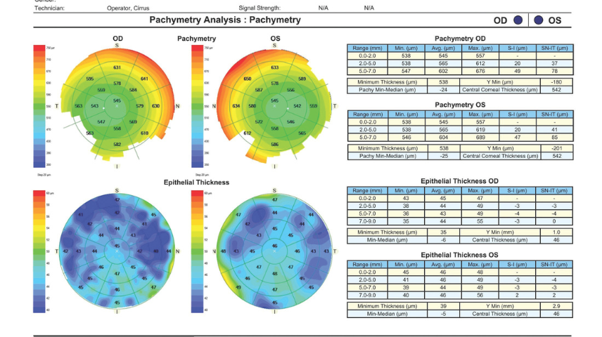
Upon presentation, the patient's uncorrected distance visual acuity is 20/100 OU, and her BCVA is 20/20 OU with a manifest refraction of -2.00 -1.00 x 175° OD and -2.00 -0.50 x 015° OS. Topography, tomography, and corneal OCT scans are shown in Figures 1 and 2.


She is interested in full distance correction for the right eye and a refractive target of -0.50 D in the left eye. How would you proceed?
— Case prepared by O. Bennett Walton IV, MD, MBA
Michael D. Greenwood, MD
The patient presents with low myopia, mild astigmatism, and a desire for blended vision. Her history is notable for transient dry eye symptoms immediately following contact lens cessation, indicating active ocular surface changes.
Corneal OCT epithelial thickness profiling confirms significant irregularity characterized by nonuniform, asymmetric remodeling across both corneas. In addition, tomography reveals bilateral inferior corneal steepening. The left eye exhibits a highly suspicious topographic pattern, with an elevated inferior-superior ratio of 2.76.
Although these inferior steepening patterns mimic early ectasia or the findings in a keratoconus suspect, the highly irregular epithelial maps and the patient's clinical history strongly indicate a confounding etiology: contact lens–induced corneal warping exacerbated by localized dry eye disease (DED) or meibomian gland dysfunction. Her initial 2-week contact lens holiday was simply insufficient to achieve structural homeostasis.
Proceeding immediately with laser vision correction would carry an unacceptable risk of an unpredictable ablation profile or permanent structural ectasia. Instead, the patient would be asked to remain out of her soft contact lenses for an additional 2 to 4 weeks to allow complete mechanical decompression of the cornea. Aggressive ocular surface therapy with preservative-free artificial tears and targeted DED management would be initiated to normalize the epithelium. In 3 to 4 weeks, tomography and epithelial mapping would be repeated. Laser treatment would be planned only once consecutive maps confirm complete anatomic stability.
If the topography completely regularizes, the patient's requested monovision strategy could be safely executed via LASIK or PRK. If, however, the inferior steepening and asymmetric elevation parameters persist despite a normalized epithelium, she would be managed as a keratoconus suspect, and PRK with close follow-up and possible CXL would be recommended.
Luke Rebenitsch, MD
The patient has compound myopic astigmatism, early presbyopia, and contact lens intolerance. She may also have DED. Because the dry eye symptoms improved after contact lens wear was discontinued, the condition is more likely to be contact lens related. Given the irregularity of the epithelial thickness maps, however, either she has not fully recovered from her use of contact lenses, or she truly has DED.
In situations like this, I usually opt for the EVO ICL (STAAR Surgical). In the United States, however, where I practice, the lowest power available is a -3.00 D spherical equivalent, which is equivalent to -2.50 D at the spectacle plane. Unfortunately, this would not allow her to have the blended vision that she desires. It would also be considered an off-label use given that her aqueous depth is less than 3 mm, although that is greater than the international standard of 2.8 mm.
I would therefore either proceed with wavefront-optimized PRK or aggressively treat the ocular surface and revisit the patient's candidacy in another 1 or 2 months. If epithelial thickness mapping demonstrates improvement, I would consider keratorefractive lenticule extraction or LASIK; of these, the former would be my preference because it disrupts the ocular surface less. I would also counsel the patient that she is likely at risk of ocular surface disease in the future and should be evaluated yearly going forward.
Evan D. Schoenberg, MD
The first question is whether laser vision correction will be safe and effective for this patient. Her topography (Figure 2) is notable for asymmetric with-the-rule bow tie astigmatism. The inferior to superior difference is 1.40 D OD and 2.60 D OS, which warrants scrutiny. The anterior and posterior elevation maps are unremarkable. The pachymetry maps show a mild temporal displacement of the thinnest zone of the cornea without focal thinning. Epithelial mapping (Figure 1) demonstrates mild relative epithelial thickening inferiorly. If the patient has early keratoconus, I would expect thinning, not thickening, in the topographically steep zone, in which case I would recommend against corneal refractive surgery.
The patient's reported success with monofocal soft contact lenses configured for only mild blended vision (-0.50 D target in the nondominant eye) suggests that she has good vision quality despite somewhat irregular astigmatism and that she is prepresbyopic. I would confirm that her refraction is stable and that she has reasonable expectations regarding future presbyopia progression. I would also discuss her vision quality because the irregular astigmatism could be causing nighttime dysphotopsias.
Although the lack of epithelial thinning points away from keratoconus as the source of inferior steepening, I think PRK would be safer for her than LASIK. The inferior to superior epithelial difference is only about 4 µm, which does not fully explain the 2.60 D of topographic difference. The approximate percent tissue altered for PRK would be only 18%, versus 27% for LASIK. Both would be well below the threshold for concern in a perfectly healthy eye, but in this case, my preference would be to minimize the percent tissue altered. I would not use a topography-guided ablation because the topography is likely to change after epithelial debridement and subsequent remodeling.
What I Did: O. Bennett Walton IV, MD, MBA
As the panelists point out, inferior steepening to this degree normally gives ophthalmologists pause when considering refractive surgery, but the cause is concern about stromal disease, namely keratoconus. Studies have evaluated inferior steepening as a risk factor for ectasia development but without the ability to optically separate epithelial and stromal changes. Modern epithelial mapping reveals that the patient's inferior steepening matches her epithelial irregularity in a distribution that can occur with contact lens use (ie, thicker epithelium where there is steepening).
I advised the patient to take a contact lens holiday and initiated ocular surface treatment with lubrication and mild topical steroid drops to be followed by repeat scans. Out of an abundance of caution, I recommended she undergo PRK rather than LASIK once the health of the ocular surface stabilized.
As this case illustrates, contact lens–induced DED is sometimes most bothersome during the initial days or weeks after lens wear is discontinued because of increased contact between the eyelids and cornea. In this situation, patients may say they experience greater comfort with contact lenses than glasses. A longer period out of contact lenses can resolve the portion of DED caused by the contact lenses and reveal the patient's baseline level of dryness.
Ready to Claim Your Credits?
You have attempts to pass this post-test. Take your time and review carefully before submitting.
Good luck!
Recommended
 October 2026 Issue
Interpreting the Preoperative Lifestyle Questionnaire in Premium IOL Selection
Ji Lee; Mitchell JacksonJi Lee; Mitchell Jackson