The Role of Anterior Segment OCT in MIGS
How this mode of advanced imaging can enhance the patient evaluation and surgical outcomes.
Anterior segment OCT (AS-OCT) has become invaluable for the diagnosis and management of glaucoma. First-generation instruments offered time-domain and spectral-domain OCT. Newer-generation swept-source OCT (SS-OCT) employs a longer wavelength of light (1,310 nm) and higher scan speeds (up to 100,000 A-scans per second) to provide improved axial resolution (5 µm). Currently available SS-OCT instruments include the Casia2 (Tomey), Anterion (Heidelberg Engineering), and Triton DRI (Topcon).
Its ability to image the angle, Schlemm canal, and trabecular meshwork gives AS-OCT a role in MIGS.1
THE PERIOPERATIVE UTILITY OF AS-OCT
Preoperative Evaluation
A comprehensive preoperative evaluation of the anterior segment is critical for identifying patients with an open angle who are suitable candidates for MIGS. The ability of AS-OCT to provide precise cross-sectional imaging of the angle facilitates the detection of structural variations in conjunction with traditional gonioscopy. AS-OCT can detect narrow angles, iridotrabecular contact, and areas of peripheral anterior synechiae. The newer-generation AS-OCT modalities use swept-source technology, giving them heightened sensitivity to detect even subtle anatomic abnormalities. The technology’s precision occasionally leads to overdiagnosis, however, making the careful interpretation of findings essential.2
Intraoperative Adjustments
AS-OCT has revolutionized MIGS by enabling real-time intraoperative imaging. With instruments such as the Artevo 800 (Carl Zeiss Meditec), surgeons can confirm an implant’s position and make immediate adjustments as needed. This precision reduces the likelihood of malpositioned devices, which can compromise surgical efficacy and postoperative IOP control. Repositioning devices during surgery reduces the risk of failed procedures.
A study found that intraoperative OCT allowed real-time visualization of an overimplanted iStent (Glaukos), with the flange observed to be flush against the trabecular meshwork.3 The same study described intraoperative OCT signs of an optimally placed Hydrus Microstent (Alcon). Specifically, the absence of the tram-track and knuckle signs indicated incorrect device positioning.
Postoperative Outcomes
AS-OCT can evaluate MIGS outcomes. In a prospective analysis, the protrusion of an iStent was associated with lower postoperative IOP compared to overimplanted devices.4
AS-OCT has also been used to evaluate the trabeculotomy opening size following Trabectome (MicroSurgical Technology) surgery. One study found no correlation between opening size and IOP reduction.5
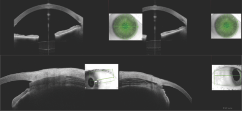
In Liverpool, United Kingdom, AS-OCT has been used postoperatively to confirm the presence of and evaluate pores created in the trabecular meshwork with an excimer laser trabeculotomy (Elios, Elios Vision; Figure 1).

All the aforementioned findings highlight the potential of AS-OCT to investigate whether specific structural changes achieved during surgery are predictive of clinical success.
In the postoperative setting, AS-OCT can be used to track gradual anatomic changes over time. This is particularly helpful for monitoring implant performance. For instance, AS-OCT can determine if a suprachoroidal implant is correctly positioned in the anterior chamber and monitor the size of the distal suprachoroidal space in correlation with clinical outcomes (Figure 2).

AQUEOUS OUTFLOW AND SCHLEMM CANAL
Aqueous outflow beyond the trabecular meshwork is critical to MIGS efficacy. New-generation SS-OCT can provide detailed images of Schlemm canal, an area of growing research interest. Studies have shown that the canal’s dimensions decrease with age and primary open-angle glaucoma.6,7
The technology’s ability to evaluate the impact of MIGS—particularly trabecular meshwork bypass and viscodilation—on the structure of Schlemm canal provides valuable quantitative insights. A prospective analysis found that properly placed iStent devices resulted in greater Schlemm canal diameters and lower postoperative IOPs.4 The dynamic, active nature of aqueous outflow, its impact on Schlemm canal measurements, and the need for precise delineation of this small structure should be central considerations in future research in this field.8
LIMITATIONS
AS-OCT provides static images of anatomic structures, which cannot capture dynamic changes in aqueous outflow in real time. Motion and blinking artifacts can compromise image quality. The high cost and limited accessibility of AS-OCT systems, moreover, may hinder widespread adoption of the technology. Advances such as automated landmark detection and enhanced intraoperative visualization may help address these challenges. The utility of AS-OCT for evaluating minimally invasive bleb-forming surgeries also warrants further exploration but is beyond the scope of this article.
CONCLUSION
AS-OCT can facilitate candidate selection for MIGS and accurate implant placement. The imaging technology can also provide important insights into surgical outcomes, including whether failure is due to incorrect technique or distal pathophysiologic/unknown factors. As it evolves, AS-OCT may come to play an integral role in advancing the science and practice of MIGS.
Ready to Claim Your Credits?
You have attempts to pass this post-test. Take your time and review carefully before submitting.
Good luck!