
Enigmatically Ultrathin, Steep Corneas
Surgeons consider how to manage this pediatric patient.
KEY TAKEAWAYS
- Surgeons consider how to manage a young girl with mysteriously ultrathin and steep corneas.
- The differential diagnosis of ultrathin corneas in pediatric patients includes diseases of extracellular matrix composition, early-onset ectatic diseases, dystrophies, and inflammatory secondary changes.
- Close observation is warranted in this population to detect disease progression promptly and monitor for amblyopia.
Case Presentation
A 7-year-old girl seeking improved vision is referred for a consultation and presents with her family. The patient's left eye has been occluded for 3 hours per day since her third birthday to treat relative amblyopia (0.3 VA OD), and this therapy has improved her vision.
On examination, her spectacle refraction is -3.00 -6.00 x 0° = 0.5 OD and -2.00 -4.50 x 0° = 0.5 OS. An automated refraction under cycloplegia is -2.75 -6.25 x 1° OD and -0.75 -4.75 x 1° OS. She has normal stereoscopic vision and normal motility without phoria. An examination of the anterior and posterior segments finds a clear cornea and lens and a healthy optic nerve and retina in each eye. Her IOP is within a normal range in each eye.
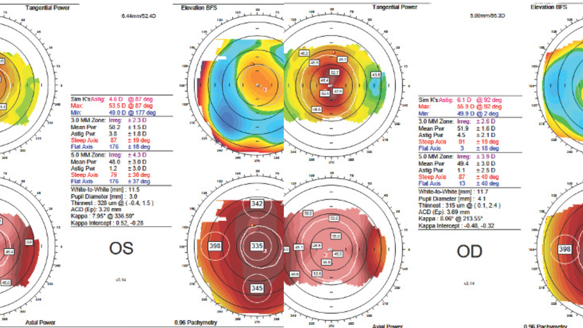
An evaluation with the Orbscan IIz (Bausch + Lomb) shows a homogeneous ultrathin and very steep cornea in each eye and regular corneal astigmatism of -6.10 and -4.60 D in the right and left eyes, respectively (Figure 1). The anterior chamber depth (measured from the epithelium) is 3.99 mm in the right eye and 3.2 mm in the left eye.


The patient has no history of ocular disease or surgery. She is generally healthy with no known systemic diseases. She has no signs or symptoms indicative of any syndrome. Her family history is unremarkable.
How would you proceed?
- Case prepared by Suphi Taneri, MD, PhD, FEBOS-CR
Claus Cursiefen, FEBO, FARVO
The history and examination show marked corneal thinning (approximately 320 µm), high regular astigmatism in each eye, and amblyopia. The differential diagnosis of very thin corneas in a pediatric patient includes the following:
- Diseases of extracellular matrix composition (eg, brittle cornea syndrome [BCS]). Other causes in this group should already have been identified because of their comorbidities (eg, Ehlers-Danlos syndrome, Down syndrome, osteogenesis imperfecta, Marfan syndrome, etc);
- Early-onset ectatic diseases (eg, keratoconus, keratoglobus, etc), which are unlikely given the regular astigmatism; and
- Dystrophies (eg, macular corneal dystrophy) or inflammatory secondary changes (eg, atopic diseases).
The most likely diagnosis here is BCS. Genetic testing and counseling in cooperation with human genetics and pediatrics colleagues would be the next step.
BCS is a rare connective tissue disease that typically presents with associated ocular, auditory, musculoskeletal, and cutaneous abnormalities. Almost all patients experience a decline in vision caused by corneal scarring, thinning, and rupture. The most common ophthalmologic findings include keratoconus, progressive central corneal thinning, high myopia, irregular astigmatism, and retinal detachment, and patients are at increased risk of spontaneous corneal or scleral rupture. My colleagues and I published a case report on a 3-year-old boy with acute hydrops, corneal perforation, and BCS.1
The use of protective eyewear, avoidance of sports that pose a particular danger to eyes, and perhaps CXL could decrease the risk of corneal rupture and ameliorate hydrops.2 Additionally, I would take care in the future not to overlook falsely low IOP readings due to the thin corneas, which could mask the development of glaucoma.
Farhad Hafezi, MD, PhD, FARVO
My first instinct would be to request an extensive corneal evaluation, including biomechanics and OCT/Placido-based tomography. The Orbscan is a useful device but offers limited insight in extreme cases like this one; the corneas appear to be uniformly thin, yet I cannot assess peripheral thickness.
Given the patient's age and relatively regular astigmatism, I would rule out classic early-onset keratoconus because of the unusual, uniform thickness distribution. The absence of localized central corneal irregularity, moreover, argues against the central ectasia typical of individuals who rub their eyes. The patient's profile instead suggests early keratoglobus, which belongs to the ectasia family and often presents initially with relatively regular astigmatism. One differential diagnosis worth considering is BCS, but the scleral thinning in that condition typically produces a blue sclera, which is not described here.
My group was the first to propose immediate CXL in pediatric ectasia instead of waiting for documented progression.3 The recently published Second Global Consensus on Keratoconus reached consensus support (defined as ≥ two-thirds expert agreement) for this approach.4 We also developed the sub400 protocol for ultrathin corneas in which the stroma is treated to a depth of approximately 214 µm,5 and we have applied the technique to eyes with keratoglobus.6
After a complete examination of the patient, CXL would be performed to stabilize her condition. Visual rehabilitation would be challenging. Although specialty contact lenses would be the best first option, this modality might be difficult for the patient because of her age. If she cannot be fitted with specialty contact lenses, her vision would be corrected to the greatest extent possible with spectacles, and she would be closely monitored by a cornea specialist and a pediatric ophthalmologist for potential amblyopia.
George Kymionis, MD, PhD
The patient has symmetric, homogeneously thin corneas (335 µm OD and 326 µm OS centrally), steep keratometry readings, and high but regular myopic astigmatism. These findings do not align with the focal, asymmetric, irregular steepening typical of keratoconus or pellucid marginal degeneration and are instead more suggestive of congenital diffuse corneal thinning or steepening, with keratoglobus high in the differential diagnosis.
Baseline Scheimpflug tomography, epithelial thickness mapping, anterior segment OCT, and corneal biomechanics would be documented. Additionally, the patient would be evaluated for blue sclera syndromes, such as Leber congenital amaurosis, Ehlers-Danlos syndrome, osteogenesis imperfecta, and other connective tissue disorders.
Management would be conservative. Initially, the full cycloplegic correction would be prescribed. A shift from spectacles to rigid gas permeable or scleral contact lenses would be advisable to regularize the anterior optical surface and achieve the patient's BCVA.
Because keratoglobic corneas are fragile and at increased risk of rupture from minor trauma, polycarbonate lenses are suitable for protection and correction. All corneal refractive and ablative surgery must be avoided in eyes with ultrathin and steep corneas as well as in patients of this age.
Tomography would be used to monitor disease stability. If progression is documented, CXL with modified protocols suited to the patient's thin corneas could be considered. Any future refractive surgical correction should be an intraocular procedure, such as phakic IOL implantation, and occur only after the eye is stable and if contact lens correction is insufficient.
What I Did: Suphi Taneri, MD, PhD, FEBOS-CR
Like the panelists, I considered the patient's bilateral symmetric homogeneously ultrathin and steep corneas to be suggestive of congenital diffuse corneal thinning or steepening, with keratoglobus high in the differential diagnosis. She did not, however, have the large corneas typically associated with this disorder. Because focal, asymmetric, irregular steepening typical of keratoconus or pellucid marginal degeneration was not present, I ruled out these conditions. Similarly, because the appearance of the sclera in each eye was unremarkable, blue sclera syndromes such as Leber congenital amaurosis, Ehlers-Danlos syndrome, osteogenesis imperfecta, and other connective tissue disorders seemed unlikely and were excluded clinically.
I recommended close observation, including tomography, rather than immediate CXL because no protocols suited to the patient's extremely thin corneas were available at the time of her initial presentation in 2016 (when the Orbscan measurements were taken). The findings have remained stable for the past 10 years (Figure 2).


In May 2026, the patient's spectacle refraction was -5.50 -7.50 x 3° = 0.67 OD and -4.00 -6.00 x 2° = 0.5 OS. A slit-lamp examination revealed normal scleras; fully transparent, thin corneas and crystalline lenses; and no iris transillumination defects. Central endothelial cell density was normal (2,561 cells/mm2 OD and 2,417 cells/mm2 OS) with unremarkable pleomorphism and polymegathism. Axial lengths were also normal (23.6 mm OD and 23.3 mm OS). Epithelial thickness was rather low in each eye (approximately 40 µm), but no other systemic signs or symptoms were present. Genetic testing has not been performed because the results would not influence therapy.
In the past, the patient, her family, and I discussed the option of rigid gas permeable or scleral contact lens wear to regularize the anterior optical surface and optimize her BCVA. Because her vision was satisfactory, however, I did not recommend this form of correction, which could traumatize her potentially brittle corneas and carries the risk of infection. When she reaches 18 years of age, phakic IOL implantation could be an option to reduce the distortion caused by high-cylinder glasses. The enigmatically ultrathin and steep corneas are a contraindication for corneal refractive and ablative surgery.
Ready to Claim Your Credits?
You have attempts to pass this post-test. Take your time and review carefully before submitting.
Good luck!
Recommended
 August 2026 Issue
Preventing Infection After Cataract Surgery
Mark F. Pyfer, MD; Edward E. Manche, MDMark F. Pyfer, MD; Edward E. Manche, MD- August 2026 Issue
Distorted Vision After Cataract Surgery
Cristos Ifantides, MD, MBA; Sunee Chansangpetch, MD, BPHCristos Ifantides, MD, MBA; Sunee Chansangpetch, MD, BPH