
Distorted Vision After Cataract Surgery
Surgeons discuss how to improve the patient’s result and level of satisfaction.
KEY TAKEAWAYS
- Surgeons consider how to manage a patient’s distorted vision in one eye after cataract surgery with the implantation of a monofocal toric IOL.
- Astigmatism caused by prior filtration surgery led to inconsistent preoperative measurements, which cast doubt on the target refraction.
- Filtration surgery can induce a large amount of posterior corneal astigmatism that might not be evident on topography.
- The implantation of a Light Adjustable Lens (RxSight) can achieve high levels of astigmatism correction beyond the US FDA-approved amount.
Case Presentation
A 69-year-old man presents for a cataract surgery evaluation. The patient has difficulty reading fine print and sees poorly in dim lighting conditions. Glare makes daytime and nighttime driving difficult and causes blurry vision indoors.
The patient's medical history is significant for glaucoma, and he has a family history (father and brother) of the disease. His highest measured IOP is 22 mm Hg OD and 25 mm Hg OS. Eight years ago, after these readings were recorded, his left eye received an Ex-Press Glaucoma Filtration Device (Alcon). The medical regimen for his right eye consists of latanoprost administered at bedtime and brimonidine instilled twice daily.
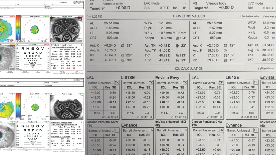
On examination, the patient's UCVA is 20/20 OD and 20/60+2 OS. His BCVA is 20/30- OS with a manifest refraction of +0.75 +3.25 x 005°. Glare testing reduces his visual acuity to worse than 20/800 OS. A slit-lamp examination of the left eye shows a low-lying bleb. Anterior corneal astigmatism measurements obtained with corneal topography and the IOLMaster 700 (Carl Zeiss Meditec) are similar, but total keratometry is higher with the latter technology (Figures 1 and 2).


The left eye undergoes uncomplicated cataract surgery with a monofocal toric IOL. Three weeks postoperatively, the patient's UCVA is 20/50 OS, and his manifest refraction is -2.25 +2.00 x 005° = 20/30- OS. The vision in that eye is distorted, and the patient reports ghosting. After several coaching visits, he remains unhappy with his vision.
How would you proceed?
- Case prepared by Cristos Ifantides, MD, MBA
Sunee Chansangpetch, MD, BPH
Preoperative corneal topography showed superior corneal flattening, probably related to the low-lying filtering bleb. A visible iridotomy and a shallow anterior chamber suggest underlying primary angle-closure glaucoma. Following monofocal toric IOL implantation, the patient developed distorted vision and monocular diplopia with approximately 2.00 D of residual astigmatism, likely accounting for both symptoms. The iridotomy is a less likely contributor because it is not particularly large, although eyelid alignment may still induce glare or diplopia.
The main issue appears to be residual astigmatism. The first step would be to exclude malrotation of the toric IOL and then reassess keratometry. Although a bleb-related keratometric change after phacoemulsification is theoretically possible, such a change would likely be minimal given the long-standing bleb, which would generally be expected to have a relatively mature and stable morphology.
Regarding astigmatism management, partial excision of the overhanging corneal bleb with limbal compression suturing would be technically feasible, but the refractive effect might be unpredictable and require several months to stabilize, with residual astigmatism potentially persisting afterward. Given the relatively early postoperative timing, an IOL exchange would be a reasonable option. The implantation of a secondary sulcus IOL (AddOn, 1stQ) could also be considered, but a careful anterior segment assessment would be necessary because of the angle-closure anatomy. Corneal refractive surgery such as PRK would be another alternative but might be less desirable due to its effect on the accuracy of IOP assessment.
If the iridotomy appears to be contributing to glare or diplopia, closure with a Siepser sliding knot could be considered, potentially in conjunction with an IOL exchange or secondary sulcus IOL implantation.
Florian T.A. Kretz, MD, FEBO
The IOL power is not stated, which makes it difficult to determine whether it is the cause of the patient's problem. Because the remaining astigmatic axis is the same as the preoperative refraction axis, an undercorrection of astigmatism seems likely. The only preoperative measurement that took the posterior cornea into account was obtained with the IOLMaster, and it indicated high total corneal astigmatism. It would be interesting to compare this result to measurements obtained with corneal tomography using Scheimpflug imaging rather than topography.
I suspect that the lens sits more anteriorly than expected due to the bleb and possibly anatomic changes caused by the Ex-Press shunt. This would explain the myopic shift if emmetropia was targeted. Also, the anterior chamber is smaller than usual.
If additional information on the IOL supports the hypothesis of undercorrection, residual astigmatism and sphere would be addressed with classical PRK. Central corneal thickness is slightly greater in the left versus right eye, moreover, which might indicate modest chronic stromal edema due to endothelial cell loss after placement of the glaucoma shunt.
An IOL exchange or implantation of an AddOn IOL could increase the risk of glaucomatous progression or damage the cornea.
What I Did: Cristos Ifantides, MD, MBA
Topography showed 1.89 D of anterior corneal astigmatism, which was similar in magnitude and axis to the keratometry readings of the IOLMaster 700. My initial IOL of choice was a +2.75 D enVista Toric IOL (Bausch + Lomb), which produces +1.93 D of cylinder correction at the corneal plane. A postoperative refraction showed an undercorrection of astigmatism due to the 1.50 D of additional posterior corneal astigmatism.
After a discussion of his options, the patient elected to undergo an IOL exchange for a Light Adjustable Lens (RxSight). I recommended this lens both because of the variability in astigmatism measurement and to minimize the potential for a second IOL exchange in a glaucomatous eye if the target refraction were not achieved once again.
One day after surgery, his UCVA was 20/40 OS. After the final lock-in treatment, his UCVA was 20/25 OS, and he was highly satisfied with his result. The patient's glare and ghosting symptoms resolved, and he was able to see in dim light and read fine print with reading glasses.
The main lesson I learned is to appreciate the posterior corneal astigmatism induced by glaucoma surgery. Another lesson was that the Light Adjustable Lens can achieve a large amount of astigmatism correction beyond its on-label intended use. Finally, I learned that, when the reliability of astigmatism measurements in an eye with an abnormal cornea is in doubt, a preoperative refraction can serve as a tiebreaker.
Ready to Claim Your Credits?
You have attempts to pass this post-test. Take your time and review carefully before submitting.
Good luck!
Recommended
 August 2026 Issue
Where Pharmacologic Presbyopia Correction Fits
Eric Donnenfeld, MD; Taylor LinaburgEric Donnenfeld, MD; Taylor Linaburg