
Sponsored By

For many years, the IOLMaster has been the gold standard in optical biometry. This technology has undergone many technological developments throughout its history, the most recent of which is the integration of swept-source frequency-domain OCT, available on the IOLMaster 700 (Carl Zeiss Meditec; Figure 1). This technology, which provides a full–eye length tomography that depicts the anatomical details on a longitudinal cut through the entire eye, allows the practitioner to detect unusual eye geometries such as tilt or decentration of the crystalline lens (Figure 2). It also performs a 1-mm central retinal scan to ensure safety.

Figure 1. The IOLMaster 700.
Courtesy of Carl Zeiss Meditec
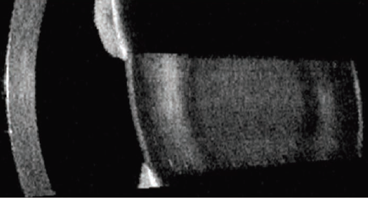
Figure 2. Suspected tilted lens, imaged with the IOLMaster 700.
Courtesy of Walter Sekundo, MD
The IOLMaster 700 is one component of the ZEISS Cataract Suite, a group of products designed to work together for markerless toric IOL alignment. In addition to the IOLMaster 700, the suite consists of the CALLISTO eye, FORUM, and the OPMI LUMERA family of microscopes. When these products are used in combination, the ZEISS Cataract Suite allows practitioners to skip many of the manual steps associated with toric IOL implantation, including pre- and intraoperative marking and manual data transfer, and to achieve highly accurate refractive outcomes postoperatively.
HIGHLY REPEATABLE AND ACCURATE MEASUREMENTS
The IOLMaster 700 is the entry point to the ZEISS Cataract Suite. Intended to reduce the risk of refractive surprises, this device provides highly repeatable and accurate lens thickness, anterior chamber depth, and corneal thickness measurements of the eye. In short, the swept-source OCT is capable of measuring everything but the anterior corneal surface, and it does so at a depth of 44 mm and with six scan lines (0º, 30º, 60º, 90º, 120º, and 150º). The entire process is completed in about 50 seconds, with a total of 2,000 scans taken per second. During the process, the IOLMaster 700 also detects abnormal lens geometry and performs a 1-mm central retinal scan. Furthermore, the unique Fixation Check software detects poor fixation by visualizing the foveal pit.
KEY FEATURES
Swept-source OCT. Being able to visualize the anatomical details of the eye using swept-source OCT is advantageous for many reasons. By and large, the biggest advantage is that it can reduce the number of refractive surprises by revealing abnormal shapes or structures, including tilt or decentration of the crystalline lens. This is true in normal eyes, but it is even moreso in abnormal ones, as measurements are traditionally difficult to obtain in these cases. Second, the IOLMaster 700 provides the surgeon with visual verification of the ocular structures that have been measured (Figure 3).
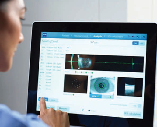
Figure 3. The IOLMaster 700 provides visual verification of the ocular structures.
Courtesy of Carl Zeiss Meditec
Telecentric keratometry. The software’s telecentric keratometry provides accurate corneal readings regardless of pupil size, and, with its 950-nm light source, it penetrates the sclera as well. It is independent of focus, which makes it easy to use. Another thing I like about the IOLMaster 700’s telecentric keratometry is that I get to see the refractions right on the printout, enabling me to detect immediately which are good and which are not. The visual assessment of raw data with telecentric keratometry is crucial.
B-scan.In comparison to A-scans, which do not provide a true image of the ocular structure being measured, B-scans generate true images of intraocular structures that can be measured. Therefore, the information provided by B-scans is more objective to that of A-scans, and it can be used to evaluate the entire intraocular anatomy, including the position of the crystalline lens and to determine sources that may affect the biometric readings. The IOLMaster 700 provides an incredibly robust B-scan of almost any eye, with the exception of white mature cataracts. Furthermore, if a cloudy image is detected, the eye can immediately be re-measured with a subsequent B-scan. In some eyes, it is occasionally possible for the IOLMaster 700 to detect abnormalities of the retinal surface and macular pathologies; however, one must not rely on this device to do so.
IN CLINICAL USE
I trust in the accuracy, repeatability, and consistency of measurements taken with the IOLMaster 700. Given the fact that it makes 2,000 scans/sec, the repeatability of the IOLMaster 700 is absolutely outstanding.
When I think of the success of biometry in my office and how much I depend on it, I first have to think of the total ease of use, the distance-independent measurements, and the up to four times faster reading capability of the IOLMaster 700 compared with other optical devices. The test time with this device can be less than 50 seconds, which minimizes chin- and chairtime for my patients and increases the workflow for my office. Each day, my tech performs so many preoperative tests, and therefore I appreciate anything that is helpful to her in decreasing the time and aggravation associated with these exams.
The composite signal analysis of the IOLMaster 700 does a great job of penetrating dense cataracts, and it pretty much eliminates the need to review, manipulate, and delete individual readings. This is an especially great feature for surgeons who do not take their own axial length measurements. Furthermore, because the full-length OCT image shows anatomical details on a longitudinal cut throught the entire eye, I can easily visualize unusual eye geometries and tilt or decentration of the crystalline lens.
Both the axial length and keratometry readings (which uses telecentric optics, as they are less affected by vertex distance) provide a measurement workflow that is easy and consistent, even on patients who are typically difficult to measure, such as more elderly patients with tremors and fixation problems, including those with Parkinson disease.
I use the Haigis-L suite for all of my post-LASIK and post-PRK patients. It is a great formula to use when there is no prior refractive history, and, with my large post-refractive practice, this is a must. The Holladay 2 formula is integrated as well, and the Holladay 1 and Barrett formulas will be integrated soon. This gold standard in optical biometry helps me to improve my refractive outcomes.
Finally, the User Group for Laser Interference Biometry website database (http://ocusoft.de/ulib/czm/index.htm) has hundreds of IOLs included with optimized lens constants, and this can be used as a starting point for further on-board optimization of personalized lens constants. This is absolutely unique in the ophthalmic industry.
CONCLUSION
The IOLMaster 700, the entry point to the ZEISS Cataract Suite, is a new technology that combines optical biometry with swept-source OCT, allowing clinicians to image and measure the entire length of the eye. The device can be linked to the CALLISTO eye and the OMPI LUMERA microscope through ZEISS’ FORUM software. These elements of the suite will be discussed in greater detail in subsequent issues of CRST.
The statements of the healthcare professionals giving this presentation reflect only their personal opinions and experiences and do not necessarily reflect the opinions of any institution with whom they are affiliated.
The healthcare professionals giving the presentation may have a contractual relationship with Carl Zeiss Meditec, Inc., and may have received financial compensation.


