


In the 1850s, surgical loupes grew in popularity due to their relatively lightweight nature and 2x to 3x magnification capability.1 Around the same time, surgical microscopes began establishing roots, but adoption stalled for nearly 100 years while the optics and working distance were developed and optimized. Although the superiority of surgical microscopes was demonstrated almost 50 years into their development, surgeons’ preferences and the popularity of loupes delayed their widespread adoption. With time and continued evolution—including the development of coaxial illumination, hands-free design, and extended depth of focus—modern surgical microscopes have become an irreplaceable commodity.1,2
And now, after 15 years of development, another viewing format is available for routine use in ophthalmic surgery.3,4 Stereoscopic high-definition visualization systems were quickly adopted by posterior segment surgeons due to their increased depth of focus, higher magnification, and precise focus, all of which augment the measured and delicate maneuvers required in posterior segment surgery.
Anterior segment surgeons have been slower to adopt digital microscopes for various reasons, including their price, discomfort caused by the headsets, inconvenient camera box location, requirements for efficient and high-volume setups, and concerns regarding visual processing lag of early models.
The improved ergonomics, smaller size, progressive teaching capabilities, synchronized intraoperative OCT, built-in digital corneal marking and phaco/fluidics overlays, and improved efficiency provided by these microscopes, however, indicate that 3D digital visualization platforms will be the next evolutionary step forward in anterior segment surgery. (For more on ergonomics, see Ergonomics and Digital Display Systems.)
ERGONOMICS AND DIGITAL DISPLAY SYSTEMS
By Robert J. Weinstock, MD
One of the most beneficial aspects of operating heads-up with a 3D visualization system is being able to sit in a comfortable position during surgery.
Ophthalmic surgeons are well aware of the challenges that can make getting comfortable during surgery a difficult task. Often, the positioning required to see through the oculars of a traditional microscope results in chronic or acute neck, back, and shoulder pain, which can lead to acute injuries, short-term disability, or the inability to perform one’s daily work. Long-term chronic pain and disability may also occur and potentially shorten a surgeon’s functional career in the OR.
The Ngenuity (Alcon) and Artevo 800 (Carl Zeiss Meditec) systems both employ a 3D camera attached to a conventional microscope. The camera displays the image of the surgical field on a high-definition display screen that can be placed either directly across from the surgeon, on the other side of the patient, if the surgeon is operating temporally, or close to the foot of the bed if he or she is operating superiorly.
When operating with the Beyeonics One (Beyeonics) system, the surgeon views the surgical field on a display screen inside a wearable headset rather than on a display screen. For this reason, the Byeonics One system likely provides greater freedom than the other digital display systems because the surgeon can still move his or her head in any direction without losing sight of the surgical field. This capability allows the surgeon slightly greater freedom in finding the most ergonomically comfortable sitting position. On the other hand, the headset of this system may make communication with OR staff difficult, and some may find the limitations in peripheral vision while wearing the headset a challenge.
The Artevo 800 and Ngenuity systems offer similar ergonomic benefits. The display screen must be positioned across from the surgeon, and a slight tilt of the head is often required while viewing the display screen in order to see around the head of the microscope.
All 3D digital display systems allow the operating surgeon to sit back in a comfortable position and change position freely during surgery. This freedom means the surgeon can shift position to alleviate small discomforts, pain, or muscle cramps that can set in as a result of maintaining a single position for a long period.
There is hope that all three of these systems will allow surgeons to enjoy a more comfortable operating environment and avoid short- and long-term pain and disabilities. Additionally, it is my opinion that digital display systems will also reduce the risk of intraoperative complications. It stands to reason that, if a surgeon is operating in an uncomfortable position, he or she could be more likely to rush through a procedure to end the discomfort, which could increase the likelihood of surgical errors.
I hope that as the 3D digital ophthalmic surgery space continues to broaden, and more surgeons across the globe begin to embrace 3D visualization, the result will be better care and surgical outcomes for patients and increased ergonomic health for surgeons.
In this article, we provide an overview of the attributes of three available or soon-to-be available 3D visualization platforms in order of their emergence in the market: (1) the Ngenuity (Alcon), (2) the Artevo 800 (Carl Zeiss Meditec), and (3) the Beyeonics One (Beyeonics).
NGENUITY 3D VISUALIZATION SYSTEM
The Ngenuity 3D Visualization System (Figure 1) features four primary components: a 3D high dynamic range (HDR) camera; a 3D 4K 55-inch 60 frame per second organic light-emitting diode (LED) surgical display; an ultrahigh-speed image processor; and passive, polarized 3D glasses. The Ngenuity HDR camera offers true stereoscopic 3D imaging with two full HD 1920 x 1080 complementary metal-oxide-semiconductor (CMOS) sensors that do not require alignment, focus, or synchronization (Figure 2). Additionally, the CMOS sensors provide low-noise, high-sensitivity imaging.
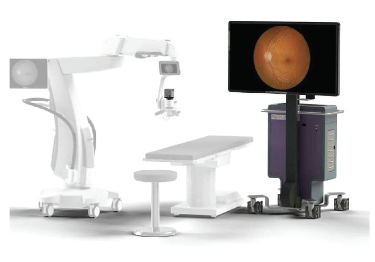
Figure 1. The components of the Ngenuity 3D Visualization System.
Courtesy of Alcon
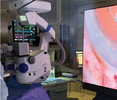
Figure 2. The Ngenuity’s HDR camera. The attachment of the camera can be fixated to most surgical microscopes.
The Ngenuity employs an advanced stereoscopic display to recreate binocular disparity, targeting the delivery of visual information to the right and left eyes. Notably, this kind of depth perception is not reproducible with standard analog oculars. When this is combined with passive, polarized 3D glasses, the system provides a dynamic and immersive 3D visualization experience not only for the surgeon but also for all others in the OR, including residents, fellows, medical students, and cosurgeons (Figure 3).
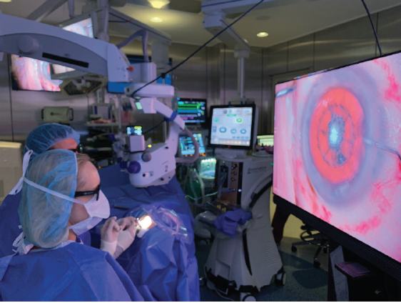
Figure 3. Dr. Rosenberg operates the Ngenuity while Kimberly C. Sippel, MD, proctors via the digital display.
The Ngenuity system has several key attributes.
Mobility and adaptability. The Ngenuity offers the ability to move the system between ORs and microscopes. The Ngenuity’s HDR camera attachment can be fixated to most surgical microscopes, including those available from Carl Zeiss Meditec, Leica, and Luxor, with ease. Additionally, all components of the Ngenuity can be incorporated into the cart that houses both the image processing system and the 55-inch monitor, which can then be wheeled to any OR within a facility. Along the same lines, the HDR camera box can be moved to a microscope with a slimmer design profile, thereby increasing surgeon visibility around physical structures.
If the operating microscope begins to malfunction during surgery, simply moving the Ngenuity to another microscope can preserve the surgeon’s HD view. The Ngenuity 3D Visualization System provides unparalleled detail on all of our operating microscopes.
Real-time 3D HDR surgical image optimization. Proprietary image-processing technology optimizes the Ngenuity’s 3D HDR surgical images in real time. This processing elevates the intensity of the imagery, adding contrast, sharpness, and color. The resulting ability to appreciate subtleties offers many advantages to the surgeon: There is no light saturation with automatic gain, elevating the intensity of the imagery, and allowing fine details to become visible in very dim light.
Anecdotally, we have found that when as much ambient light is eliminated from the room as possible, up to threefold less coaxial light is required intraoperatively (Figure 4). Remarkably, when we need an enhanced view of surrounding structures, instead of turning on the microscope surrounding field illumination we instruct the nursing team to turn on the overhead lights. Ultimately, this leads to a more comfortable patient and less potential light-induced macular toxicity. Although advanced processing comes at a price of time, with lag reported in earlier models, we have not found this to be noticeable during surgical cases.
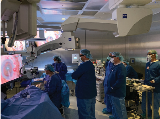
Figure 4. Eliminating as much ambient light as possible from the OR, as shown in this photograph, requires up to threefold less coaxial light intraoperatively.
Figures 2-4 courtesy of Eric Rosenberg, DO, MScEng
Future applications and open architecture. The Ngenuity’s smart 3D platform enables the addition of future applications and provides open architecture for multiple digital inputs. In the not-too-distant future, we believe that we will start seeing integrated intraoperative aberrometry and fluid dynamics.
With digitalization and open architecture, the Ngenuity offers a modular platform that can change along with the evolving nature of our ophthalmic specialty. This platform has the good fortune of having been on the scene the longest. It is user friendly and has an ergonomic setup that is highly customizable to surgeon preferences.
ARTEVO 800
The Artevo 800 (Figure 5) is the first 3D-capable digital ophthalmic microscope, according to the manufacturer. It introduces the novel DigitalOptics technology, which produces higher resolution optics and provides better light transmission than traditional optical microscopes. These features allow surgery to be performed using lower illumination levels, which translates to greater patient comfort during surgery.
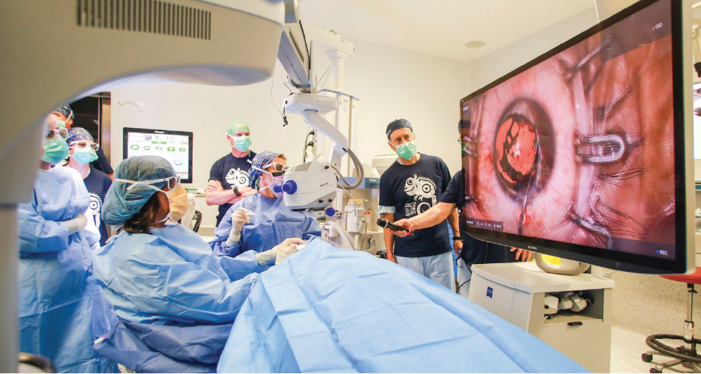
Figure 5. Drs. De Francesco and Ahmed operate using the Artevo 800 3D-capable digital microscope.
Courtesy of Iqbal Ike K. Ahmed, MD, FRCSC
There are several key attributes of the Artevo 800.
Hybrid mode. Hybrid mode allows the surgeon to view the surgical field using 3D imaging on the screen or through the oculars. If the surgeon wants to use the microscope oculars at any point, the surgical team in the OR can continue to view the image on the monitor, helping to maintain an immersive experience for the surgical team and supporting cooperation among OR staff members. This feature also provides greater flexibility in the OR than other 3D visualization platforms for which switching between monitor and ocular visualization is time-consuming, can affect workflow, and could lead to mechanical failures.
Viewability and image quality. The stereoscopic 3D images of the Artevo 800 are viewed on a 55-inch 4K resolution monitor that provides outstanding depth of field and high-resolution images with natural colors. The monitor should be positioned approximately 1.2 meters from the surgeon to help him or her maintain a comfortable position and the rest of the team to access a clear view.
To eliminate the signal delay present in earlier 3D visualization systems, the optical system of the Artevo 800 has two three-chip 4K cameras that produce remarkably clear images with no lag perceivable to human eyes.
Intraoperative OCT. Equipped with intraoperative OCT, the Artevo 800 streamlines surgical decision-making and helps to ensure correct tissue placement during surgical procedures, increasing safety and optimizing outcomes.
Integration. Cloud connectivity with the Zeiss Cataract Suite (Carl Zeiss Meditec) allows the surgeon to easily access patient data remotely and at any stage in the pre-, intra-, or postoperative periods, improving OR workflow and efficiency. Intraoperatively, the patient’s identification, IOL specifications, and phaco parameters can be overlaid onto the real-time image without blocking the surgical view, thereby enhancing efficiency and safety during surgery.
Integration with the Callisto eye markerless alignment system (Carl Zeiss Meditec) offers further benefits, with information about incision, capsulorhexis, and astigmatism alignment displayed directly in the surgeon’s eyepiece. This technology can help cataract surgeons enhance the accuracy and precision of toric IOL alignment, optimizing refractive results.
AutoAdjust. This feature is designed to anticipate the surgeon’s workflow and optimize visualization by automatically adjusting settings without the need for additional interaction.
The Artevo 800 provides ophthalmic surgeons with enhanced connectivity, viewability, and workflow in the OR, optimizing surgeon experience, OR efficiency, and patient outcomes.
BEYEONICS ONE
The Beyeonics One is a 3D visualization system for anterior and posterior segment surgery. It includes a surgical microscope, computer system, and wearable helmet headset with a built-in 3D display (Figure 6). The technology and design of the system are modeled after those of military fighter pilot headsets that provide pilots with digital and graphic information about the cockpit and the airspace around them. The head position of the wearer can control modification of settings and display changes.
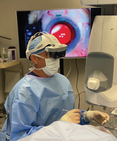
Figure 6. Dr. Weinstock operates using the Beyeonics One wearable helmet headset with a built-in 3D display.
Courtesy of Robert J. Weinstock, MD
There are several key attributes of the Beyeonics One system.
Optimized surgeon experience. During surgery, the headset allows the surgeon to use head gestures, such as tilting the head up or down and left or right, to control multiple functions of the microscope view in various software applications. The headset displays images in a quality equivalent to the resolution of 4K display monitors and similar to that provided by other current 3D visualization platforms. The projected image size on the headset’s display screen is equivalent to a 55-inch monitor placed at a distance of 1.5 meters.
The design of the Beyeonics One headset allows the surgeon to have situational awareness during surgery because it enables peripheral vision and because the headset’s transparency setting can be controlled. If the surgeon uses spectacles, including those with multifocal optics, these can be worn to support the best possible viewing.
Although the headset is currently tethered to the computer system by wire, it will ultimately be wireless, and use of up to three headsets per system will be possible.
Exceptional imaging. The system is designed with two optical channels with a resolution greater than 100 megapixels, which is better than the resolution of an 8K display. With LED illumination, the camera delivers exceptional stereoscopic imaging of the surgical field. The LED illumination supports a widefield view as well as red reflex capability for cataract surgery.
Customizable head gesture controls. Depth of field, image focus, image zoom, x-y axis movement, and illumination can all be controlled with head gestures. These are customizable functions. Head movements can also control magnification and zoom. This functionality eliminates the awkward maneuvers required by conventional microscope systems, often using the left foot to control these operations. The surgeon thus gains finer control over illumination, magnification, and zoom than is achievable with other platforms.
Additional features such as intraoperative OCT can be incorporated into the microscope system during surgery, and this can also be controlled using head gestures.
Automated 3D camera positioning. The Beyeonics One 3D cameras are suspended on a five-axis motorized arm that can follow the surgeon’s head movements and place the camera in the optimal position for the surgeon’s view. The latency of the processing chain, from the camera grabbing the image through the imaging process and fusion to the surgeon’s display, is less than 20 milliseconds, allowing seamless viewing with minimal to no latency.
Beyeonics One is undergoing FDA regulatory review processes, and the manufacturer hopes the device will be available commercially soon. It will be exhilarating to see yet another 3D visualization system enter the ophthalmic surgery space. The evolution in 3D visualization will continue to improve surgical outcomes and the surgeon’s experience in the OR.
CONCLUSION
Cataract surgery is one of the oldest documented surgical procedures, dating back to ancient Egypt and the couching technique. Continual evolution in technology and surgical prowess over the past 300 years has elevated the procedure to one of the most successful treatments in medicine. With each iteration of technique, a unique skill, apparatus, or scientific development can be identified as the driving force behind the changes in how cataract surgery is performed, ultimately improving safety and outcomes for patients.
The shift to the use of 3D digital visualization platforms for anterior segment surgery seems inevitable.
Each of the available platforms has unique attributes. Ophthalmologists should welcome this digital awakening in the surgical arena, as it will allow us to provide the best possible care to our patients. Innovation can be fraught with challenges, but it will ultimately refine our processes and allow us to deliver optimum outcomes with the most successful procedure in medicine.
1. Keeler R. The Evolution of the Ophthalmic Surgical Microscope. Historia Ophthalmologica Internationalis. 2015:35-66.
2. Cionni RJ, Pei R, Dimalanta R, Lubeck D. Evaluating red reflex and surgeon preference between nearly-collimated and focused beam microscope illumination systems. Transl Vis Sci Technol. 2015;4(4):7.
3. Weinstock RJ, Diakonis VF, Schwartz AJ, Weinstock AJ. Heads-up cataract surgery: complication rates, surgical duration, and comparison with traditional microscopes. J Refract Surg. 2019;35(5):318-322.
4. Skinner CC, Riemann CD. “Heads up” digitally assisted surgical viewing for retinal detachment repair in a patient with severe kyphosis. Retin Cases Brief Rep. 2018;12(3):257-259.