
They present to eye care practices across the country—patients with cataracts so dense that what is happening behind them is a mystery. I would like to assume that their vision will improve dramatically once the cataract is removed. I want to tell them how much better they will see after surgery, but what if there is hidden pathology? If all is healthy behind the cataract, these patients could be great candidates for a premium lens, but possible retinal problems could render the technology a poor option.
What I need is quality preoperative information.
THE TEST
Full-field flicker electroretinography (ffERG) allows me to evaluate patients’ retinal health even in the presence of a dense media opacity cataract. My ffERG device uses a mini-Ganzfeld stimulus that produces high-frequency flash stimuli. A sensor placed on the patient’s lower eyelid records the retinal response, demonstrating the health of the cone photoreceptors. The magnitude of the signal reflects the function of the cones, and the phase is a measure of the timing of the response. The variance of these measurements represents the consistency of the strength and speed of the signal.
The test is conducted on an eye with a nondilated pupil. If patients have dense cataracts, I order ffERG testing to be performed when they come back for their scheduling appointment. The test is only a few minutes long, and the results tell me if patients have normal or abnormal retinal function.
BENEFITS
The ffERG results help me in two ways. First, they enable me to set realistic expectations for my patients. If their retinas are healthy, cataract surgery will likely greatly improve their vision. If ffERG testing shows problems, cataract surgery may only restore some central vision, but the procedure will allow me to better visualize the retina and determine if the other pathology is treatable. Second, ffERG results help me to decide if a patient could benefit from a premium lens.
CASE EXAMPLES
I recently saw two separate patients who each had amblyopia in one eye and brunescent cataracts in both eyes. The visual acuity of both eyes of both patients was 20/200 or worse. In one case, ffERG testing showed normal retinal function in both eyes, and I was able to tell the patient that cataract surgery would greatly improve his vision (Figure 1). The patient ended up with 20/40 BCVA in the amblyopic eye and 20/20 BCVA in the other eye. The second patient had a very similar profile, but ffERG showed decreased macular function in the amblyopic eye (Figure 2). I was able to warn the patient ahead of time. Postoperatively, he had a BCVA of 20/200 in the amblyopic eye and 20/20 in the other eye.
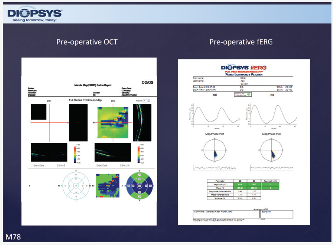
Figure 1. Prior to cataract surgery, ffERG testing was normal in both eyes.
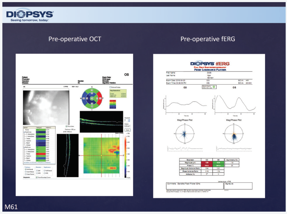
Figure 2. In this patient, preoperative ffERG testing was abnormal in the right eye and normal in the left eye. This information allowed Dr. Jackson to set realistic expectations for the patient about his postoperative visual recovery.
CONCLUSION
As a surgeon, I never want to overpromise and underdeliver. I want to be confident and provide patients with realistic expectations. In my experience, ffERG provides me with preoperative information I need to achieve those goals.