Ophthalmology is a fast-growing technologic field, and I try to be an early adopter of new devices. I am particularly excited about corneal inlays, especially the Raindrop Near Vision Inlay (ReVision Optics). In this month’s featured Eyetube video, Jeffrey Whitman, MD, shares his technique for inserting the implant.
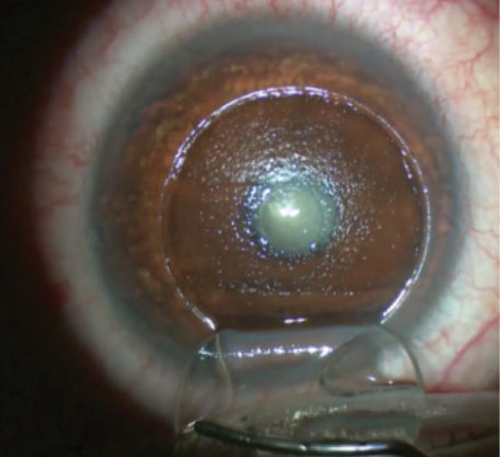
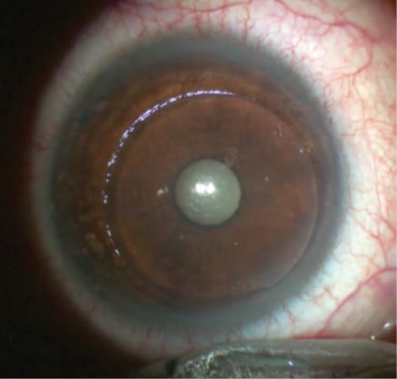
Figure 1. After centering the inlay, Dr. Whitman waits approximately 60 seconds for it to dimple like the skin on an orange before proceeding.
Dr. Whitman begins by making a laser flap at 30% of the eye’s central corneal thickness. To prevent striae, when lifting the flap, he is careful not to fold or “taco” it. He places the insertion device firmly on the corneal stroma, deposits the inlay on the cornea, and holds the implant in place with the back side of the Sinskey spatula while he removes the insertion device. Dr. Whitman then gently nudges the inlay to center it and waits about 60 seconds for the device to dimple like the skin of an orange (Figure 1). During this interval, he continues to wet the epithelial and stromal sides of the flap with balanced salt solution about every 20 seconds to prevent the formation of striae. The flap is exposed for longer than it is during LASIK, so keeping it moist is a must.
Dr. Whitman checks the inlay and moistens the hinge and stromal side of the flap before reflecting it back in place. He then relifts the flap and performs irrigation to remove any debris, while being careful not to touch the stoma or the inlay. Next, he brushes the flap into place with a wet sponge. He comments that a large gap or inferior gutter from the displacement of the inlay is not a cause for concern, because it will disappear by the first postoperative day (Figure 2). Dr. Whitman removes excess fluid with a flap applanator, administers a drop of antibiotic and a nonsteroidal drop, and places a contact lens. His careful technique serves as a model for ophthalmologists who are ready to embrace this exciting new technology.

