TRANSEPITHELIAL VERSUS EPITHELIUM-OFF CORNEAL CROSS-LINKING FOR THE TREATMENT OF PROGRESSIVE KERATOCONUS: A RANDOMIZED, CONTROLLED TRIAL
Soeters N, Wisse R, Godefrooij D, et al1
Soeters et al compared the clinical effects and safety of transepithelial (TE) versus epithelium-off (epi-off) corneal collagen cross-linking (CXL) in a single-center randomized trial of progressive keratoconus. The TE group (n = 35 patients) was treated with Ricrolin TE (transepithelial riboflavin; Ofta hi-tech; not available in the United States), and the epi-off group (n = 26 patients) was treated with isotonic riboflavin. The primary outcome was maximum keratometry 1 year postoperatively, and the secondary outcomes included distance BCVA and complications 1 year postoperatively.
According to the study, the epi-off group experienced significant flattening of 1.20 to 1.50 D from the 3-month follow-up through the 12-month mark. In comparison, maximal keratometry was stable at all visits for the TE CXL group. Keratoconus in 23% of the TE CXL group continued to progress compared to no patients in the epi-off group. BCVA was better in the TE CXL group, and there were no complications. Four eyes (15%) in the epi-off group developed complications, including herpes simplex virus, corneal infiltrate, and delayed epithelial healing.
The investigators concluded that, although TE CXL is a safe procedure, the continued progression in 23% of eyes demonstrates a lack of clinical efficacy. They recommended against replacing epi-off CXL with TE CXL for the treatment of progressive keratoconus.
DISCUSSION
CXL has been shown to halt the progression of keratoconus but requires adequate penetration of riboflavin into the corneal stroma to do so.2,3 Epithelium-on (epi-on) CXL has shown one-fifth the corneal biomechanical rigidity compared to standard epi-off CXL in human eyes.4 Alternative approaches to epi-on protocols such as the Ricrolin-assisted formulation using ethylenediaminetetraacetic acid have produced mixed clinical results.5-7 The study by Soeters et al is the first randomized, prospective, controlled trial to compare this formulation to epi-off protocols.
That TE CXL was shown to have less clinical efficacy than epi-off CXL is consistent with prior studies that reported that various TE riboflavin formulations result in some penetration but less than the standard protocol.6,7 The corneal steepening that occurred in TE CXL patients (23%) is of concern, because it indicates continued progression of keratoconus. There are distinct disadvantages of epi-off CXL such as pain, risk of infection, inability of the epithelium to heal, scarring, and haze.8 The lack of efficacy of TE CXL protocols, however, suggests that the method may not be useful in eyes with progressive keratoconus.
There may be some cases in which the risks associated with epi-off CXL outweigh the benefits. Monocular patients, uncooperative patients, or those with a history of nonhealing epithelium may not do well with epi-off CXL. The opportunity to re-treat patients who do not succeed with epi-on CXL is interesting, because the results in this study and others show a second procedure removing the epithelium can result in flattening.9 Perhaps there is a subgroup of patients in whom epi-on CXL is the protocol of choice with the option to re-treat with epi-off CXL if keratoconus progresses.
Additionally, there may be alternative methods of epi-on CXL that would provide better results such as iontophoresis or permeability enhancers.10 Patients should be counseled on the advantages and disadvantages of the two treatment protocols, and surgeons should consider using the epi-off protocol when possible for the treatment of progressive keratoconus.
At a Glance
• Although transepithelial corneal collagen cross-linking is a safe procedure, the method lacks clinical efficacy: keratoconus continued to progress in 23% of eyes.
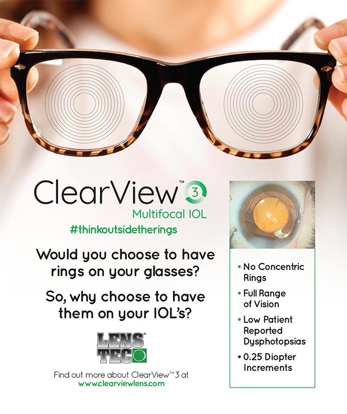
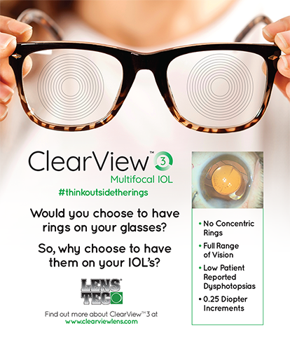
• The inlay group was best for contrast sensitivity, but the multifocal groups performed best for near vision.
COMPARISON OF CONTRAST SENSITIVITY AND THROUGH FOCUS IN SMALL-APERTURE INLAY, ACCOMMODATING INTRAOCULAR LENS OR MULTIFOCAL LENS SUBJECTS
Vilupuru S, Lin L, Pepose JS11
Vilupuru et al compared monocular and binocular mesopic contrast sensitivity and through focus after monocular implantation with the Kamra inlay (AcuFocus) versus binocular implantation with an accommodating or multifocal IOL. The investigators placed the Kamra inlay in the nondominant eye of 507 phakic patients with no surgery performed on the dominant eye. Three predetermined arms included 327 subjects who underwent contrast sensitivity testing, 114 subjects who underwent defocus curve testing, and 78 subjects randomized to undergo bilateral cataract surgery with implantation of the Crystalens Advanced Optics (Bausch + Lomb), the AcrySof IQ Restor +3.0 D (Alcon), or the Tecnis +4D Multifocal (Abbott Medical Optics) IOL.
In the Kamra inlay group, intermediate and near vision improved as expected, with no improvement in distance vision in that eye. That group also had better contrast sensitivity compared to the multifocal lens patients. The Crystalens group had better intermediate UCVA than the Kamra group but worse near vision. The multifocal group performed the worst in terms of intermediate vision but the best with regards to near vision.
The authors concluded that the decision of which device to use should be based on a careful discussion of patients’ goals and the degree of crystalline lens dysfunction.
DISCUSSION
Despite the advent of new lenses, devices, and techniques, presbyopic correction remains a challenge. The Kamra inlay represents the newest device in the quest. It is implanted in the nondominant eye into a lamellar pocket and blocks unfocused peripheral light rays, thereby reducing the size of the blur circle. The goal of the inlay is to extend intermediate to near vision, while the nonoperated dominant eye maintains distance vision.12
The Kamra’s superiority to the multifocal lenses in regard to contrast sensitivity is to be expected. Multifocal lenses use concentric circles to obtain multiple focal points. A reduction in contrast sensitivity and photoptic phenomena is a concern for patients and surgeons.13 The Crystalens has shown less optical scatter compared to multifocal lenses, but its ability to accommodate to distance, intermediate, and near is limited.14
The Kamra and Crystalens groups had the best outcomes with respect to intermediate vision. The two multifocal lenses studied, however, are designed for distance and near vision; intermediate vision is not in the design of the defocus curve. Newer “low-add multifocals” such as the ZKBOO and ZLBOO (both from Abbott Medical Optics) and the ReStor SN6AD1 (Alcon) have defocus curves set for distance and intermediate with excellent clinical results.15,16 These lenses would likely provide a better comparison to the Kamra inlay and Crystalens with regard to intermediate vision.
This study highlights the limitations of presbyopia-correcting lenses as well as those of the Kamra inlay, and it also emphasizes what physicians must acknowledge: there is still no “perfect” treatment for presbyopia. n
1. Soeters N, Wisse R, Godefrooij D, et al. Transepithelial versus epithelium-off corneal cross-linking for the treatment of progressive keratoconus: a randomized controlled trial. Am J Ophthalmol. 2015;159(5):821-828.
2. Wittig-Silva C, Chan E, Islam FM, et al. A randomized, controlled trial of corneal collagen cross-linking in progressive keratoconus: three-year results. Ophthalmology. 2014;121:812-821.
3. Raiskup F, Theuring A, Pillunat LE, Spoerl E. Corneal collagen crosslinking with riboflavin and ultraviolet-A light in progressive keratoconus: ten-year results. J Cataract Refract Surg. 2015;41:41-46.
4. Wollensak G, Iomdine E. Biomechanical and histological changes after corneal crosslinking with and without epithelial debridement. J Cataract Refract Surg. 2009;35(93):540-546.
5. Filippello M, Stagni E, Buccoliero D, et al. Transepithelial corneal collagen crosslinking: bilateral study. J Cataract Refract Surg. 2012;38(2):283-291.
6. Taneri S, Oehler S, Lytle G, Dick HB. Evaluation of epithelial integrity with various transepithelial corneal cross-linking protocols for treatment of keratoconus. J Ophthalmol. 2014;2014:614380.
7. Alhamad TA, O’Brart DP, O’Brart NA, Meek KM. Evaluation of transepithelial stromal riboflavin absorption with enhanced riboflavin solution using spectrophotometry. J Cataract Refract Surg. 2012;38:884-889.
8. Randleman JB, Rocha KM. CXL complications and their management. In: Hafezi F, Randleman JB, eds. Corneal Collagen Cross Linking. Thorofare, NJ: Slack; 2013.
9. Hafezi F, Tabibian D, Richoz O. Additive effect of repeated corneal collagen cross-linking in keratoconus. J Refract Surg. 2014;30(10):716-718.
10. Khandelwal SS, Randleman JB. Current and future applications of corneal cross-linking. Curr Opin Ophthalmol. 2015;26(3):206-213.
11. Vilupuru S, Lin L, Pepose JS. Comparison of contrast sensitivity and through focus in small-aperture inlay, accommodating intraocular lens, or multifocal lens subjects. Am J Ophthalmol. 2015;160(1):150-162.
12. Seyeddain O, Bachernegg A, Riha W, et al. Femtosecond laser-assisted small-aperture cornea inlay implantation for cornea compensation of presbyopia: two-year follow up. J Cataract Refract Surg. 2013;39(2):234-241.
13. de Vries NE, Nuijts RM. Multifocal intraocular lenses in cataract surgery: literature review of benefits and side effects.
J Cataract Refract Surg. 2013;39(2):268-278.
14. Pepose JS, Qazi MA, Chu R, Stahl J. A prospective randomized clinical evaluation of 2 presbyopia correcting intraocular lenses after cataract extraction. Am J Ophthalmol. 2014;158 (3):436-446.
15. Hayashi K, Manabe S, Hayashi H. Visual acuity from far to near and contrast sensitivity in eyes with a diffractive multifocal intraocular lens with a low addition power. J Cataract Refract Surg. 2009;35(12):2070-2076.
16. Kretz FT, Gerl M, Gerl R2, et al; ZKB00 study group. Clinical evaluation of a new pupil independent diffractive multifocal intraocular lens with a +2.75 D near addition: a European multicenter study [published online ahead of print May 18, 2015]. Br J Ophthalmol. doi:10.1136/bjophthalmol-2015-306811.
Section Editor Edward Manche, MD
• director of cornea and refractive surgery at the Stanford Eye Laser Center
• professor of ophthalmology at the Stanford University School of Medicine, Stanford, California
• edward.manche@stanford.edu
Sumitra S. Khandelwal, MD
• assistant professor of ophthalmology, Baylor College of Medicine, Cullen Eye Institute, Houston, Texas
• sumitra.khandelwal@bcm.edu
• financial disclosure: none acknowledged