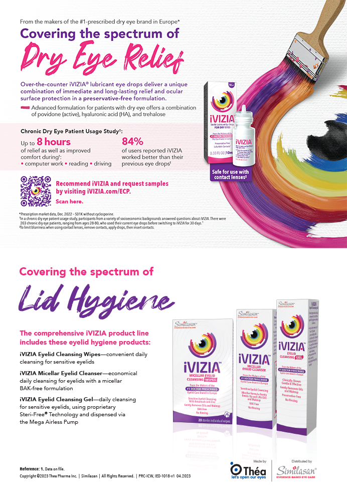
Modifications to cataract surgery occur at a number of different stages of the operation when using the laser versus a manual technique. This article discusses some changes I have made to my procedure with my integration of the Catalys Precision Laser System (OptiMedica Corporation).
ANESTHESIA
It is important that surgeons become comfortable with conscious sedation during the laser cataract procedure, as patients will be more alert than if they were to receive a block. I perform my cases under topical anesthesia, and although patients sometimes will receive a bit more sedation during the lens removal and the IOL's implantation, I administer just 0.5 mg of oral alprazolam.
Patients are therefore more alert under conscious sedation and can see me and hear the staff giving them instructions during the procedure. I interact with my patients a fair amount and let them know what they are going to experience. I cover their other eye with a shield so that they can fixate with the operative eye.
Catalys has an integrated bed that enables stable patient positioning, which is important for imaging and the treatment. The device compensates well for lens tilt with its Integral Guidance system; however, the patient is more comfortable if the corneal apex is as perpendicular as possible to the laser system before docking to minimize extra lateral forces on the docking device.
PATIENT INTERFACE
I inform patients that they will feel a bit of pressure when I first touch the eye, and I position the nonapplanating suction ring portion of the two-piece Liquid Optics Interface. The interface's alignment bar adjusts to my astigmatic marks when I use a toric IOL or create arcuate incisions with the laser. The system will automatically align with the marks on the suction ring at 90º and 180º. Automatic compensation for cyclorotation leads to more precise results and is an important and easy-to-use feature of the Catalys.
I fill the suction cup of the interface with saline and place the patient under the laser. Then, I bring the second piece, the disposable lens optic that is attached to the laser, together into the suction device and activate secondary suction. Less pressure is applied with the Liquid Optics interface than with applanation patient interfaces that other laser cataract surgery platforms employ. In fact, two independent studies from H. Burkhard Dick, MD, and colleagues and from Brendan Vote, MD, et al validated that the IOP rise after suction with the Liquid Optics Interface was about 10 mm Hg.1,2 The patient does not experience much pressure so it is safe for those with glaucoma, and the interface is secure by its design.
I ask the patient to relax and look straight ahead. The Catalys employs a lateral force sensor that monitors the patient and detects any asymmetry in lateral force. This superb safety feature gives me plenty of time to coach the patient to adjust his or her position. It is the first system that I am aware of that has a real-time feedback mechanism to tell the surgeon and the team how the patient is doing while docked so that the case does not fall apart without warning in the middle of imaging, or more importantly, treatment. The time under dock is 2.5 to 4 minutes to complete imaging and laser treatment of capsulotomy, lens fragmentation, arcuate incisions, and a primary cataract incision with paracenteses.
The Catalys uses a proprietary integrated optical coherence tomography system called Integral Guidance that applies sophisticated algorithms designed to ensure that the laser pulses are delivered precisely to the intended location.
THE CAPSULOTOMY
The system ensures the capsulotomy's precision and centration and it also provides the surgeon with the ability to place the capsulotomy in relation to the system-calculated pupillary center or the center of the capsular bag. Specifically, for an accommodating lens like the Crystalens (Bausch + Lomb), I prefer to center the treatment on the bag. This strategy provides sufficient clearance for the lens, and with a 5.5-mm capsulotomy, the edge tends to run over both hinges of the IOL. It is difficult to achieve this every time with handheld instruments, yet doing so has a great impact on the lens' performance.
It only takes about 1.5 seconds to create the capsulotomy. This is a huge advantage of the laser procedure in terms of its safety and precision. Such a small amount of energy is used that few bubbles and little heat are created.
I have completed about 50 cases at Surgisite Boston, including difficult ones such as a black cataract and a corneal transplant patient, and I have yet to see a capsulotomy that has not been completely free. Some will float, and others sit right on top of the lens cortex. If the capsulotomy is not floating well up into the anterior chamber, I will carefully fill the chamber with a dispersive viscoelastic like Viscoat (Alcon Laboratories, Inc.) to ensure that the capsule is stable. Then, I will use a bent cystotome or a capsulorhexis forceps and ensure the edge is free.
In patients with intraoperative floppy iris syndrome or other small pupil situations where the laser treatment is close to the iris, miosis may occur. In those cases, I instill a drop of 10% phenylephrine into the eye after the laser is applied, especially if there will be a delay before completing the surgery. Alternatively, I will use intracameral epinephrine intraoperatively. The use of the laser is a major advantage in complex cataract cases and makes difficult maneuvers easier.
HYDRODISSECTION AND I/A
The biggest difference between manual surgery and the use of the laser centers on my initial approach to the lens nucleus and then hydrodissection and I/A of the cortex. If there is very little gas in the capsular bag following capsulotomy and lens fragmentation, hydrodissection can proceed as in manual surgery. When I use lens softening, the capsular bag can become more distended due to the gas created with the laser cavitation bubbles, making initial hydrodissection less effective. As a result, I have modified the order of my steps to free the cortical lens attachments. Instead of starting with hydrodissection, I first rock the lens slightly to attempt to release gas bubbles from around the softened lens. I may also push apart the nucleus at the center along the lens fragmentation lines to allow gas bubbles to come forward. If these maneuvers do not free the lens, I will perform hydrodissection.
With Catalys, it is not necessary to hydrodissect as aggressively as with standard phacoemulsification to mobilize the lens. During phacoemulsification, I use somewhere between 10% and 50% power. In fact, the laser softens the nucleus so effectively that many of these lenses can be removed using aspiration alone.
The cavitation bubbles created during lens softening will press the cortex, creating a sheet that is flush with the anterior leaflet of the capsulotomy in some cases. I use a bent, round-ended I/A tip. For surgeons to succeed at laser cataract surgery, they should be comfortable using bimanual I/A. I create two sideport incisons with the Catalys, which makes it easy to get under the capsular leaflet, mobilize the cortex, and strip it away in an efficient and controlled fashion.
Surgeons who use traditional, straight I/A tips may be used to having the capsulorhexis farther away from the entry wound compared to its location when centered with the laser. Therefore, they need to adjust to a split approach in which the primary incision is used and I/A proceeds through the main handpiece. One can utilize I/A functions via two smaller cannulas through the two sideport incisions. All of the lasers may require this adaptation, owing more to the position of the capsulotomy than the design of a particular platform.
CONCLUSION
There are numerous advantages to laser cataract surgery with Catalys that help improve surgical technique and patients' outcomes and experiences. These include the safety, precision, reproducibility, and speed of the laser-delivered capsulotomies as well the system's ability to execute highly precise corneal incisions and soften the nucleus so effectively that little to no ultrasound energy is needed. The laser has taken cataract surgery to a new level, and I believe that the future portends advances that we cannot even imagine today.
Jonathan H. Talamo, MD, is the medical director of Surgisite Boston, LLC, founding partner of Talamo Hatch Laser Eye Consultants, and an associate clinical professor of ophthalmology at Harvard Medical School in Boston. He is a member of OptiMedica's Medical Advisory Board. Dr. Talamo may be reached at (781) 890-1023; jtalamo@lasikofboston.com.
- Schultz T, Conrad-Hengerer I, Hengerer FH, Dick HB. Intraocular pressure variation during femtosecond laserassisted cataract surgery using a fluid-filled interface. J Cataract Refract Surg. 2013;39(1):22-27.
- Kerr NM, Abell RG, Vote BJ, Toh TY. Intraocular pressure during femtosecond laser pretreatment of cataract [published online ahead of print January 8, 2013. J Cataract Refract Surg. doi: 10.1016/j.jcrs.2012.12.008.