Sponsored By


In the past few years, there has been an increasing emphasis for cataract surgeons to nail our refractive results. However, uncorrected astigmatism often keeps our patients from achieving their optimum visual outcomes after cataract surgery. Although arcuate incisions are moderately effective in correcting astigmatism, in my opinion, and in the opinion of many cataract surgeons, toric IOLs are the low-hanging fruit in our quest of turning cataract surgery into a refractive procedure. When surgeons incorporate toric IOLs into their armementarium, they can expect their patients to achieve better optical correction, including better uncorrected vision.
With that said, the adoption of toric IOLs, especially in the United States, has been surprisingly low. One of the reasons that many surgeons have shyed away from toric IOLs has been the lack of ability to precisely plan treatments and align the lens during surgery, as a well-oriented toric IOL is integral to reducing postoperative cylinder. Without a reliable way to do this, many times patients fail to achieve the refractive results that they have paid for with the upgrade to the toric IOL.
THE NECESSARY TOOLS
So what technologies exactly are needed to achieve precise results with toric IOLs? It has been shown that intraoperative guidance with anatomic registration reduces the refractive cylindrical error,1 with intraoperative pseudophakic aberrometry performing better than traditional toric IOL placement with a markered technique.2 However, I strongly believe that, today, we should be placing great emphasis on the advantages of a markerless alignment system. In my practice, we have upgraded from a traditional marking system to the CALLISTO eye (Carl Zeiss Meditec).
CALLISTO eye is one component of the ZEISS Cataract Suite, a family of technologies designed to work together seamlessly to achieve excellent results in cataract surgery, including markerless toric IOL alignment. This device is an innovative computer-assisted cataract surgery system that uses biometry data to improve clinical workflow efficiency and surgical precision.
Because of the built-in eye registration (Figure 1), the CALLISTO eye eliminates the need to manually mark the eye and ensures proper alignment of overlays (Figure 2), regardless of eye movement and rotation. It also accounts for anterior chamber depth and the patient’s visual axis to provide accurate centration of both the capsulorrhexis and the IOL.
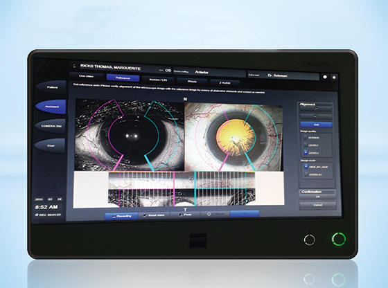
Figure 1. Built-in eye registration of the CALLISTO eye.
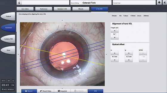
Figure 2. Overlays can be properly aligned with the CALLISTO eye.
CALLISTO eye has given me the peace of mind that all of the data I need for toric IOL implantation has been measured, analyzed, and interpreted perfectly. It transfers all preoperative data from the IOLMaster (Carl Zeiss Meditec), both reducing procedure time and eliminating transcription errors.
In many cases, I combine the use of the CALLISTO eye with intraoperative aberrometry, as I like the idea of having continuous monitoring during cataract surgery. With that said, CALLISTO eye is really the only technology that a surgeon needs to achieve perfect lens alignment during toric IOL implantation, completely markerless.
STUDY
We recently conducted a study to report the residual refractive astigmatism and the mean absolute error after cataract surgery and toric IOL implantation when the CALLISTO eye system was used and when intraoperative aberrometry with ORA+ With Verifeye (Alcon) was used. In order to determine the differences in measurements, our prospective, double-armed, contralateral eye study compared the refractive correction achieved using these two different methods of toric IOL alignment. A total of 70 eyes were included in the study, 34 in the CALLISTO group and 36 in the ORA group. Both groups had identical preoperative workups, and all treated eyes in both groups received the Tecnis Toric lens (Johnson & Johnson Vision). All eyes had regular corneal astigmatism treated with more than 0.61 D of keratometric astigmatism and previously calculated residual refractive astigmatism of less than 0.50 D. All follow-up were conducted approximately 1 month postoperatively. The outcome measures were residual refractive astigmatism and difference vectors.
We designed this study as a typical validation study that we do for all of the technologies we use in clinical practice. Each patient’s eye was individualized, and the measurements were then applied to each system, so that the information provided by CALLISTO eye could be compared to the intraoperative aberrometric measurements taken with the ORA. We also used this information to determine which technology provided the better mean absolute error.
RESULTS
Figure 3 shows the 1-month postoperative visual acuity. In the CALLISTO eye group, 97% of eyes had a visual acuity of 20/20 or better, compared with 83% in the ORA group. When we looked at postoperative spherical equivalent refraction with the CALLISTO eye and ORA systems (Figure 4), what we found is that results were similar for 1.00, 0.75, and 0.50 D. However, for 0.25 D, the CALLISTO eye performed better (74% vs 61%). The same was true of postoperative residual refractive cylinder (Figure 5), with the CALLISTO eye really outperforming the ORA in that 0.25 D range (71% vs 36%).
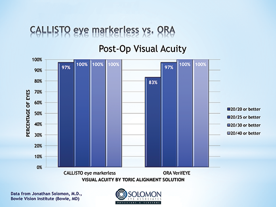
Figure 3. One-month postoperative visual acuity with the CALLISTO eye and with the ORA system.
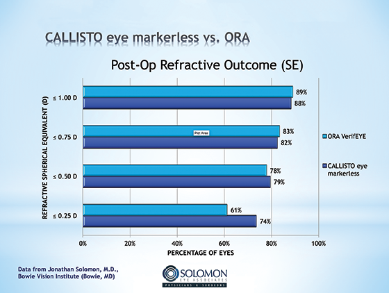
Figure 4. One-month postoperative spherical equivalent refraction with the CALLISTO eye and with the ORA system.
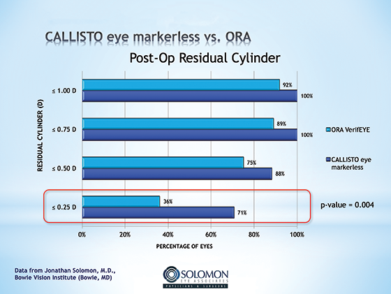
Figure 5. One-month postoperative residual refractive cylinder with the CALLISTO eye and the ORA system.
I also wanted to break the residual cylinder category down to the correction of high and low cylinder. What we found is that the CALLISTO eye did a better job at lining up low-cylinder toric IOLs than it did to the higher-power toric IOLs. The majority of patients that we see require low-cylinder corrections.
CONCLUSION
There is no doubt that intraoperative toric alignment tools can optimize the refractive outcomes of cataract surgery. What this study shows is that, when the most advanced techniques for toric IOL alignment are compared—the CALLISTO eye and the ORA With Verifeye—the CALLISTO eye system is superior to intraoperative aberrometry.
By using the CALLISTO eye and then taking intraoperative aberrometry measurements thereafter, we were able to determine that the toric IOLs implanted during our study were perfectly aligned. We were also able to determine that there was no movement of the IOL after surgery was complete.
From the results of the 70 eyes that were included in our prospective, double-armed, contralateral eye study, we concluded that markerless toric IOL alignment with the ZEISS Cataract Suite provided comparable refractive outcomes to intraoperative aberrometry, with possible superiority in its ability to achieve no more than 0.25 D of residual cylinder. We also determined that the ZEISS Cataract Suite enhanced workflow compared with a manual marking alignment technique and intraoperative aberrometry. Therefore, in my practice, the ZEISS Cataract Suite and intraoperative aberrometry will likely work in synergy to increase patient outcomes.
1. Potvin R, Gundersen KG, Masket S, Osher RH, Snyder ME, Vann RR, et al. Prospective multicenter study of toric IOL outcomes when dual zone automated keratometry is used for astigmatism planning. J Refract Surg. 2013;29(12):804-809.
2. Hatch KM, Woodcock EC, Talamo JH. Intraocular lens power selection and positioning with and without intraoperative aberrometry. J Refract Surg. 2015;31(4):237-242.
The statements of the healthcare professionals giving this presentation reflect only their personal opinions and experiences and do not necessarily reflect the opinions of any institution with whom they are affiliated.
The healthcare professionals giving the presentation may have a contractual relationship with Carl Zeiss Meditec, Inc., and may have received financial compensation.
