
Microinvasive glaucoma surgery (MIGS) procedures provide an alternative or adjunctive surgical treatment to the medical management of mild to moderate glaucoma without precluding the future use of a trabeculectomy or tube shunt, because most procedures offer an ab interno approach. Despite a current lack of head-to-head studies comparing the safety and efficacy of MIGS procedures to medical management alone or to traditional glaucoma surgeries, early evidence suggests a lower risk of major surgical complications such as endophthalmitis, hypotony, and choroidal hemorrhage. The downside to many of the MIGS options is their inability—without the use of additional medication—to achieve an IOP in the low teens or single digits, which is often needed in cases of advanced glaucoma.1
AT A GLANCE
• Microinvasive glaucoma surgery (MIGS) is an alternative or adjunct to the medical management of mild to moderate glaucoma that does not preclude the future use of a trabeculectomy or glaucoma drainage device.
• An advantage of MIGS is its safety compared with traditional filtration surgery, but many of the former procedures generally decrease IOP less.
• MIGS procedures can be categorized based on their mechanism for aqueous filtration.
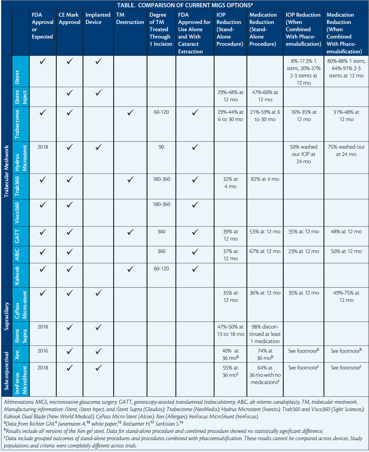
MIGS procedures can be categorized based on their mechanism for aqueous filtration (Table). Until recently, the only FDA-approved MIGS procedures focused on reducing aqueous resistance at the trabecular meshwork (TM). In addition to requiring the patient to have a clear cornea and the ophthalmologist to have experience with gonioscopy-assisted surgery, these minimally invasive TM procedures are often combined with cataract extraction and IOL placement. In general, the IOP-lowering effect of TM-based procedures is restricted by a patient’s episcleral venous pressure and postoperative scarring at the TM or Schlemm canal. On July 29, 2016, the FDA approved the »CyPass Micro-Stent (Alcon), making it the first supraciliary MIGS device for use in the United States. The potential for decreasing IOP through this route is dictated by the highly vascularized and metabolically active choroidal tissue. The third category of MIGS procedures is subconjunctival, which is the route for traditional glaucoma surgery. (Editor’s note: the categorization of subconjunctival procedures as MIGS is in flux, with some calling these hybrids.)
TM- AND CANAL-BASED PROCEDURES
iStent
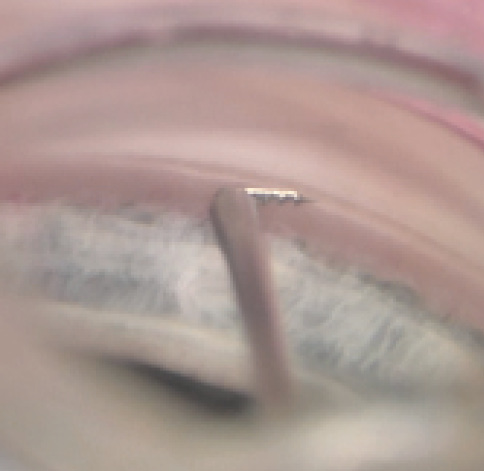
Figure 1. Approach for implanting the iStent in the TM.
This heparin-coated, nonferromagnetic titanium stent resembles a snorkel and is placed into the nasal angle (Figure 1). The pointed tip penetrates the TM, and the device is inserted into Schlemm canal with a sideways motion, after which three retention arches hold the stent in place. With a height of 300 µm and a length of 1 mm, the »iStent (Glaukos) is the smallest FDA-approved implantable device. Its efficacy is likely related to the iStent’s placement near one of the 25 to 30 aqueous collector channels, which are more abundant in the inferonasal angle.2 Rare complications include iris touch, stent malpositioning, and postoperative stent obstruction with iris or pigment. (For more information on this device, see Catch up With Glaucoma Today Journal Club, and visit beye.com/a125 and beye.com/600.)
iStent Inject
This second-generation titanium device is 360 µm long and 230 µm wide. The »iStent Inject (Glaukos; not FDA approved) features four outlets connected to a central lumen open to the anterior chamber (AC). The inserter comes preloaded with two stents, and its simplified design makes insertion easier compared with the original iStent based on users’ experience. IOP reduction has been greater with the iStent Inject versus the original iStent; 12 months out in a head-to-head trial, the iStent Inject was at least as efficacious as two IOP-lowering medications, latanoprost and timolol.3 Unlike its predecessor, the iStent Inject has the potential to be used as a stand-alone procedure.
Trabectome
The »Trabectome (NeoMedix) consists of a disposable 19.5-gauge handpiece with an insulating footplate bent at 90º containing bipolar electrocautery; irrigation and aspiration functions are controlled by a three-stage foot pedal. The device uses plasma-mediated ionization (not true electrocautery) to ablate the TM, creating a reduced-resistance pathway for aqueous outflow. On postoperative day 1, IOP can spike, and there is a slightly increased rate of cystoid macular edema after the combined procedure compared with cataract extraction and IOL implantation alone (2.2% vs 1.9%). Intraoperative blood reflux and postoperative (78%) and delayed (4.5%) hyphemas are common, creating a relative contraindication in patients on anticoagulation and antiplatelet medications.4-6 (For more information on this device, see Watch It Now, and visit beye.com/587.)
Hydrus Microstent
This crescent-shaped, elastic nitinol alloy stent is 8 mm long and has multiple micro-openings. The »Hydrus Microstent (Ivantis; not FDA approved) is placed using a preloaded injector. A 1-mm inlet portion resides in the AC to drain aqueous into an expanded (four to five times its natural width) Schlemm canal. The Hydrus’ mechanism is similar to that of the iStent devices, but the former has the advantage of covering a larger area of the canal in addition to holding the canal open. Focal peripheral synechiae at the stent area have been shown, but whether this translates to a clinical change in efficacy is uncertain. (For more information on this device, see Catch up With Glaucoma Today Journal Club, and visit beye.com/602.)
Trab360 and Visco360
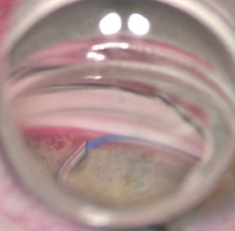
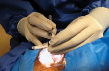
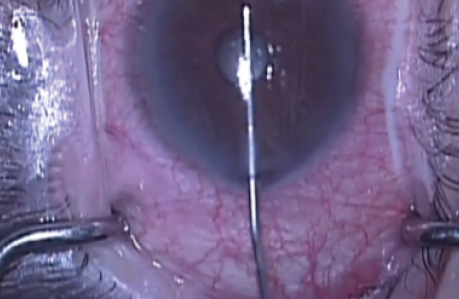
Figure 2. The Trab360 device unroofing the TM.
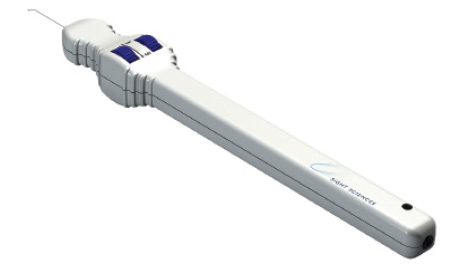
The »Trab360 (Sight Sciences; Figure 2) consists of a stainless steel trabeculotome body that has a polypropylene catheter inside. Once threaded 180º around Schlemm canal, the catheter is pulled toward the surgeon, creating a tear in the TM. The process can then be repeated on the other 180º of TM. (For more information on this device, see Catch up With Glaucoma Today Journal Club.)
Similarly, the »Visco360 (Sight Sciences) involves 180º threading of a catheter into Schlemm canal. Instead of tearing the TM, however, the Visco360 injects viscoelastic into the canal in a controlled manner through the catheter as it is pulled out of the canal. The intention is to stretch the TM, clearing the canal of debris and opening up any collapsed portions of TM. The advantages of the Visco360 procedure are its dilation of more distal areas of outflow resistance such as Schlemm canal and the collector channels and its lack of TM destruction.
Gonioscopy-Assisted Transluminal Trabeculotomy
During gonioscopy-assisted transluminal trabeculotomy (GATT), the surgeon threads a suture through 360º of Schlemm canal via a small TM incision. The two ends of the suture or catheter are then pulled tight and toward the surgeon, creating a 360º tear through the TM. Postoperative complications include hyphema, AC shallowing, and choroidal folds, all of which resolve by 1 month after surgery.7 The GATT procedure is likely the most economical approach to removing the TM.8
Ab Interno Canaloplasty
Ab interno canaloplasty (ABiC) bears some similarities to GATT: in both procedures, the surgeon performs a goniotomy and cannulates Schlemm canal with a microcatheter. In ABiC, however, the microcatheter is primed with viscoelastic. After threading the catheter through 360º of the canal through a small incision, the surgeon ties a suture to the microcatheter’s tip. The catheter is withdrawn via the suture while viscoelastic is delivered to widen the canal and collector channels. The iTrack 250 Microcatheter (Ellex Medical Laser) has an illuminated tip for easy tracking as it is threaded through Schlemm canal. The advantage of ABiC over the Visco360 is that the microcatheter is threaded 360º in the former versus two 180º canalizations with the latter procedure. (For a demonstration of ABiC, see Watch It Now, and visit beye.com/586 for more information on the iTrack 250.)
Kahook Dual Blade
The »Kahook Dual Blade (New World Medical) is another TM device used to perform an ab interno trabeculectomy. The instrument engages and smoothly cuts the TM with a unique blade without damaging adjacent angle tissue. Clinical data on this device are minimal. (For more information on this device, see Watch It Now.)
SUPRACHOROIDAL PROCEDURES
CyPass Micro-Stent
The most recent US addition to the MIGS armamentarium consists of a polymide implant that is placed between the ciliary body and the sclera for a controlled cyclodialysis (Figure 3). The CyPass Micro-Stent has a length of 6.35 mm, an outer diameter of 510 µm, and an inner diameter of 300 µm. The implant is loaded onto a retractable guidewire and inserted via blunt dissection between the ciliary body and sclera to gain access to the supraciliary space. Along the length of the stent are microholes that allow the egress of aqueous from the AC. Retention rings hold the device in place at the ciliary body face.
Studies have shown no choroidal or retinal detachments after the CyPass’ implantation. The device’s efficacy is thought to relate to the amount of subscleral lake that is maintained and can be seen with anterior segment imaging. Complications include transient (less than 1 month) hypotony (13.8%); postoperative IOP elevation (10.5%), with some cases requiring additional surgical intervention; postoperative inflammation (4.4% lasting longer than 1 month); partial obstruction; increased cataract progression (12.2% at 12 months); and rare hyphemas.4,9 (For more information on suprachoroidal procedures, see the article by Cynthia Mattox, MD, on p. 53. For more on the CyPass, visit beye.com/604.)
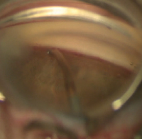
Figure 3. Implantation of the CyPass Micro-Stent.
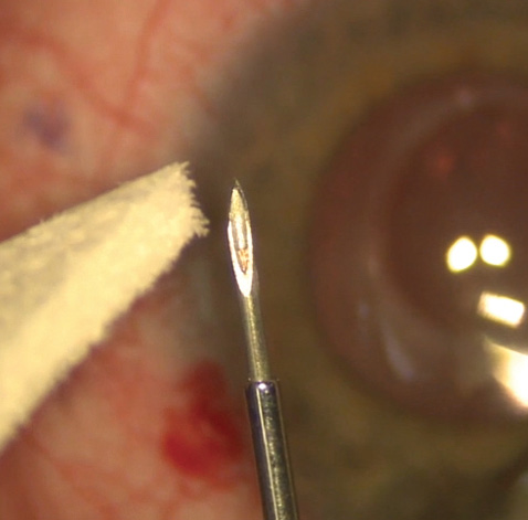
Figure 4. A dehydrated Xen45 gel stent inside the 27-gauge needle inserter.
iStent Supra
The »iStent Supra (Glaukos; not FDA approved) is a 4-mm ridged polyethersulfone and titanium tube with a 160- to 170-µm lumen. Like the CyPass, the iStent Supra is placed between the ciliary body and sclera to allow aqueous to flow into the supraciliary space. Studies thus far have shown no adverse events after the device’s placement. Most patients have been able to discontinue at least one medication, and 98% of patients experienced at least a 20% drop in IOP.10
SUBCONJUNCTIVAL PROCEDURES
Xen45
Watch it Now
Constance O. Okeke, MD, MSCE, demonstrates hand positioning during a routine Trabectome procedure.
Mark J. Gallardo, MD, demonstrates ab interno canaloplasty using the iTrack microcatheter, also known as the ABiC procedure.
Malik Kahook, MD, describes the safety, efficacy, and economics of the Kahook Dual Blade.
The »Xen45 gel stent (Allergan) is 6 mm long and 220 µm thick when fully hydrated—slightly thicker than a human hair. The implant is composed of a hydrophilic porcine gelatin cross-linked with glutaraldehyde (Figure 4). The device is placed via an inserter through the angle and then sclera to create outflow to the subconjunctival space. The Xen remains stiff during implantation; as the collagen gelatin hydrates, the stent enlarges to its intended dimensions (an inner diameter of 45-55 µm) and becomes soft and flexible. Ideally, the device ends up about 3 mm posterior to the limbus and 2 mm outside of the external sclera.
The Xen was designed using Poiseuille’s model of laminar flow to create enough resistance through the stent to prevent hypotony. The procedure results in a low-lying bleb without conjunctival dissection or the need for patch grafting, because the stent’s flexibility prevents erosion (0.4% rate of conjunctival erosion [data on file with Allergan]). The device may be a treatment option for patients with all stages of glaucoma and also for those with narrow-angle and angle-closure glaucoma. Disadvantages of the Xen include potential failure from bleb fibrosis or encapsulation; the best results are achieved with the application of antimetabolites at the site of the device to prevent scarring. (For more information on this device, see Catch up With Glaucoma Today Journal Club for videos with Dr. Sheybani and Herbert Reitsamer, MD, and visit beye.com/599.)
LISTEN UP
Gary Wörtz, MD; Ike Ahmed, MD; and John Berdahl, MD, discuss the exciting evolution of microinvasive glaucoma surgery.
Listen here.
InnFocus MicroShunt
Made of poly(styrene-b-isobutylene-b-styrene) and 9.5 mm long, the »InnFocus MicroShunt (InnFocus; not FDA approved) is implanted via an ab externo approach through conjunctival and scleral dissection after a 3-minute application of a sponge soaked in mitomycin C. The device is intended to treat all stages of glaucoma. Early data have shown transient hypotony and choroidal effusions, both resolving by 3 months.11 A head-to-head trial comparing the safety and efficacy of the InnFocus MicroShunt to trabeculectomy in 412 patients is underway. (For more information on this device, visit beye.com/601.)
1. The Advanced Glaucoma Intervention Study (AGIS): 7. The relationship between control of intraocular pressure and visual field deterioration. The AGIS Investigators. Am J Ophthalmol. 2000;130(4):429-440.
2. Ascher KW. The Aqueous Veins: Biomicroscopic Study of the Aqueous Humor Elimination. Springfield, IL: Thomas; 1961:269.
3. Fea AM, Belda JI, Rekas M, et al. Prospective unmasked randomized evaluation of the iStent inject versus two ocular hypotensive agents in patients with primary open-angle glaucoma. Clin Ophthalmol. 2014;8:875-882.
4. Richter GM, Coleman AL. Minimally invasive glaucoma surgery: current status and future prospects. Clin Ophthalmol. 2016;10:189-206.
5. Ahuja Y, Malihi M, Sit AJ. Delayed-onset symptomatic hyphema after ab interno trabeculotomy surgery. Am J Ophthalmol. 2012;154(3):476-480.e2.
6. Luebke J, Boehringer D, Neuburger M, et al. Refractive and visual outcomes after combined cataract and Trabectome surgery: a report on the possible influences of combining cataract and Trabectome surgery on refractive and visual outcomes. Graefes Arch Clin Exp Ophthalmol. 2015;253(3):419-423.
7. Grover DS, Godfrey DG, Smith O, et al. Gonioscopy-assisted transluminal trabeculotomy, ab interno trabeculotomy: technique report and preliminary results. Ophthalmology. 2014;121(4):855-861.
8. Grover DS, Smith O, Fellman RL, et al. Gonioscopy assisted transluminal trabeculotomy: an ab interno circumferential trabeculotomy for the treatment of primary congenital glaucoma and juvenile open angle glaucoma. Br J Ophthalmol. 2015;99(8):1092-1096.
9. Hoeh H, Vold SD, Ahmed IK, et al. Initial clinical experience with the CyPass Micro-Stent: safety and surgical outcomes of a novel supraciliary microstent. J Glaucoma. 2016;25(1):106-112.
10. Jünemann A. Twelve-month outcomes following ab interno implantation of suprachoirodal stent and postoperative administration of travopost to treat open angle glaucoma. Poster presented at: XXXI Congress of the ESCRS; October 5-9, 2013; Amsterdam, The Netherlands.
11. Batlle JF, Fantes F, Riss I, et al. Three-year follow-up of a novel aqueous humor microshunt. J Glaucoma. 2016;25(2):e58-65.
12. White paper: Ab-interno canaloplasty—the minimally invasive glaucoma surgery that keeps its promise. Ellex Medical Pty Ltd. http://bit.ly/2bnUftK. Published 2016. Accessed August 17, 2016.
13. Reitsamer H. Ab interno approach to subconjunctival space: first 567 eyes treated with new minimally invasive gel stent for treating glaucoma. Paper presented at: ASCRS/ASOA Symposium & Congress; April 17-21, 2015; San Diego, CA.
14. Sarkisian S. New way for ab interno trabeculotomy: initial results. Poster presented at: ASCRS/ASOA Symposium & Congress; April 17-21, 2015; San Diego, CA.
Joel Palko, MD
• resident, Department of Ophthalmology and Visual Sciences, Washington University School of Medicine in St. Louis
• financial interest: none acknowledged
Arsham Sheybani, MD
• assistant professor, Department of Ophthalmology and Visual Sciences, Washington University School of Medicine in St. Louis
• (314) 362-3937; sheybaniar@vision.wustl.edu
• financial disclosure: has received consulting fees from Allergan
Catching up with Glaucoma Today
JOURNAL CLUB
Check out recent coverage of microinvasive glaucoma surgery in this video series moderated by Steven Vold, MD, GT’s chief medical editor.
Steven Sarkisian, MD, suggests how to implement the iStent Trabecular Micro-Bypass Stent in practice.
Herbert Reitsamer, MD, shares a European perspective on microinvasive glaucoma surgery.
Thomas Samuelson, MD, provides an overview of study data on the Hydrus.

Arsham Sheybani, MD, discusses his experience with the Xen45 and where the gel stent fits into glaucoma management.


