CASE PRESENTATION
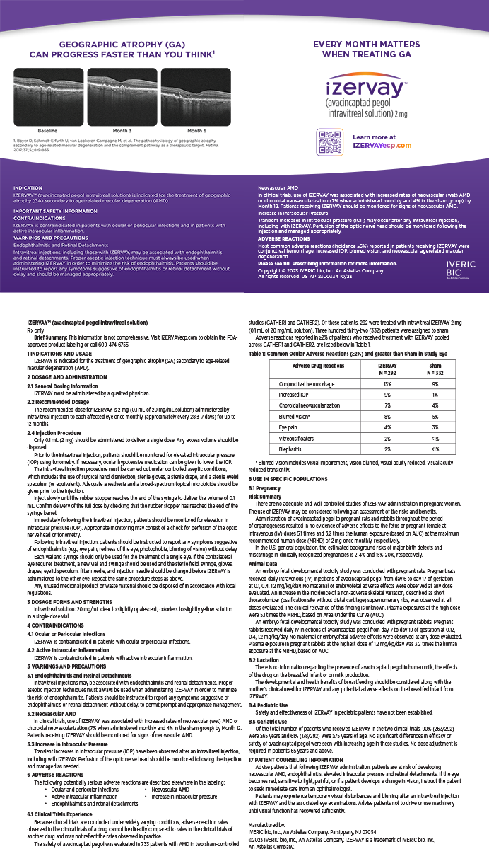
A white male patient in his early 50s presented with bilateral cataracts. Additionally, he had a history of ocular trauma to the right eye that occurred approximately 10 years prior to the formation of his cataracts when a racquetball struck the eye. The preoperative clinical examination revealed posterior subcapsular 3 to 4+ cataracts that were similar in appearance. I detected nothing abnormal about the right eye, including phacodynesis.
I proceeded with cataract surgery in the patient's right eye. Because he was fairly young and had a soft cataract, I planned to prolapse the lens out of the bag and aspirate it using NeoSoniX (Alcon Laboratories, Inc., Fort Worth, TX). For harder cataracts, I normally perform in-the-bag chopping. To begin, I created a temporal, 3-mm, clear corneal incision. As I made the capsulorhexis, I began to suspect a problem with the superior zonules. While extracting the patient's lens, however, I noticed that all of the superior zonules were absent (Figure 1). Using NeoSoniX, I extracted the lens nucleus at the iris plane without incident. Removing the epinucleus and cortex, however, posed a problem because the superior portion of the capsular bag collapsed, wrinkling downward like a raisin to where it was supported by the inferior zonules.
HOW WOULD YOU PROCEED?
1. Would you attempt manual or automated I/A?
2. Would you leave or remove the remaining capsular bag?
3. Which IOL would you choose, and how would you implant it?
4. How would you prevent vitreous prolapse?
SURGICAL COURSE
I did not have access to a capsular tension ring, so I elected to employ iris retractors (Alcon Laboratories, Inc.) to engage, tighten, and support the capsular bag (Figure 2). I created four paracentesis incisions, one each at the 10:30-, 11:30-, 12:30-, and 2-o'clock positions and placed the iris retractors through them. Next, I removed the residual inferior cortex with the I/A handpiece. Superiorly, I used a combination of manual stripping with a 27-gauge cannula and the I/A handpiece to remove the cortex.
The next challenge was to determine how to support an IOL in the bag. I chose to suture a foldable, acrylic MA60AC lens (Alcon Laboratories, Inc.). It was necessary to enlarge the incision to 3.5 mm in order to accommodate the IOL. I then made a conjunctival peritomy over the area where the needle would exit the sclera and created a half-thickness, triangular, scleral flap sized approximately 2 X 2 mm with a crescent blade angled with its base toward the limbus. I positioned the flap approximately 3 mm posterior to the limbus, at the 12-o'clock position. Next, I passed the needle with a PROLENE Polypropolene Suture (ETHICON INC, Somerville, NJ) through the inferior limbus of the cornea, the anterior chamber, the anterior capsulorhexis, and the capsular bag at its superior equator. I moved the needle out under the base of the iris, through the wall of the sclera, and under the scleral flap. Using a Lester lens hook, I retrieved the suture end through the anterior capsulorhexis and drew it out of the incision. I tied the loose end of the suture to the IOL's superior haptic.
With a folding forceps, I placed the lens in a mustache fold and maneuvered it into the capsular bag. As the IOL unfolded, I pulled the suture tight and drew the lens up to the proper position. I then used the needle to take a small bite under the scleral flap in order to secure the suture, after which I sealed down the flap with a nylon suture. After removing the iris retractors, I performed thorough I/A and injected Miochol (CIBA Vision, Duluth, GA). I finished the case in the usual fashion, and the corneal incision closed without a suture.
OUTCOME
The patient was satisfied with his outcome in the right eye. His postoperative UCVA was 20/50, and his BCVA was 20/25. I later removed the cataract in the patient's left eye and implanted an SA60AT IOL (Alcon Laboratories, Inc.) without incident.
Robert Arleo, MD, is in private practice at the Arleo Eye Institute in Ithaca, New York. He holds no financial interest in any product or technology mentioned herein. Dr. Arleo may be reached at (607) 257-5599; Rarleo@aol.com.