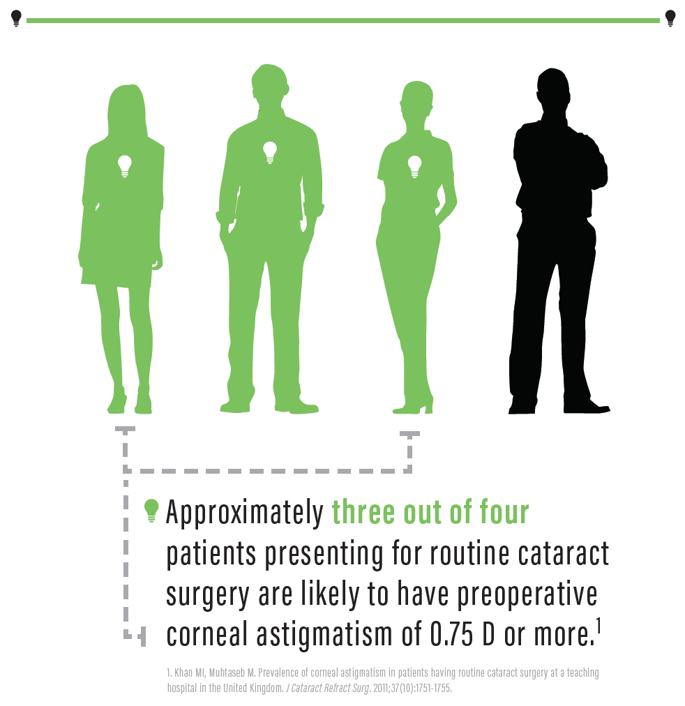
Astigmatic correction with toric IOLs has proven to be one of the most effective strategies by which to improve the refractive outcomes of cataract surgery. Selecting an appropriate lens for an individual patient, however, requires accurate methods to predict the required toric cylinder power and axis as well as alignment of the IOL with the desired meridian at the time of surgery. Manual keratometry and freehand marking have been considered adequate to achieve these aims, but these simple measures are insufficient if one hopes to achieve less than 0.50 D of residual astigmatism in the majority of patients.
AT A GLANCE
- Astigmatic correction with toric IOLs has proven to be one of the most effective strategies by which to improve the refractive outcomes of cataract surgery.
- Selecting an appropriate lens for an individual patient requires accurate methods to predict the required toric cylinder power and axis as well as alignment of the IOL with the desired meridian at the time of surgery.
- Using multiple instruments to measure the cornea and image-guided alignment are helpful, but surgeons can achieve excellent astigmatic outcomes without investing in additional adjunctive technology.
Improved methods have been introduced to measure the corneal surface, and the use of multiple instruments for keratometry is now widely practiced. Similarly, image-guided technology and intraoperative aberrometry have been proposed as more precise methods to achieve accurate alignment. The need for adjunct technology can be daunting to surgeons contemplating using toric IOLs, but fortunately, excellent results can be achieved without a large expenditure or expensive instrumentation.
MEASURING THE CORNEA
There is widespread agreement that the posterior cornea must be taken into account when predicting the correct toric IOL power required for an individual patient.
The posterior cornea can be measured using Scheimpflug-based topographers or swept-source optical coherence tomography biometers. In a comparative study, however, my colleagues and I found that a toric calculator incorporating a theoretical model for predicting posterior corneal astigmatism was as accurate as direct measurements.1
Perhaps more important is the concept of using multiple devices to measure the cornea, because each device measures the cornea in a different manner. Warren E. Hill, MD, has popularized the concept of considering multiple instruments as secondary devices to validate the measurement of a primary or preferred device for the magnitude and axis of astigmatism. This process is subjective, however, and somewhat of an art form that develops with experience. Furthermore, because astigmatism is a vector with magnitude and direction, it is invalid to combine the axis of astigmatism from one device with the magnitude of the cylinder from another, as commonly practiced.
To simplify the process of combining multiple instruments, I have incorporated a “K Calculator” into the online Barrett Toric Calculator (www.ascrs.org/barrett-toric-calculator). This method allows the user to select the keratometry (K) readings of up to three devices and provides an integrated K using appropriate vector mathematical calculation. If two devices are selected, the integrated K is the mean of the two devices, and if three devices are selected, then the integrated K is the median of the three devices. The median as a measure of central tendency de-emphasizes outliers, and it proved to be the most accurate, with the lowest prediction error of residual astigmatism, in a personal series of 128 cases of toric IOL implantation using preoperative Ks and the measured postalignment of the IOL.2
The three devices used in this study were the Lenstar (Haag-Streit), the IOLMaster 500 (Carl Zeiss Meditec), and the anterior surface measurement of the Pentacam (Oculus Optikgeräte). The percentage of cases predicted to be within 0.50 D of residual astigmatism was 71% using the Lenstar and improved to 78% with the integrated K.

The integrated K proved to be as accurate as the keratometry of a single device in predicting spherical outcome. The percentage of cases predicted within 0.50 D of spherical equivalent error was 87.8% with the integrated K compared to 83% with Ks from a single biometer. Using a combination of Ks obtained from a biometer (Lenstar or IOLMaster 500), topographer (Pentacam), and manual Javal keratometer also proved to be more accurate than Ks from a single device.
The results of this study support the concept that multiple devices are helpful in predicting astigmatic outcome. Three devices will suffice, and many surgeons already have these instruments in their practices.
DEVICES TO ASSIST WITH THE ALIGNMENT OF IOLs
I do not have personal experience with intraoperative aberrometry to assist with the alignment of toric IOLs, but literature supporting this methodology is lacking. Data suggesting that a greater percentage of patients achieve 0.50 D of residual astigmatism or less after IOL alignment with intraoperative aberrometry than with manual marking and prediction of the required axis have been published.3 The method of calculation in this study, however, was a legacy calculator, which did not incorporate the posterior cornea; the estimation of surgically induced astigmatism was lacking; and the number of low-powered toric IOLs was restricted. Intraoperative aberrometry may be useful in locating the correct meridian in eyes with high levels of cylinder, but the technology is unlikely to be as reliable with low toric IOL cylinders. In a more recent comparison, only 10% of cases had an axis measured by the aberrometer within 10º of the axis measured by postoperative refraction.4
The image-guided location of the axis for toric IOL alignment is more promising. The technology uses limbal vessel registration to identify landmarks that are used to determine a reference axis in relation to the desired location for the toric IOL. In concept, this method will work best if the desired axis is the steep axis measured by the same device where the image capture occurs. In practice, however, this is rarely the case. The desired axis is often derived from keratometry obtained from other instruments or a combination of instruments, and the method therefore depends on ensuring that the patient’s head is completely level when the image is captured for registration. Inattention to this detail or erroneous registration could explain errors I have noted when comparing image-guided alignment to the toriCAM approach to determine the actual axis of manually placed reference marks. Although the workflow of image-guided systems is attractive, I believe the app-guided (toriCAM) method is more accurate and less prone to error; in my opinion, adjunctive technology is helpful but not obligatory for accurate alignment.
RELAXING INCISIONS
Limbal relaxing and laser arcuate incisions have been suggested as an alternative to toric IOLs for low levels of astigmatism. These incisions can be a source of irritation, however, and they are less stable than toric IOLs. Most importantly, the astigmatic effect of corneal incisions, even when performed with the precision of a femtosecond laser, is less predictable than that of toric IOLs.5 In particular, low-powered toric IOLs are a preferable method for correcting low levels of preexisting astigmatism, but these cylinder powers are not available in all markets (see Dr. Waring’s article).
CONCLUSION
The requirements for using toric IOLs effectively are relatively modest. Using multiple instruments to measure the cornea and image-guided alignment are helpful, but surgeons can achieve excellent astigmatic outcomes without investing in additional adjunctive technology.
1. Abulafia A, Hill WE, Francina M, Barrett GD. Comparison of methods to predict residual astigmatism after intraocular lens implantation. J Refract Surg. 2015;31(10):699-707.
2. Barrett GD. Plotting the right course for toric IOLs: the Barrett K Calculator. Paper presented at: Australian Society of Cataract and Refractive Surgery Meeting; August 2-5, 2017; Hamilton Island, Queensland, Australia.
3. Woodcock MG, Lehmann R, Cionni RJ, et al. Intraoperative aberrometry versus standard preoperative biometry and a toric IOL calculator for bilateral toric IOL implantation with a femtosecond laser: one-month results. J Cataract Refract Surg. 2016;42(6):817-825.
4. Huelle JO, Druchkiv V, Habib NE, et al. Intraoperative aberrometry-based aphakia refraction in patients with cataract: status and options. Br J Ophthalmol. 2017;101(2):97-102.
5. Wang L, Zhang S, Zhang Z, et al. Femtosecond laser penetrating corneal relaxing incisions combined with cataract surgery. J Cataract Refract Surg. 2016;42(7):995-1002.


