A physician's perspective

Several years ago, I was knocked unconscious after I fell off a ladder. When I awoke, I was unable to move my extremities for a (long and terrifying) minute. Subsequent imaging detected advanced spinal stenosis, which accounted in part for my temporary quadriplegia. Neck surgery—anterior cervical discectomy and fusion, to be exact—followed, and as did headaches that occurred after long days in the OR.
I needed to protect my spine and neck from long-term injury, but still needed to provide the expert care my patients expected in the OR. That is when I turned to the ZEISS ARTEVO 800.
Use of the ZEISS ARTEVO 800 heads-up 3D visualization system has improved my ergonomics...but benefits to using [it] aren't limited to posture and musculoskeletal protection. My surgical efficiency and precision have improved, as has my ability to communicate with surgical staff.
Use of the ZEISS ARTEVO 800 heads-up 3D visualization system has improved my ergonomics, assuring me that I am doing what's best for my body during some of my most intense hours. But benefits to using the ZEISS ARTEVO 800 aren't limited to posture and musculoskeletal protection. My surgical efficiency and precision have improved, as has my ability to communicate with surgical staff. Further, ZEISS ARTEVO's interoperability with other ZEISS platforms in my surgery center have enhanced my experience with heads-up 3D visualization, helping turn my OR into a center of technologic harmony that ultimately improves patient experiences and outcomes.
Surgeons seeking to push their OR into the next era should consider how any or all of these elements could positively affect their surgical practice.
Ergonomics. Surgeons using the ZEISS ARTEVO 800 sit in a relaxed position during surgery, visualizing patient anatomy on a large monitor while wearing a set of 3D glasses. Traditional platforms, which require the surgeon to conform to the physical positioning of microscope oculars, result in strained posture and may place surgeons at risk of musculoskeletal damage. With a heads-up 3D display, surgeons can focus less on their body's positioning and more on the patient case at hand.
Improved visualization. 3D visualization significantly improves the depth of field during surgery. This is particularly useful during MIGS procedures, in which spatial context and exact placement are keys to success. Further, my ability to visualize the angle irrespective of patient, microscope, or physical positioning has improved my efficiency with MIGS procedures and has enabled me to perform the most precise surgery possible.
The enhanced depth of field also makes cataract surgery more precise, particularly in patients with a shallow anterior chamber. When I am better able to understand the distance between visualized elements—say, a surgical instrument and the endothelium—I perform safer surgery.
Use of the ZEISS RESCAN iOCT platform during glaucoma surgery has allowed me to visualize anatomic structures like never before. For example, iOCT has enabled me to confirm the efficacy of viscodilation in the canal of Schlemm by showing in real time the interaction between OVD and canal tissue. It has also increased my precision when placing MIGS devices in the superciliary and subconjunctival spaces, as a clear depiction of a device's location relative to nearby tissue is easily obtained. My experience: safer, more precise glaucoma surgery and increased surgeon confidence.
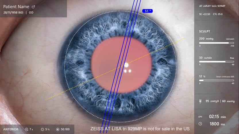
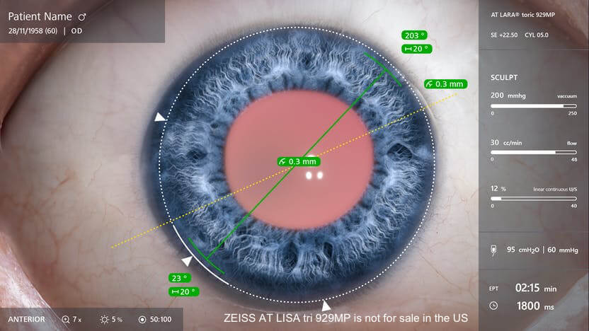
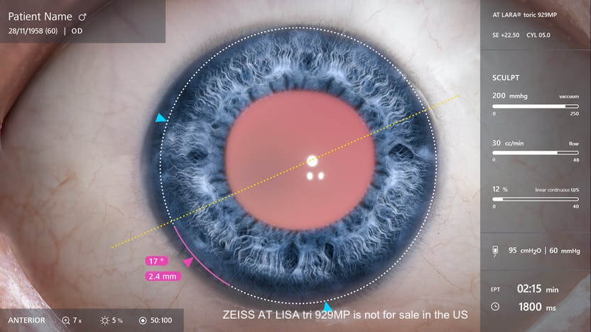
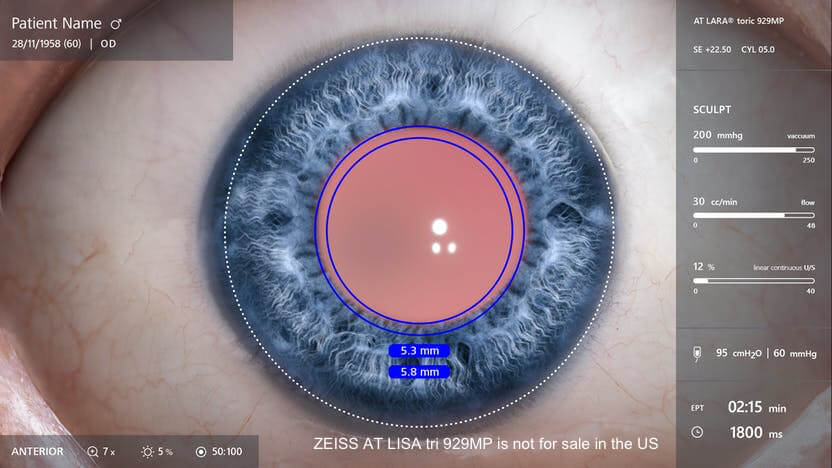
Communication with other ZEISS platforms. ZEISS ARTEVO's potential is at its highest when used simultaneously alongside other ZEISS technologies. Consider, as an example, cataract surgery. During cataract surgery, preoperative alignment parameters captured on the ZEISS IOLMaster 700 are presented alongside patient demographic data, all of which is synthesized into a digital overlay via the ZEISS CALLISTO eye. This overlay, placed atop the 3D image on the screen the surgeon uses in the OR, keeps relevant data in a single place within the surgeon's view. Such technology is uniquely useful during toric IOL placement, and my confidence that I have placed the IOL in the position determined during the preoperative period is highest when using this digital overlay.
Communicating with staff. Before heads-up 3D visualization was a part of my OR, my staff's ability to anticipate my needs was limited. Now that they can see what I see by merely donning a pair of 3D glasses, they are in sync with my surgical movements and can stay one step ahead of me. This has led to increased surgical efficiency and has improved the capacity of my surgical staff, who now has a more direct ability to learn the reasonings behind my protocols and procedures.
Increasing the efficiency of and my confidence in cataract and glaucoma surgery has led to, in my estimation, improved patient satisfaction—the ultimate measure of a new technology’s successful integration into a surgeon’s workflow. Add in improved physical comfort, decreased risk of long-term musculoskeletal damage, enhanced visualization, and a staff who can anticipate my needs, and I can confidently say that the inclusion of ZEISS ARTEVO 800 into my surgical suite has improved my capacity for running a cutting-edge OR.
Inder Paul Singh, MD
Dr. Singh is a consultant for Carl Zeiss Meditec, Inc.
References:
1. Measured comparing the number of vertical TV lines of a 4K 3D monitor with polarization using a resolution test chart ISO 12233 from Esser. Comparing the ZEISS ARTEVO 800 to a competitor’s system on the same monitor which yields 1000 TV lines resolution for the ZEISS ARTEVO 800, and 800 TV lines resolution for the competitor’s system.
2. Depth of field depends on magnification.
3. Based on transmission calculation. Data on file. Increased optical transmission together with light sensitive ZEISS ARTEVO 800 cameras result in light reductions of up to 85% (according to Peter Stalmans, MD).
4. Hansraj KK. Assessment of stresses in the cervical spine caused by posture and position of the head. Surg Technol lnt. 2014 Nov;2s:2n-9. PMID: 25393825.
5. Weinstock RJ, Ainslie-Garcia MH, Ferko NC, Qadeer RA, Morris LP, Cheng H, Ehlers JP. Comparative assessment of ergonomic experience with headsup display and conventional surgical microscope in the operating room. Clin Ophthalmol. 2021 Jan 29;15347-356. doi: 10.2147/0PTH.5292152. PMID: 33542618; PMCID, PMC7854362.
The statements of the doctor in this insert reflect only his personal opinions and do not necessarily reflect the opinions of any institution with which he is affiliated. The doctor shown in this insert has a contractual or other financial relationship with Carl Zeiss Meditec, Inc. and has received financial support.
Not all products and services are available in all countries.
CAP-en-US_32_025_0223II

Sponsored by ZEISS


