
Eric D. Rosenberg, DO, MScEng: While cataract surgery has become one of the safest and most common surgical procedures performed worldwide, it requires a sequence of well-orchestrated surgical steps in conjunction with proper patient selection. Here, we share our preferred tools and techniques for making complex cases less complex based on our own personal experiences to ensure that we can achieve the best possible outcomes in anterior segment surgery.
STEP NO 1: WOUND CONSTRUCTION BASICS
Dr. Rosenberg: Dr. Fintelmann, what is the most important thing you’re focused on when creating your clear corneal incisions for cataract surgery?
Robert Fintelmann, MD, FACS: Creating a reproducible and self-sealing incision.
Dr. Rosenberg: What difficulties and/or complications have you faced when creating incisions in the past, and how did those difficulties affect the outcomes with those patients?
Dr. Fintelmann: Two major things come to mind. Sometimes, when the blade was too sharp, I would make a small scratch with the side of the blade and then create a tri-planar incision. If you’re not careful and go full thickness, you can unintentionally create a limbal relaxing incision. Fortunately, they’re self-sealing and can be fixed fairly easily. What’s more frustrating is if the tip of the blade is not sharp enough and you can’t get a nice inner edge, it can create a little tag that can lead to problems with sealing. Additionally, I get frustrated with tips that have a bur on them because basically that makes the knife unusable.
Dr. Rosenberg: In your experience, how do BVI’s Beaver® Knives (Figure 1) perform compared to other knives you’ve used?
Dr. Fintelmann: I can count on the sharpness of the Beaver knives being consistent. I also like that the Beaver safety knife has a guard on it. I am handed these knives with the guard down. I open and close it before I hand it back to my surgical tech to prevent injuries in the OR. The only time it’s exposed is right before I use it. I also like the little reference line etched on the blade, indicating that I’m far enough into the cornea.
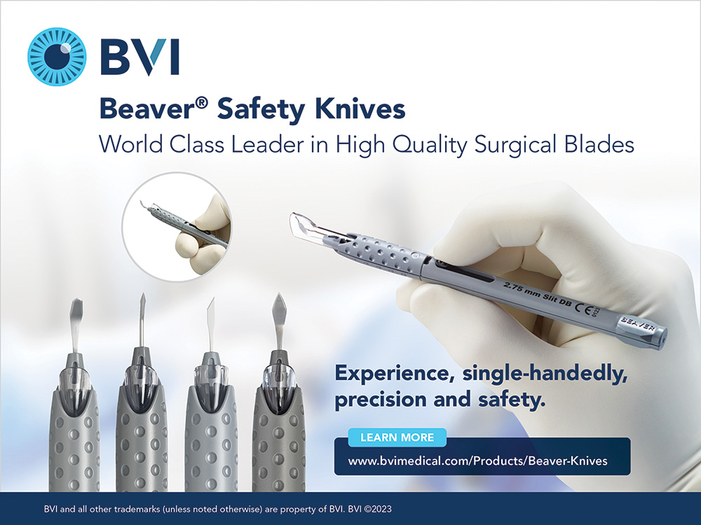
Figure 1. BVI’s knives and blades have safety features to prevent accidents.
Dr. Rosenberg: For those who have needed to make scleral tunnels in difficult cataract cases, have you used the Beaver Knives?
Dr. Fintelmann: I see quite a lot of post–radial keratotomy (RK) eyes with a high number of cuts. It’s hard to fit an incision in between them. I’ve never had an issue using the Beaver knife, knock on wood, with creating a scleral tunnel; they always seal. It’s similar with cataracts and corneal transplants. I like scleral tunnels, but without a good blade you’re lost.
Deborah Ristvedt, DO: Recently, I did two mission trips to Guatemala and Africa and had some of the hardest cases I’ve ever done. To be a skilled manual small-incision cataract surgeon, self-sealing incisions of the wound are crucial. I was so thankful to BVI Medical for supplying me with their beautiful blades for these complex cases.
Jeffrey Whitman, MD: It’s also important to mention how deflating it is for patients to experience a wound leak at 1-day postoperative. Whether it is a post-RK or a routine case, a shaggy incision will need help sealing. Sometimes, these patients have to go back to the OR for a suture to be placed. It’s not fun for the surgeon or the patient. With a good blade, that rarely happens, if ever.
Dr. Rosenberg: Why is the right knife so important in cataract refractive surgical procedures, which we’re doing so commonly now?
Dr. Fintelmann: Reproducibility is key to getting a good idea of what impact the wound has on the postoperative outcome. I’ve found that my femtosecond laser cases are a little bit more variable. What I like about BVI’s blades is you can go onto the limbus and be as peripheral as possible. This is harder to do with a laser. Additionally, the self-sealing nature of wounds with a blade, again, is more reproducible for me.
Dr. Rosenberg: Do you have any pearls for surgically induced astigmatism (SIA)? What have your years of practice taught you about the right blades allowing your calculations to be a little bit more on par? I imagine repeatability has something to do with that.
Dr. Fintelmann: Correct. My SIA number is 0.10 D. If you look at that on a vector diagram, it’s very disheartening what 0.10 D of astigmatism does to outcomes. I wish there was a more precise way of predicting the impact of a wound on surgical outcome.
Dr. Whitman: Research has shown that SIA usually ends up being about 0.10 to 0.20 D. The thing is, if you’re doing a refractive procedure, you and your patients want good results early. Waiting 6 or 12 months isn’t ideal.
Dr. Rosenberg: Dr. Fintelmann, any pearls for how to create the best wound architecture?
Dr. Fintelmann: One of my mentors always used to say, “A little whiter makes it tighter.” I try to get as close to the limbus as possible without nicking the conjunctiva. Additionally, I like to make a small groove to help create a nice-looking outside edge, acting as a little line that I can use to pre-plan the start of my incision. I tunnel up, avoiding entering straight in to get a nice interior straight edge as opposed to a dimple edge if you push down too much in the entry.
Dr. Rosenberg: Does anybody use a femtosecond laser to create their wounds?
Dr. Whitman: I do about 35% to 40% of the time. I still prefer to take a Beaver blade to open the incisions rather than poke through them. A femtosecond laser is a good guideway to get a perfect nonleaking incision, but I still think using a nice, sharp blade makes all the difference, particularly for refractive patients.
Dr. Fintelmann: A laser takes away a little bit of tissue and doesn’t go as far peripheral. I recently had a case for which it would’ve been better not to use a femtosecond laser for the incision because it went from the surface of the LASIK flap and straight into the eye.
Jonathan Solomon, MD, FACS: I gave up on clear corneal incisions probably 8 years ago. We’d put a fair amount of work into creating instrumentation to assist in opening femtosecond laser incisions. We modified the incisions slightly by bringing them further from the limbus. Ultimately, we learned that femtosecond lasers just don’t do well that far out at the limbus where you really want to be.
And whether you intend to go through the peripheral vessels, keep in mind that a femtosecond laser can only cut what it can optically see through. In my experience, it isn’t worth the extra effort. At this stage, nothing beats a good, clean, reproducible blade. BVI is known to be the upper echelon of incisional instrumentation.
Dr. Rosenberg: I completely agree. I don’t think there’s a substitute for creating excellent self-sealing wounds, and BVI’s blades are the highest quality.
STEP NO. 2: SMALL PUPIL MANAGEMENT
Dr. Rosenberg: Dr. Whitman, what are some leading indicators, preoperatively or in the OR, of a complex cataract case?
Dr. Whitman: I look for miotic pupils, synechiae, dense and mature cataracts, and any other condition or pathology that affects my ability to visualize ocular structures and complicates my ability to perform surgery safely. A small pupil makes every step of the surgical procedure more difficult compared to routine surgery. In these cases, I like to use a pupil-expanding device such as the BVI I-Ring® (BVI Medical; Figure 2). When in doubt, I reach for a BVI I-Ring. I never regret using it.
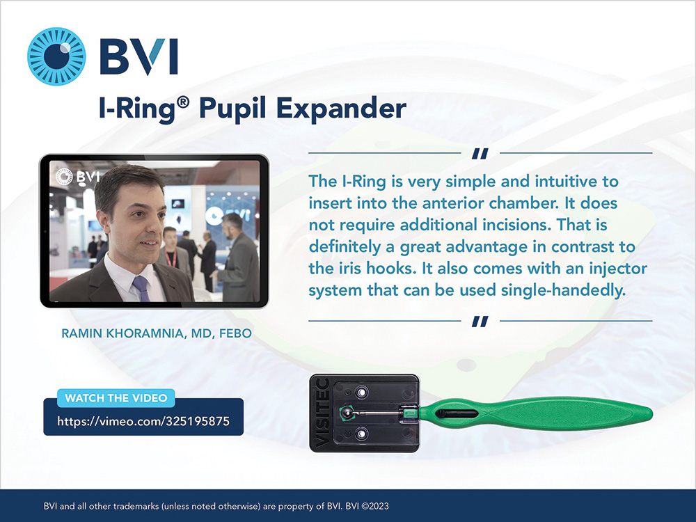
Figure 2. The I-Ring is made of a bright green polyurethane and is easy to visualize inside of the eye.
What’s nice about the I-Ring is that you can put it in at any time. It can even be used after a femtosecond laser capsulotomy by placing some OVD between the iris and the capsulotomy. You may think that it would make the case take a little bit longer, but the few seconds it takes to implant is worth it so you aren’t sweating through the case.
Dr. Rosenberg: What steps are required to dilate the pupil with the I-Ring?
Dr. Whitman: The I-Ring is made of a bright green polyurethane, so it’s very easy to visualize inside of the eye. In my hands, it’s much gentler to the eye compared to the Malyugin Ring (MicroSurgical Technology). It comes preloaded. The injector requires an incision size of about 2.4 mm. Once inserted, the I-Ring creates a pupil size of about 6.2 to 6.5 mm. I find that, postoperatively, the pupil is rounder. It can be used in both regular and complex cases.
There are several techniques to remove it. I prefer a one-handed, one-step technique. I enter the eye, push the inserter back out of the injector, hook onto the ring that is distal from me, and pull it toward me. By bringing it back in, the I-Ring generally goes right into the inserter and can be pulled out of the eye.
The I-Ring is useful for hard cases that we get involved in. I used it just recently in an eye with a 1-mm pupil and almost 360º synechiae and plateau iris. For a mature cataract, I paint the capsule with trypan blue dye under an OVD. Alternatively, the OVD can be removed before the capsule is painted.
Dr. Rosenberg: Iris preservation is a great point. My father-in-law, who is an ophthalmologist too, loves stretching the iris mechanically. I’ve seen some of those pupils on day 1 and week 1 postoperatively, and I like the way it looks with the ring in the eye. Everything comes back down to normal. Are there additional advantages to using the I-Ring over other devices?
Dr. Whitman: I think there are two primary advantages. First, it’s easy to get it into the eye and remove from the eye. Second, there is less trauma to the iris. To me, that’s what it’s all about. It’s nice when the pupil is nice and round after surgery in the presence of a dense cataract and synechiae. I started practicing ophthalmology in a time when either two instruments or a Beehler pupil dilator would be used to stretch the iris hoping that it would rebound and stay dilated. Now, we have better tools that work consistently and effectively.
One other thing I’d like to mention is that just because you may not be comfortable with another pupil expansion device doesn’t mean you shouldn’t try the I-Ring design. I recommend that surgeons talk to their BVI rep and watch some surgical videos from colleagues because sometimes that’s the difference between succeeding with a device and not having a good experience. The I-Ring is a great advancement in pupil expansion techniques.
STEP NO. 3: IOL SELECTION
Dr. Rosenberg: Another way to make complex cases less complex and provide the best visual outcomes is our choice in monofocal IOLs. We speak a lot about premium IOLs, but according to various reports, about 80% of IOLs implanted today in the United States are monofocal IOLs. So for the 80% of patients where a monofocal is the right choice, we still should think about which monofocal lens is best for our patients.
When it comes to monofocal IOLs, I prefer the IPure® IOL for multiple reasons. The B1PC is a one-piece hydrophobic IOL, and the B3PC is a three-piece IOL. Both are aspherically neutral in the central 2.4-mm zone and have -0.18 µm of spherical aberration in the peripheral zone. I find patients experience next to no dysphotopsias and a low posterior capsular opacification rate with these lenses.
Interestingly, if you look at the aspheric optic profile of the B1PC and the B3PC compared to the Eyhance (Johnson & Johnson Vision), the power distribution as it goes over the lens is similar. Do you find that you get an added range with these designs?
Dr. Solomon: Going back about 15 years when new technology IOLs hit the market, the goal was to improve night vision. Before then, all IOLs had some positive spherical aberration. A small subset of patients with these IOLs also achieved good intermediate vision. Fast forward to today, the concept of manipulating the spherical aberrations across an IOL to provide some degree of depth of focus has caught on. What was in vogue then is coming back again with improvements to those types of lenses. The IPure lense is designed to give patients a blend of high-quality, high contrast night vision by way of its negative asphericity in the peripheral zone with some degree of depth of field by maintaining a neutral asphericity in the central zone. In my experience, I’ve found that the IPure IOL really does offer a range of vision, certainly on par as you said, as compared with some of the other enhanced monofocal optics. The IPure IOLs are the premiere lens we offer in our practice for those who are looking to truly enhance single-optic vision.
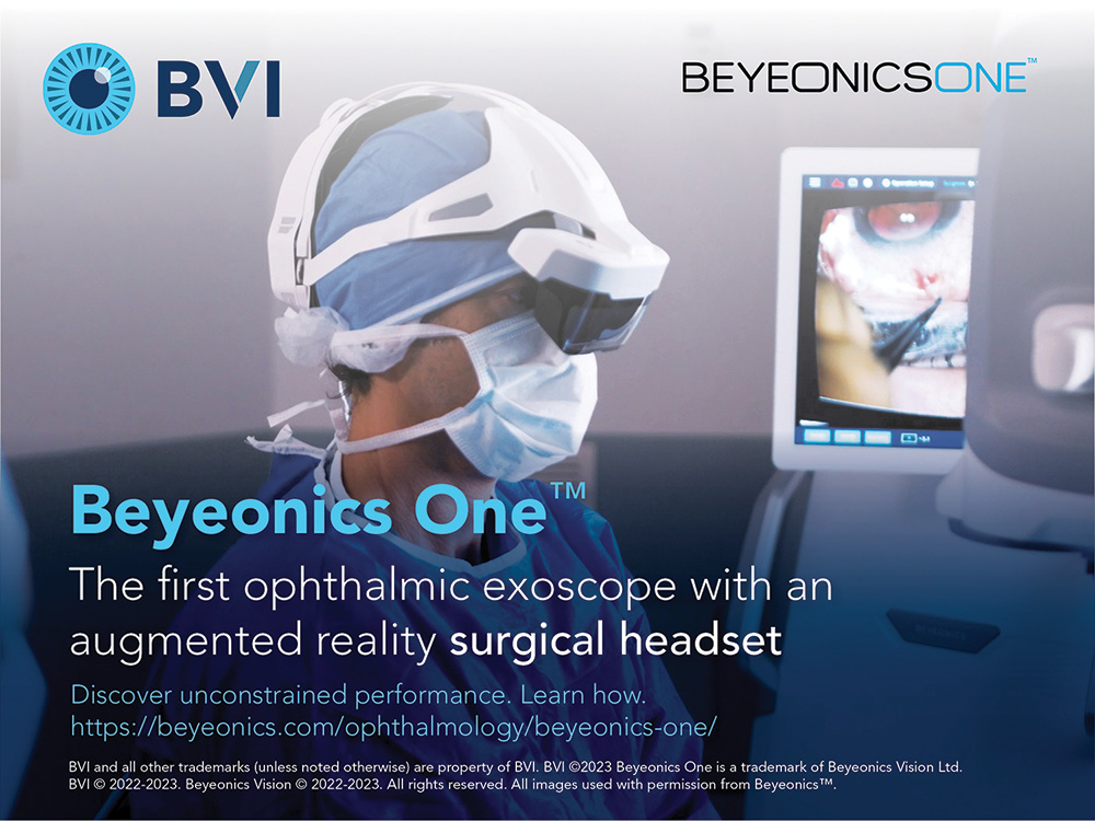
Figure 3. The Beyeonics One provides a realistic image of the surgical field.
Dr. Whitman: Because the B1PC and the B3PC are neutral aspheric centrally, patients get sharp distance vision. At the end of the day, patients want to drive. With these lenses, they’re often gaining distance vision without experiencing the drawbacks associated with some premium lenses. If you can give them that—and they even may get some intermediate vision—you’ve made them happy. A happy patient is going to tell their friends about their experience at your practice.
Dr. Solomon: I want to interject a word of caution. Patients with low myopia may lose a little bit of intermediate vision postoperatively with monofocal IOLs when the target is emmetropia for high-quality distance. As long as you manage expectations so that patients understand the goal is high-quality single vision, they tend to be pleasantly surprised and satisfied to gain some intermediate vision with these types of lenses.
Dr. Rosenberg: As an additional benefit, it goes without saying that preloaded lenses—especially three-piece preloaded lenses—increase efficiency, especially at ambulatory surgery centers and even moreso in academic and hospital systems. What advantages for your complex surgeries have you noted having a three-piece preloaded IOL that can go through a normal incision without having to enlarge it?
Dr. Whitman: For complex cases, a three-piece lens can be dialed into the sulcus. Even if I have to perform a limited anterior vitrectomy, I don’t have to start from the beginning and get a new lens out. I don’t have to use a lens cutter. Also, being preloaded is a big advantage that I don’t believe any other IOL company offers. The IPure three-piece IOL is lathe cut from a chemically bonded, crosslinked button into either a three- or one-piece IOL. So the three-piece can go through about a 2.5- or 2.6-mm incision and is prepped in the same way as the one-piece IOL with the benefit that, while off-label as is any IOL, it can be placed in the sulcus.
Dr. Rosenberg: I don’t need to tell anybody here why it’s disadvantageous in a case that goes sideways to have to ask for a three-piece IOL that our technicians aren’t used to loading, and additionally, then have to enlarge a wound to 3 mm. You’re already encountering vitreous, and you’re praying for stability of the anterior chamber and everything else that goes with it. With the IPure three-piece, you don’t have to enlarge the wound; I can maintain better stability and avoid putting in a suture. The preloaded three-piece IPure IOL can be used with the same or similar incision size, the wound closes and self-seals, and the lens unfolds just where it needs to be. I feel pretty safe without suturing the wound at the end of the case.
Dr. Whitman: When I have to explain to a patient after cataract surgery that a suture will need to be removed in 6 weeks, it’s a downer for them, and it takes more time for us in a case that’s already taken longer than routine surgery. We have an alternative with a lens with a superb optic. We’re not making a compromise, and more importantly, we’re providing our patients with better outcomes.
STEP NO. 4: VISUALIZATION AND SURGEON COMFORT IN THE OR
Dr. Rosenberg: Related to making complex cases not so complex, I think it’s important to talk about new technologies in the field of digital visualization and surgeon comfort in the OR. Dr. Solomon, you are a huge advocate of the importance of ergonomics in the OR to improve surgical outcomes. I know you were one of the first surgeons in the United States who replaced your traditional microscope with the Beyeonics One™ visualization platform (BVI Medical; Figure 3) in your OR. How is the Beyeonics One platform different from the other heads-up devices, and what were decision making factors for your adoption of this platform?
Dr. Solomon: Better visualization improves your skillset and surgical outcomes. The 3D heads-up guidance that the Beyeonics One provides is a more realistic image of the surgical field, and more importantly, gives me the ability to better visualize the true anterior chamber dimensions, which I think is a major step up from other surgical microscopes. It gives me what was initially described as an enhanced depth of the field.

Figure 4. The Endo Optiks® ECP device addresses the inflow and natural outflow pathways.
The ergonomic advantages are plentiful. It’s remarkable what happens at the end of the day when you’re not feeling back and neck strain. Earlier in my career, I remember thinking, “I’m pretty limber. I stay in good shape. There is no way I’m going to have back and neck problems.” But, it creeps up on you. And when it does, it’s very difficult to reverse.
Dr. Rosenberg: What recommendation would you give young surgeons who are coming out of training?
Dr. Solomon: I suspect everyone thinks that they are invincible when they’re getting started in their career as an ophthalmologist. I don’t know if there’s something else you could say or do other than to have a company like BVI offer a platform like the Beyeonics One that’s open and accessible to the young surgeons in training programs where they’re getting firsthand experience with the technology. I’m a believer that we’ll change the way we function in the OR in large part because of what Beyeonics One offers.
Dr. Rosenberg: What was the learning curve like for you when you brought this technology into your OR?
Dr. Solomon: I had experience with some other 3D systems prior to trialing Beyeonics One. For me, it was an easy transition. But to add context for a novice surgeon, our fellow operated with Beyeonics One on the same day I did and he didn’t miss a beat. That tells you just how easy and intuitive the adaptation is with this technology.
Dr. Rosenberg: Is there less manipulation of the settings with digital microscopes?
Dr. Solomon: The user has the option to rely on head movement to switch focus and magnification or use the footpedal. The depth of field offers a unique experience. Oftentimes, an experienced surgeon taps the pedals to stay in sharp focus and optimal magnification. The Beyeonics One system doesn’t require as much manipulation because you’re able to rise up in the anterior chamber and then potentially even dive down into the endocapsular space without changing the focus or the zoom. That’s not something I could do with the other 3D systems in surgery.
Dr. Rosenberg: I see heads-up, 3D surgery as the last and final component to a truly interoperable system for ophthalmology. I am excited about these solutions, Beyeonics One particularly, because I think it’s going to change how we practice and operate from end to end.
STEP NO. 5: GLAUCOMA TECHNOLOGIES
Dr. Rosenberg: MIGS encompasses a new paradigm that both glaucoma and cataract surgeons can offer their glaucoma patients to lower IOP and reduce the burden of multiple medications. Many cataract surgeons don’t know enough about the Endo Optiks® endoscopic cyclophotocoagulation (ECP) device (BVI Medical; Figure 4), which many ECP surgeons refer to as the best kept secret in surgical glaucoma. Deb, as a glaucoma- and cataract-trained specialist, you manage a lot of glaucoma patients and have experience with ECP. Can you share how ECP fits into your glaucoma treatment paradigm?
Dr. Ristvedt: One in five cataract patients has glaucoma. To treat this growing population, many glaucoma specialists have adopted an interventional mindset, intervening earlier for IOP control, to address the economic burden of drops, and to focus on quality of life issues from taking multiple medications. Even when IOP is under control, a patient’s visual field is changing. My belief is that we as surgeons need to have multiple tools in our toolkit when it comes to MIGS to address aqueous inflow, outflow, and the subconjunctival space. ECP is an awesome tool because not only does it address the inflow pathway, but in conditions like Plateau Iris Syndrome you can take it one step further and address the natural outflow pathway as well (see the QR code for a related video).
What I love about ECP is that it can be done as a standalone procedure as well as in conjunction with cataract surgery and another MIGS device. It can be done in pseudophakic patients to address the multiple needs of inflow and outflow. Additionally, ECP has been a game changer for patients on multiple medications. It can support optimizing the natural outflow pathway by reducing aqueous production to reduce IOP as much as possible.
My colleagues did a study looking at cataract surgery with an iStent (Glaukos) versus cataract surgery and iStent combined with ECP.1 We found an additional 14% reduction in IOP by adding ECP and a 26% reduction in pressure versus cataract surgery alone. Additionally, most patients had a medication reduction of at least one drop.
As cataract surgeons, especially those who love to do complex cases, ECP is a wonderful tool because not only do you get light illumination, but you see direct visualization on your screen of what’s going on behind the iris. This can really help determine how to manage the case. Additionally, the diode laser can be used to address the inflow of aqueous fluid.
Dr. Rosenberg: Glaucoma is a multifaceted ideology. Most comprehensive surgeons already possess the skills for ECP but haven’t tried it. What advice would you give to the comprehensive surgeon considering doing ECP?
Dr. Ristvedt: Cataract surgeons have the tools to treat glaucoma. I tell those who are interested in performing MIGS to start with one or two techniques and continue to build as they get comfortable; focusing on treating IOP through the ciliary body, the angle, and the subconjunctival space allows us to individualize care.
Individualized care is so necessary when we’re talking about glaucoma. Patients with open-angle, narrow angle, and secondary glaucomas should be treated differently. We must come up with a plan for each patient. Just as we look at the corneal topography, retinal health, lifestyle goals, etc., of someone who needs cataract surgery, we have to take a holistic approach to glaucoma management.
ECP is intuitive to perform, and the more you use it, the better you get at it. I think the procedure is not only safe for patients but also for surgeons because you see directly what you’re doing at all times. I was convinced by everyone today to use a three-piece IOL. Is anyone convinced to do ECP now?
Dr. Fintelmann: Absolutely. ECP seems like a natural way to treat IOP at the time of cataract surgery.
Dr. Rosenberg: I’ve used ECP a couple of times just for the visualization with the scope. Deb, do you find the endoscopic view useful to verify the placement of an IOL, especially in complex cases?
Dr. Ristvedt: In northern Minnesota, I see a lot of pseudoexfoliation. It can be hard to know when the patient has pseudoexfoliative glaucoma with high pressure or if I need to take out the IOL. To have a tool that allows you to go in and look at the positioning and placement of the lens and get more diagnostics on what’s happening behind the iris, it is helpful. It’s almost crucial in complex cases.
Dr. Rosenberg: Feeding back exactly to your point, Deb, visualization is everything, and identifying the problem is the first step in fixing the problem. It is crucial to have tools that allow us to do that.
CLOSING REMARKS
Dr. Rosenberg: With the right tools and surgical management techniques, we can provide our patients with the best possible outcomes after both cataract and glaucoma surgeries. We are lucky to work with innovative companies that provide various technologies for our armamentarium, including BVI’s offerings such as the Beyeonics One, Beaver blades, IPure, IRing, and Endo Optiks ECP system.
1738298-01
1. Ferguson TJ, Swan R, Sudhagoni R, Berdahl JP. Microbypass stent implantation with cataract extraction and endocyclophotocoagulation versus microbypass stent with cataract extraction for glaucoma. J Cataract Refractive Surg. 2017;43(3):377-382.


