With the beginning of 2018 comes the end of doubts about the possible value of microinvasive glaucoma surgery (MIGS). Surgeons are mastering angle-based surgery and frequently training to perform several different procedures. Most have become aware of or are already performing ab interno canaloplasty (ABiC). This procedure, performed with the illuminated iTrack microcatheter (Ellex), has been shown to reduce IOP and patients’ dependence on medications. It does so by nondestructively re-establishing normal outflow pathways at the trabecular meshwork, in Schlemm's canal, and in the collector channels. Other MIGS procedures are either destructive in their approach, address only one point of aqueous outflow obstruction, or bypass the normal outflow pathways. All of the MIGS procedures have safety profiles that are advantageous compared to traditional glaucoma surgeries. Because of the versatility of ABiC, I frequently rely on it, especially when managing advanced or challenging cases of open-angle glaucoma.
ABiC is the most versatile of the MIGS procedures. It is approved and is effective for the treatment of mild, moderate, and severe open-angle glaucoma: primary, pigmentary, and pseudoexfoliative. It can be performed in combination with cataract surgery or as a standalone procedure. ABiC functions synergistically with selective laser trabeculoplasty (SLT), whether the laser is performed before or after ABiC. Further, it can be combined with other MIGS procedures to maximize outflow facility. In the case presented here, I outline how ABiC has helped me treat a patient with primary open-angle glaucoma (POAG) who has undergone prior SLT and iStent Trabecular Micro-Bypass Stent (Glaukos).
The ABiC Procedure
Having evolved from ab externo canaloplasty, innovations of surgical technique with ab interno catheter placement have made ABiC a member of the MIGS procedure class. As with the traditional technique, ABiC is designed to access, catheterize, and viscodilate all areas that contribute to outflow resistance—the trabecular meshwork, Schlemm's canal, and the distal outflow system, beginning with the collector channels. No tensioning suture is required. Because only corneal microincisions are needed, the conjunctiva is completely spared for future procedures that might be required. Studies up to 3 years following ABiC have shown an average IOP reduction of 30% and a medication burden reduction of 50%.1
Case Study
Mrs. C is a 75-year-old woman of European descent in good overall health. She was diagnosed with bilateral POAG in 2008 with IOPs of 31/30 mm Hg. Topical therapy was initiated and advanced to three medications. Inadequate pressure control in 2012 led to SLT being performed on both eyes. By November of 2013, her IOPs had climbed back to 30/25 mm Hg on latanoprost (Xalatan; Pfizer) qHS, brinzolamide (Azopt; Alcon) BID, and timolol (Timoptic Ocudose) qD. iStent placement combined with cataract surgery was performed on both eyes in December 2013. This resulted in an 8-9 mm Hg drop in IOP but required the continuation of all three topical medications to maintain IOP at or near the therapeutic target level.
During her entire treatment course for glaucoma, Mrs. C experienced worsening dryness and ocular surface disease. She complained of a burning sensation with every instillation of her glaucoma drops. Artificial lubricants, punctal plugs, cyclosporin (Restasis; Allergan), and then serum tears were started in an attempt to alleviate chronic keratoconjunctivitis. IOPs ranged from 17-22 mm Hg but conjunctival inflammation and punctate keratopathy persisted. Switching from brinzolamide and timolol to preservative-free dorzolamide hydrochloride-timolol maleate (Cosopt; Merck) did not improve the ocular surface. A brief trial off of glaucoma medications showed significant improvement in the surface dryness and inflammation but resulted in IOP elevation to 29/26 mm Hg.
In October 2017, Mrs. C was re-evaluated for treatment options that would lower IOP and eliminate the need for topical medications, hopefully alleviating ocular surface disease. The findings at the time of this evaluation are listed in Case Study Findings below.
Mrs. C was a candidate for several MIGS procedures, as well as traditional glaucoma procedures. Trabecular meshwork bypass with a single iStent had previously yielded some decrease in IOP. Conceptually, ABiC should give further pressure lowering by dilating the entirety of Schlemm's canal, collector channel ostia, and distal collector channels for 360° of the natural outflow system. Because ABiC is nondestructive, its use would not preclude future laser, MIGS, or conjunctival/scleral surgery.
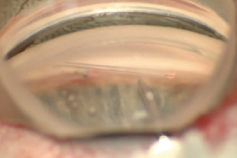
Following procedural protocol, under topical anesthesia, I performed trabeculotomy, cannulation, and viscodilation of Schlemm's canal with the iTrack microcatheter (Figure). The trabeculotomy was made beyond one end of the iStent and cannulation taken to near the other end, approximately 345°. After the illuminated end of the microcatheter had been circumnavigated, one microbolus (one injector click) of Healon GV (Johnson & Johnson Vision) per clock hour was injected as the microcatheter was slowly withdrawn. The procedure went well. Because the patient was allergic to fluoroquinolones (ciprofloxacin), tobramycin was prescribed for 1 week, and prednisolone acetate for 3 weeks. Glaucoma medications were stopped.

Figure. iTrack microcatheter in position for insertion into Schlemm's canal distal to trabecular meshwork.
Mrs. C’s eye had minimal inflammation postoperatively. IOP was 16 mm Hg at postoperative day 1, 14 mm Hg at day 7, and 14 mm Hg at day 30. If IOP remains in a therapeutic range off of medications and ocular surface disease improves, I intend to perform ABiC on her fellow eye.
Case Study findings
Age: 75 years old
Gender: Female
POH: POAG OU diagnosed in 2008; underwent SLT OU in 2012; underwent iStent combined with cataract surgery and toric IOL placement OU in 2014
Medications: Preservative-free dorzolamide hydrochloride-timolol maleate, BID, and latanoprost, qHS
BCVA: 20/20 -2 OU
SLE: Mild conjunctival injection with papillary reaction OU; moderate punctate keratopathy OU; quiet anterior chamber OU; well-positioned posterior chamber IOL OU
IOP: 20 mm Hg OD, 19 mm Hg OS
Pachymetry: 576/581 microns
Gonioscopy: Grade 4 all quadrants with well-positioned iStent nasally OU
DFE: Cup-to-disc ratio 0.7 OU; macula, vessels, and periphery normal OU
HVF: Full OU
OCT: Nerve fiber layer thinning temporal OU
ABiC Advantages
I have performed ABiC approximately 80 times in the last year, both in combination with cataract surgery and as a standalone procedure. Once practiced, the surgery typically takes 5 to 10 minutes. It is advantageous in nondestructively treating all points of obstruction in the normal outflow pathways. As mentioned previously, the procedure can be performed subsequent to SLT, stent placement, and even trabeculectomy. It is approved and reimbursable for all grades of glaucoma, whether or not performed in conjunction with cataract surgery and as a standalone procedure in phakic and pseudophakic patients.
Conclusion
ABiC is the most versatile MIGS procedure. A viable option for all stages of glaucoma, its use should also be considered when previous medical, laser, or surgical treatment has failed to adequately lower IOP. It is elegant and straightforward to perform for surgeons who are comfortable with angle-based procedures. ABiC will continue to grow in popularity as surgeons realize its value and MIGS dominates the discussion of glaucoma treatment paradigms.
ABiC and iTrack are trademarks of Ellex. © Ellex 2018. All other brand/product names are the trademarks of their respective owners
1. Khaimi MA, Gallardo MJ. 228-eye ABiC 12-month case series data. Presented at ASCRS; May 6-10, 2016; New Orleans, LA.


