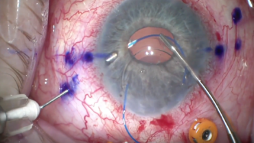
Double-Flanged Polypropylene Technique: 5-Year Results
Canabrava S, Carvalho MS1
Industry support: None
ABSTRACT SUMMARY
A prospective observational case series evaluated the outcomes of patients who underwent the flanged polypropylene scleral fixation of capsular tension segments (CTSs) and IOLs. A total of 71 eyes were divided into three groups: double-flanged CTS (n = 25), four-flanged nonfoldable IOL (n = 18), and four-flanged foldable IOL (n = 28). The sutures made of 5-0 and 6-0 polypropylene were used in 60% and 40% of the cases, respectively. Partial-thickness scleral tunnels were created in all cases to facilitate burial of the flanges. Follow-up averaged 28.24 months (range, 6–60 months). Descriptive outcome measures included patients’ visual acuity, flange position (intrascleral vs sub-Tenon vs conjunctival erosion), and complications such as IOL dislocation and retinal detachment.
Study in Brief
A prospective observational case series assessed the long-term results and complications of the scleral fixation of capsular tension segments and IOLs with the double-flanged polypropylene technique. Overall, fixation remained stable throughout the 5-year study period. Flange exposure or erosion occurred in less than 3% of cases.
WHY IT MATTERS
The double-flanged polypropylene technique has become popular for the scleral fixation of IOLs owing to its efficiency and speed. This relatively large case series demonstrated favorable long-term safety and clinical outcomes.
On average, the UCVA of patients across all three groups improved from 0.95 to 0.45 logMar. A total of 86.7% flanges were correctly positioned within the scleral tunnel. Of the malpositioned flanges, 2.89% were exposed without conjunctival coverage, 2.2% were internalized through the scleral tunnel, and 7.5% were covered with conjunctiva but located in the sub-Tenon space.
All flange-related complications—and their subsequent categorization in the study—presented within the first 4 weeks postoperatively. Patients in the flange exposure and internalization groups required a reoperation. One patient in the sub-Tenon malposition group needed a reoperation for conjunctival inflammation over the flange, but no one in this group developed a frank erosion. Three eyes (4.22%) presented with a retinal detachment 1 to 24 months after surgery, and three eyes developed cystoid macular edema. No instances of suture breakage or degradation were observed during the 5-year study period.
DISCUSSION
Shortly after Yamane et al described a double-needle technique for the intrascleral haptic fixation (ISHF) of a three-piece IOL,2 Canabrava and colleagues published a creative adaptation of the same principle in which a double-flanged polypropylene suture is used to fixate a CTS.3 The double-flanged technique has become a popular means of fixating CTSs, an assortment of secondary IOLs, dislocated IOL–capsular bag complexes, and artificial irises to the sclera. The technique’s appeal lies in the relative ease of its application and its lower expense compared to what was previously the most common approach—scleral fixation with a PTFE suture (Gore-Tex, W.L. Gore & Associates; off-label indication). Flanged fixation also avoids complications such as knot erosion and suture breakage, resulting in IOL decentration.
The most important and challenging step of the double-flanged polypropylene technique is positioning the flange within the scleral tunnel. Perfectly matching flange size with suture tension and length can be difficult because the agent of adjustment is thermocautery. Although technical guidelines for creating a flange of ideal size and length for both 27- and 30-gauge scleral tunnels have been published, the titration of flange and suture tension remains challenging even for experienced surgeons.4 The study by Canabrava and Carvalho nevertheless demonstrates the overwhelming long-term success of this technique.1
Intraocular Lens Tilt Due to Optic-Haptic Junction Distortion Following Intrascleral Haptic Fixation With the Yamane Technique
Safran JP, Safran SG5
Industry support: None
ABSTRACT SUMMARY
A case report described severe tilting of an IOL following uncomplicated double-flanged ISHF with the Yamane technique in two patients.2 The cases involved significant distortion of the optic-haptic insertion point; each haptic had rotated off axis to the plane of the optic. The IOLs (CT Lucia 602 IOL, Carl Zeiss Meditec; off-label use) had been well centered without noteworthy tilt at the conclusion of surgery.
Study in Brief
A case report presented two examples of severe postoperative IOL rotation at the optic-haptic insertion point following uncomplicated double-flanged intrascleral haptic fixation with the Yamane technique.
WHY IT MATTERS
Surgeons who use this fixation technique with a CT Lucia 602 IOL (Carl Zeiss Meditec) should be on the lookout for early postoperative structural compromise of the optic-haptic junction. When this complication arises, creative problem solving such as argon endolaser refixation is an option.
In the first case, an 81-year-old woman underwent an IOL exchange and concurrent Descemet stripping automated endothelial keratoplasty due to corneal edema from a previously placed anterior chamber IOL. One day after surgery, the anterior chamber was full of air, the cornea was edematous, and the pupil was small, making IOL assessment difficult. By 1 month postoperatively, significant IOL tilt was evident that might have been present from the first day because the patient did not report a sudden change in vision over the clinical course. She subsequently underwent an IOL exchange, and the haptics of the explanted lens were noted to have rotated approximately 45º off axis to the plane of the optic. Four weeks following the exchange, the newly placed CT Lucia 602 IOL was well positioned.
In the second case, a 75-year woman with pseudoexfoliation, a dislocated IOL–capsular bag complex, and vitreous prolapse underwent a pars plana vitrectomy and an IOL exchange with ISHF. One day after surgery, the patient presented with a severely tilted IOL that was almost perpendicular to the iris plane. An IOL exchange was performed, during which at least 60º of rotation was observed at both haptic-optic insertion points. The newly placed CT Lucia 602 IOL was well positioned at 3 months postoperatively.
DISCUSSION
The Yamane technique for ISHF has become the leading surgical approach to eyes lacking capsular bag support.2,6 The method requires extensive haptic manipulation, leading most surgeons to select a CT Lucia 602 IOL because of its PVDF haptics, which generally have excellent intrascleral stability and resistance to kinking and breaking. This case report details two examples of a newly identified mechanical compromise of the optic-haptic insertion point that led to profound IOL tilt in the early postoperative period.
The overall incidence of optic-haptic junction rotation is not well documented. Although this case report is small, the problem appears to be widespread because many high-volume surgeons, including this author (Z.Z.), have encountered the phenomenon. Most intriguing about these cases is the timing of rotation. One would expect the mechanical shifting to occur during surgery while the optic-haptic junction is actively being manipulated. Instead, rotation occurred after surgery when the mechanical forces on the junction should have been minimal. A clear explanation for optic-haptic junction instability remains elusive, but various hypotheses have been proposed, including a temperature- or fluid-induced compromise of the manufactured glue material holding the optic-haptic junction together, torque induced by early transient hypotony, and mechanical manipulation with speculum removal or eye rubbing by the patient.
Safran and Safran chose to perform an IOL exchange in both cases. In several other published case reports, argon endolaser energy was applied to expand the haptic within its insertion track in the optic, effectively locking the haptic’s position relative to the optic. Gonzalez Del Valle creatively described using laser energy to refixate an IOL optic to the haptic of a three-piece IOL in 2020.7 More recently, the AAO’s ONE Network published two videos demonstrating the successful use of argon endolaser treatment to reposition the haptic relative to the optic in eyes with a profoundly tilted CT Lucia 602 IOL.8,9 In general, 10 to 15 individual laser spots at a power of 200 to 300 mW and duration of 0.1 to 0.5 seconds are required to expand the haptic adequately and promote fixation within the optic. Utilizing laser energy in this fashion can help avoid the need for an IOL exchange.
Surgeons performing ISHF with the Yamane technique should watch for early postoperative IOL tilt due to distortion at the optic-haptic fastening zone. Although an IOL exchange is an effective management option, argon endolaser refixation should also be considered.
1. Canabrava S, Carvalho MS. Double-flanged polypropylene technique: 5-year results. J Cataract Refract Surg. 2023:49(6);565-570.
2. Yamane S, Sato S, Maruyama-Inoue M, Kadonosono K. Flanged intrascleral intraocular lens fixation with double-needle technique. Ophthalmology. 2017;124(8):1136-1142.
3. Canabrava S, Bernardino L, Batiste T, Lopes G, Diniz-Filho A. Double-flanged-haptic and capsular tension ring or segment for sutureless fixation in zonular instability. Int Ophthalmol. 2018;38(6):2653-2662.
4. Kronschläger M, Blouin S, Ruiss M, Findl O. Attaining optimal flange size with 5-0 and 6-0 polypropylene sutures for scleral fixation. J Cataract Refract Surg. 2022;48(11):1342-1345.
5. Safran JP, Safran SG. Intraocular lens tilt due to optic-haptic junction distortion following intrascleral haptic fixation with the Yamane technique. Am J Ophthalmol Case Rep. 2023;30:101845.
6. Yamane S, Inoue M, Arakawa A, Kadonosono K. Sutureless 27-gauge needle-guided intrascleral intraocular lens implantation with lamellar scleral dissection. Ophthalmology. 2014;121(1):61-66.
7. Gonzalez Del Valle F, Dominguez Fernandez MJ, Sanchez JC, et al. Running to the edge of the intraocular lens: optic transplant. Video Journal of Cataract, Refractive, and Glaucoma Surgery. March 2023;39(1).
8. Scoles D, Wolfe J. Laser to the rescue. ONE Network. December 15, 2022. Accessed February 15, 2024. https://www.aao.org/education/1-minute-video/laser-to-rescue-2
9. Chen AJ, Bui AT. Anterior endolaser for optic-haptic junction tilt. March 21, 2023. ONE Network. Accessed February 15, 2024. https://www.aao.org/education/clinical-video/anterior-endolaser-optic-haptic-junction-tilt


