

Sponsored by Bausch + Lomb
Solving the Puzzle of the 20/20 Unhappy Patient
Incorporating osmolarity testing into the presurgical workflow


With Ranjan Malhotra, MD,
and Dagny Zhu, MD


In cataract and refractive surgery, patients aren’t just asking for excellent postoperative vision, they are expecting it. Modern techniques and advanced technology have improved the odds of delivering on patients’ postoperative vision goals, but nothing should be left to chance. Consistently delivering on the promise of high-quality vision means accounting for every detail that could result in refractive surprise.
Osmolarity testing with the ScoutPro Osmolarity System (Bausch + Lomb) may be a missing piece.
Toxic hyperosmolarity is known to damage corneal cells and drive refractive surprises.1-3 Research highlights that hyperosmolarity, a core mechanism of dry eye disease, may result in 7X more patients being dissatisfied with their cataract surgery outcomes.4 And with up to 80% of cataract patients at risk for ocular surface disease,5 it has never been more urgent to evaluate for dry eye disease in surgical candidates.
Hear from Drs. Zhu and Malhotra on the role of Osmolarity testing
Hyperosmolarity and the Health of the Cornea
Optimizing the ocular surface begins early in the patient’s journey, and it starts with a thorough assessment of the quality and quantity of tears, the stability of the tear film, and the health of the ocular surface. While various tests and diagnostics may be used to discover dry eye, osmolarity testing can serve as a global indicator of the disease, regardless of its etiology.6
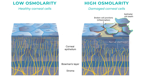
Hyperosmolarity can damage corneal nerve cells,7 cause inflammation,2 and in severe cases, result in epithelial cell death2
Hyperosmolarity creates light scatter equivalent to a grade 2 to 3 cataract1,8
1 in 6 cataract patients with hyperosmolarity risk a > 1.00 D variation in keratometry measurements3
1 in 10 cataract patients with hyperosmolarity may have a refractive miss of > 1.00 D4
Dr.’s Zhu and Malhorta discuss their osmolarity testing protocols


ScoutPro: Portable, Practical, and Precise
ScoutPro integrates seamlessly into the preoperative evaluation and is simple to interpret: Generally, abnormal results are scores of ≥ 308 mOsm/L or an intereye difference > 8.6,9
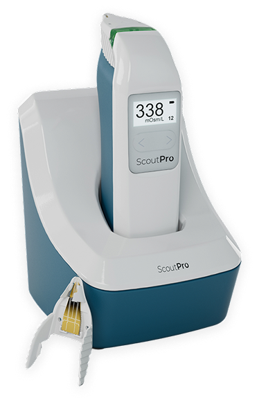
Portable: Specimen collection and analysis in one handheld device10
Practical: An end-to-end osmolarity testing method designed to meet the demands of today’s busy eye care professionals, with real-time results appearing in seconds10
Precise: 95% analytical accuracy compared to standard laboratory osmometer10
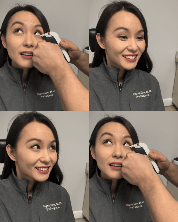

0:47
Dr. Zhu demonstrates how testing is performed
Drs. Zhu and Malhotra describe why they trust ScoutPro


Case Examples
Dry Eye Surprise on the Day of Surgery: The Role of Confirmatory Testing
By Dagny Zhu, MD
Case Presentation

- A patient was scheduled for cataract surgery with a diffractive multifocal IOL
- The patient had previously been seen by an optometrist, who did not note any dry eye disease on slit-lamp examination
- As is routine in my practice, osmolarity testing was repeated on the day of surgery as a confirmatory test
Initial Testing & Results

- Osmolarity measured 355 mOsm/L in the surgical eye, with an intereye difference of > 8 mOsm/L
- Repeat fluorescein testing revealed decreased tear breakup time
- Placido disc topography also revealed dry eye disease with an unstable tear film
Treatment and Follow-Up

- The IOL was switched from a diffractive multifocal IOL to an extended-depth-of-focus (EDOF) IOL
- The patient was informed about the presence of chronic dry eye disease and the need for long-term treatment after surgery
- Postoperatively, cyclosporine was initiated along with heated eye mask compresses
- After cataract surgery, the patient was 20/20 J2
Discussion

Sometimes dry eye can be missed on routine slit-lamp examination. Osmolarity testing provides an objective evaluation and “red flag” that prompts the surgeon to dig deeper and consider alternative IOL options as well as dry eye treatment and counseling, ideally prior to any surgery. This case is also a good reminder that if you inform a patient about dry eye prior to surgery, it’s education; but if you inform them after, it’s a complication—and it may affect the refractive outcome!
Persistent Visual Complaints Despite “20/20” Outcome: Unmasking Ocular Surface Disease
By Ranjan Malhotra, MD
Case Presentation
- A 64-year-old male presented for a second opinion 3 months after bilateral cataract surgery with monofocal toric posterior-chamber IOLs
- He had a 30-year history of soft contact lens wear and no longer required contact lenses for distance vision after surgery
- Although he accepted the need for reading glasses, he reported persistent blurry, waxy, filmy, and fluctuating vision despite being told the surgery was “perfect”
- Symptoms included glare and halos at night, ghosting and shadowing of letters while reading, and transient improvement with repeated blinking
Initial Testing & Results
- BCVA was 20/20-2 OD and 20/20-1 OS, with manifest refraction of -0.25 sphere OD and -0.25 +0.25 × 090 OS
- Tear osmolarity, measured with ScoutPro, was elevated
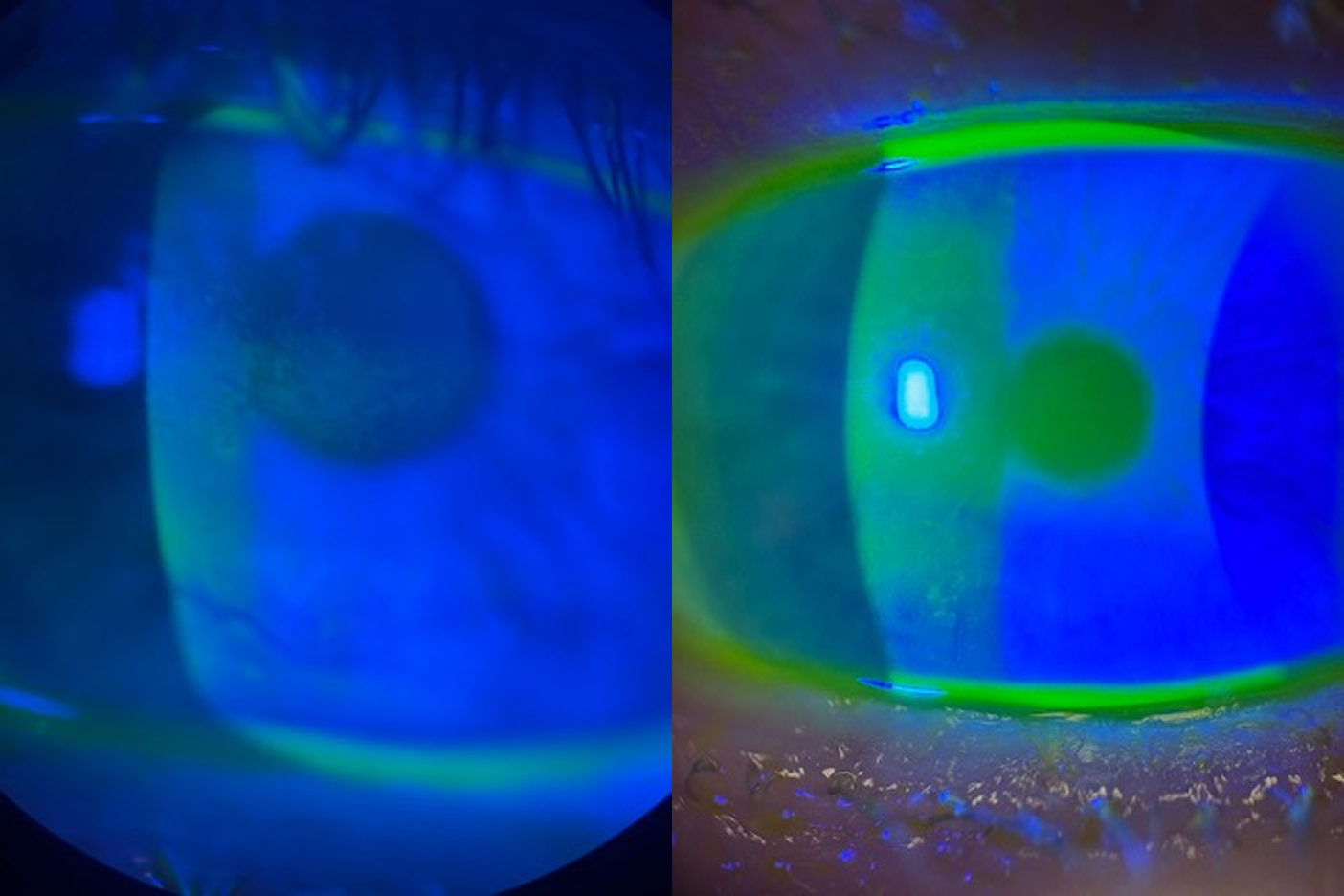
- Slit-lamp exam revealed 1+ meibomian gland dysfunction with trace collarettes, decreased tear break-up time (TBUT; ~6 sec), 2+ tear film debris, and trace inferior punctate keratitis bilaterally
+ Keep Reading
- IOLs were well-centered with no posterior capsular opacification (PCO); dilated exam and macular OCT were normal
- Findings were attributed to evaporative dry eye/MGD rather than refractive error, PCO, cystoid macular edema, or IOL malposition
- Discontinuing contact lenses postoperatively likely increased ocular surface exposure and tear evaporation, unmasking previously compensated ocular surface disease
Treatment and Follow-Up
- The patient was educated that tear film instability and evaporative dry eye were degrading the optical surface and reducing visual quality despite excellent Snellen acuity
- Discontinuation of contact lens wear after surgery may have removed a partial barrier to evaporation, unmasking previously compensated ocular surface disease
- Treatment of dry eyes was initiated with prescription and over-the-counter products
- The patient expressed relief after his symptoms were validated and explained as ocular surface disease rather than failure of the cataract surgery itself
Discussion
This case highlights the gap between visual acuity and visual quality after cataract surgery. Patients can achieve excellent Snellen acuity and have well-positioned IOLs yet remain dissatisfied because of untreated ocular surface disease. Fluctuating clarity, filmy vision, glare, halos, ghosting, and transient improvement with blinking should prompt careful evaluation of the tear film and meibomian glands.
+ Keep Reading
The patient likely had pre-existing ocular surface disease that was partially compensated while he wore contact lenses. After cataract surgery, discontinuation of contact lens wear and increased ocular surface exposure may have amplified evaporative dry eye and tear film instability. This case reinforces the value of preoperative ocular surface evaluation, including tear osmolarity, TBUT, corneal staining, and meibomian gland assessment, even in patients without classic dry eye symptoms. It also underscores the importance of validating postoperative complaints rather than relying solely on Snellen acuity or IOL positioning.
Indications and Important Safety Information for ScoutPro Osmolarity System
Indications: The ScoutPro Osmolarity System is an automated device intended to quantitatively measure the osmolarity of human tears to aid in the diagnosis of dry eye disease, in patients suspected of having dry eye disease in conjunction with other methods of clinical evaluation.
Contraindications: Do not collect tear fluid from a patient within two hours of medicinal eye drop use or use of topical medications. Do not collect or store tear fluid samples for transport or testing at a later time. Do not collect tear fluid after ocular surface staining. Do not collect tear fluid within 15 minutes of use of anesthetic or mydriatic (dilating) eye drops or after other invasive ocular diagnostic testing. Do not collect tear fluid within 15 minutes after a slit lamp examination. Do not collect tear fluid within 15 minutes from a patient who has been crying.
The ScoutPro Osmolarity System (ScoutPro) is a CLIA Waived test system for human tears. Each laboratory or testing site using the ScoutPro must have a CLIA Certificate of Waiver before starting testing.
The ScoutPro is designed for stability, reliability, and safety, and it has been developed, manufactured, and marketed under a quality management system certified to ISO 13485 (2012).
CAUTION: Federal law restricts this device to sale by or on the order of a physician.
ATTENTION: This is not all you need to know. Please refer to the User Manual for a complete listing of indications, contraindications, precautions, and use information.

- Ophthalmology Associates, St. Louis, Missouri
- RanjanMalhotraMD@gmail.com
- Financial disclosure: Speakers Bureau (AbbVie, Alcon, Bausch & Lomb, Harrow, Lenz, Ocular Sciences, Tarsus)
- Cornea, cataract, and refractive surgeon; Medical Director; and Partner, NVISION Eye Centers, Rowland Heights, California
- Member, CRST Editorial Advisory Board
- dagny.zhu@gmail.com; Instagram @DZEyeMD; X @DZEyeMD
- Financial disclosure: Financial disclosure: Consultant (Bausch + Lomb)
© 2026 Bausch + Lomb. SCP.0041.USA.26
1. Sullivan BD, Palazón de la Torre M, Yago I, et al. Tear film hyperosmolarity is associated with increased variation of light scatter following cataract surgery. Clin Ophthalmol. 2024;18:2419-2426.
2. Huet E, Vallée B, Delbé J, et al. EMMPRIN modulates epithelial barrier function through a MMP-mediated occludin cleavage: implications in dry eye disease. Am J Pathol. 2011;179(3):1278-1286.
3. Epitropoulos AT, Matossian C, Berdy GJ, et al. Effect of tear osmolarity on repeatability of keratometry for cataract surgery planning. J Cataract Refract Surg. 2015;41(8):1672-1677.
4. Kursite A, Laganovska G. Effect of tear osmolarity on postoperative refractive error after cataract surgery. Ukr J Ophthalmol. 2023;2(511):11–15.
5. Gupta PK, Drinkwater OJ, VanDusen KW, et al. Prevalence of ocular surface dysfunction in patients presenting for cataract surgery evaluation. J Cataract Refract Surg. 2018;44(9):1090-1096.
6. Sullivan BD, Whitmer D, Nichols KK, et al. An objective approach to dry eye disease severity. Invest Ophthalmol Vis Sci. 2010;51(12):6125-6130.
7. Hirata H, Mizerska K, Marfurt CF, Rosenblatt MI. Hyperosmolar tears induce functional and structural alterations of corneal nerves: electrophysiological and anatomical evidence toward neurotoxicity. Invest Ophthalmol Vis Sci. 2015;56(13):8125-8140.
8. Artal P, Benito A, Pérez GM, et al. An objective scatter index based on double-pass retinal images of a point source to classify cataracts. PLoS One. 2011;6(2):e16823.
9. Lemp MA, Bron AJ, Baudouin C, et al. Tear osmolarity in the diagnosis and management of dry eye disease. Am J Ophthalmol. 2011;151(5):792-798.e1.
10. ScoutPro Osmolarity System. Instructions for use.