

Interesting and Artistic
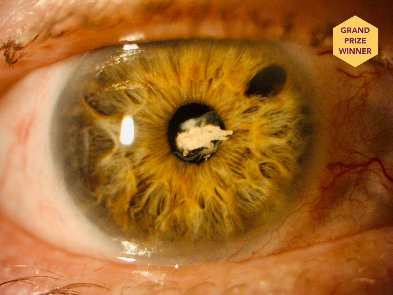
Capsular Bag in Anterior Chamber
This image shows the capsular bag in the anterior chamber of an eye with a history of trauma and surgery.
Beatrice Knowles Uchôa, MD
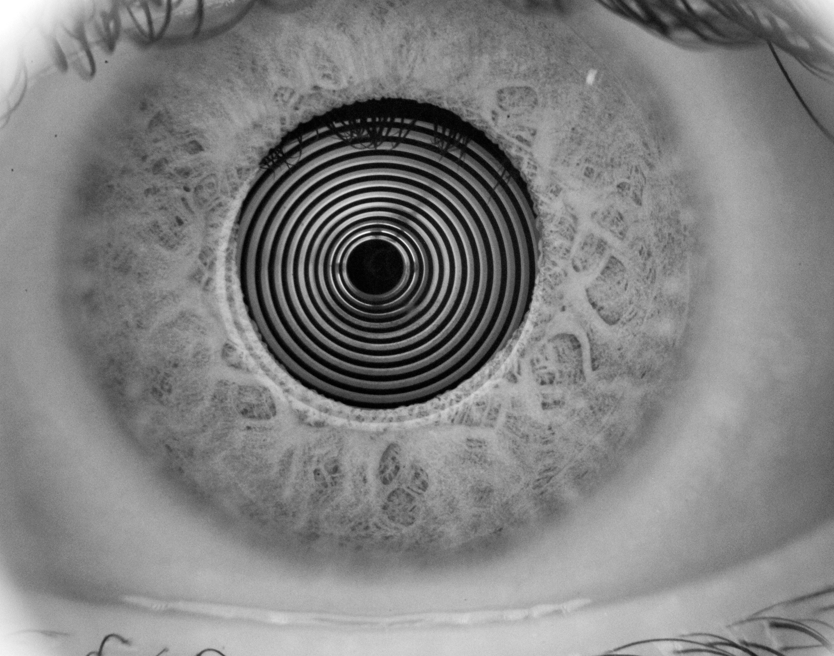
Life in Circles
This is a photograph of the eye of a patient who presented for a dry eye evaluation with a dry eye analyzer.
Aarti Heda, MD, DNB, MRCS, FCRS, FICO
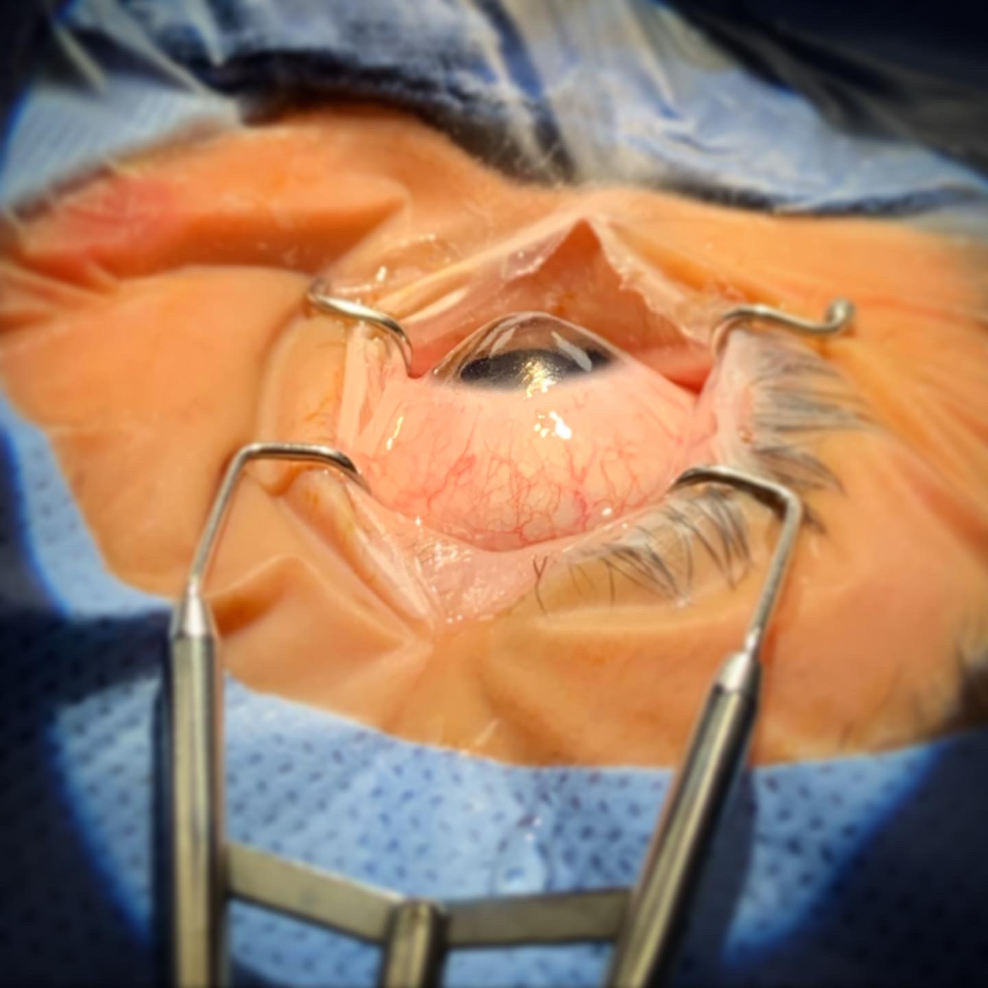
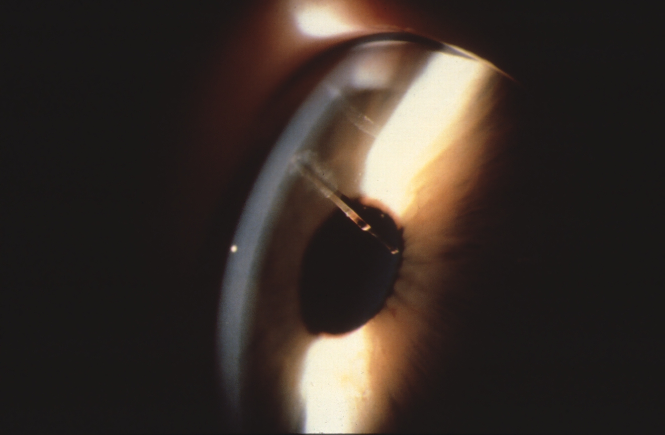
The Last Moments of Keratoconus
This photograph of an eye with keratoconus was taken just before corneal transplantation.
Daniel Wasilewski, MD, PhD
Rare and Unusual Diseases
Corneal Stromal Deposits in a Presumed Ehlers Danlos Syndrome Variant
A slit-lamp photograph of the left eye of a patient with a presumed Ehlers Danlos syndrome variant shows superior refractile whitish-brown corneal deposits—a finding that has never been reported to the contributing physicians’ knowledge—and a temporal pseudopterygium from the 2 to 8 clock positions.
Abdelrahman M. Elhusseiny, MD, MSc, and Hajirah N. Saeed, MD
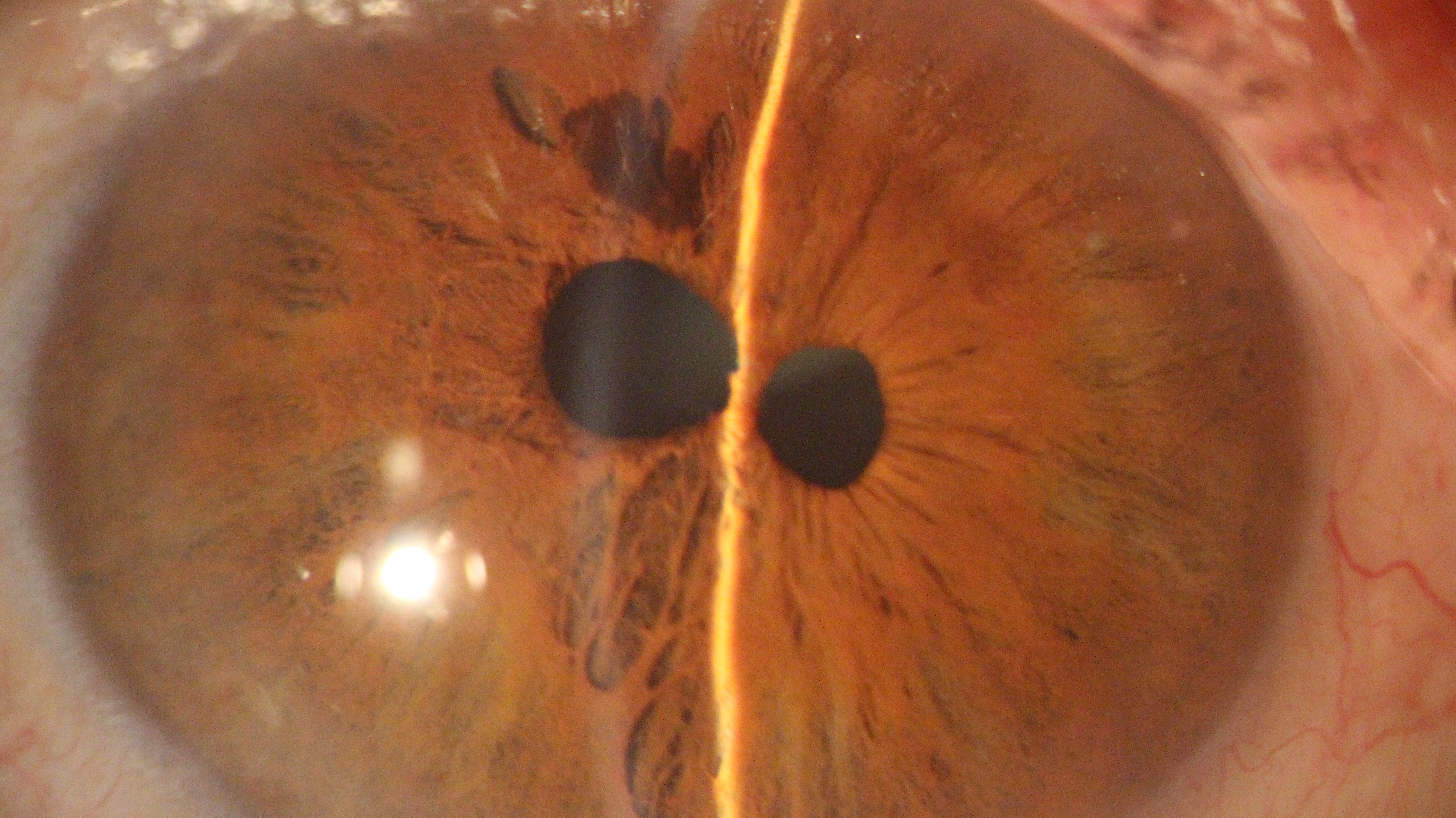
True Polycoria
This photograph shows an intact sphincter in the two pupils of an eye with polycoria.
Zahra Karjou, MD
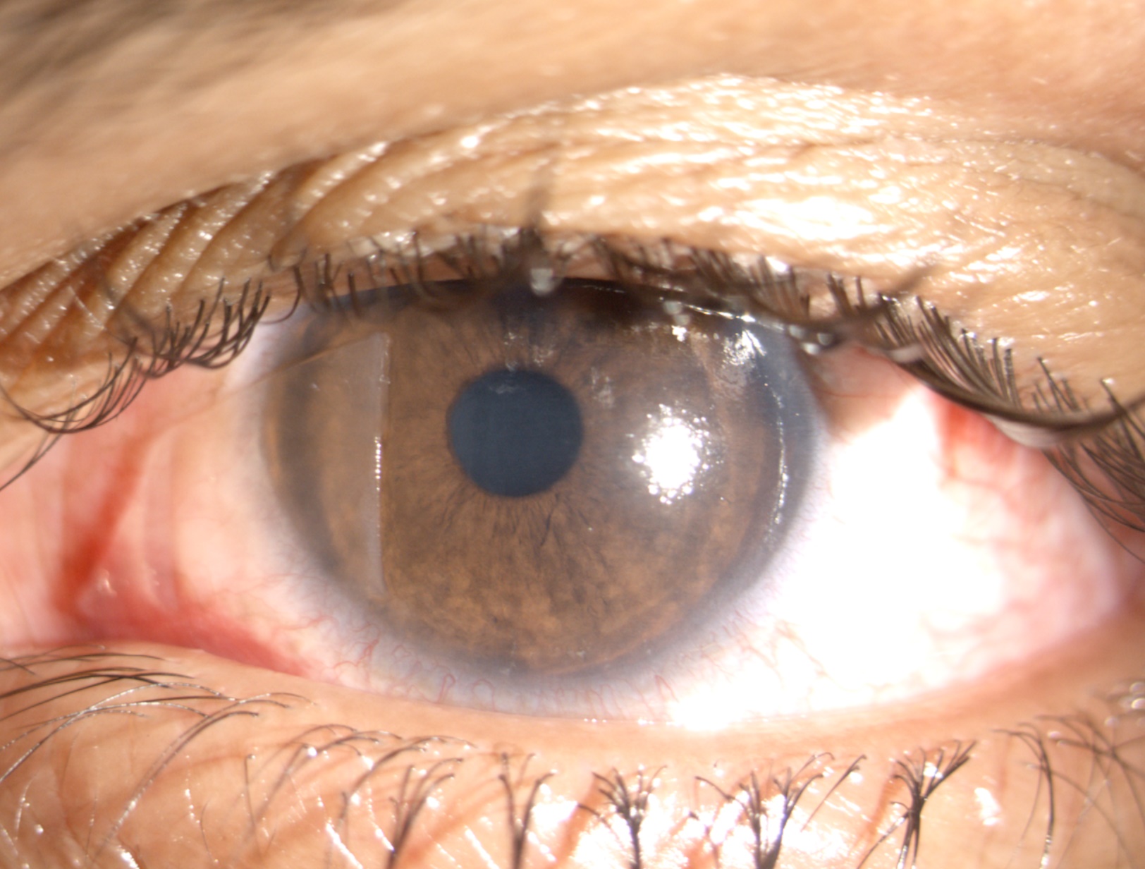
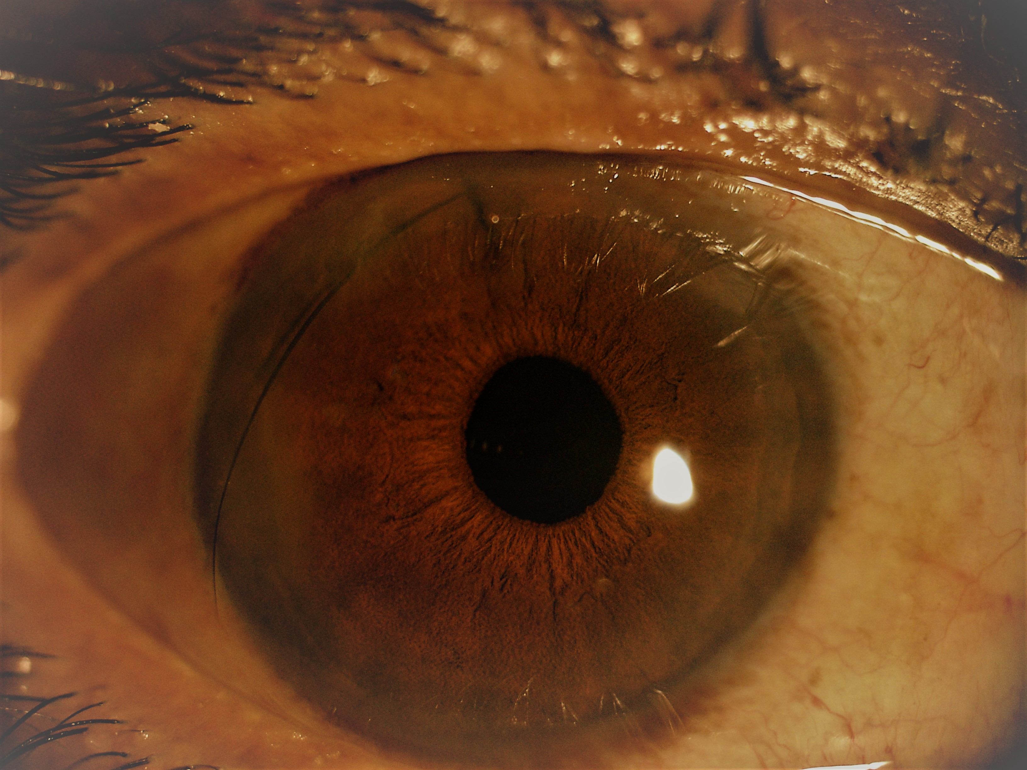
Iris Melanocytoma
This image shows an incidental finding of melanocytoma of the iris in a 14-year-old patient who presented for a routine eye exam. A raised cobblestone appearance and slight pupillary distortion are evident.
Amit Mishra, MD
Slit Lamp
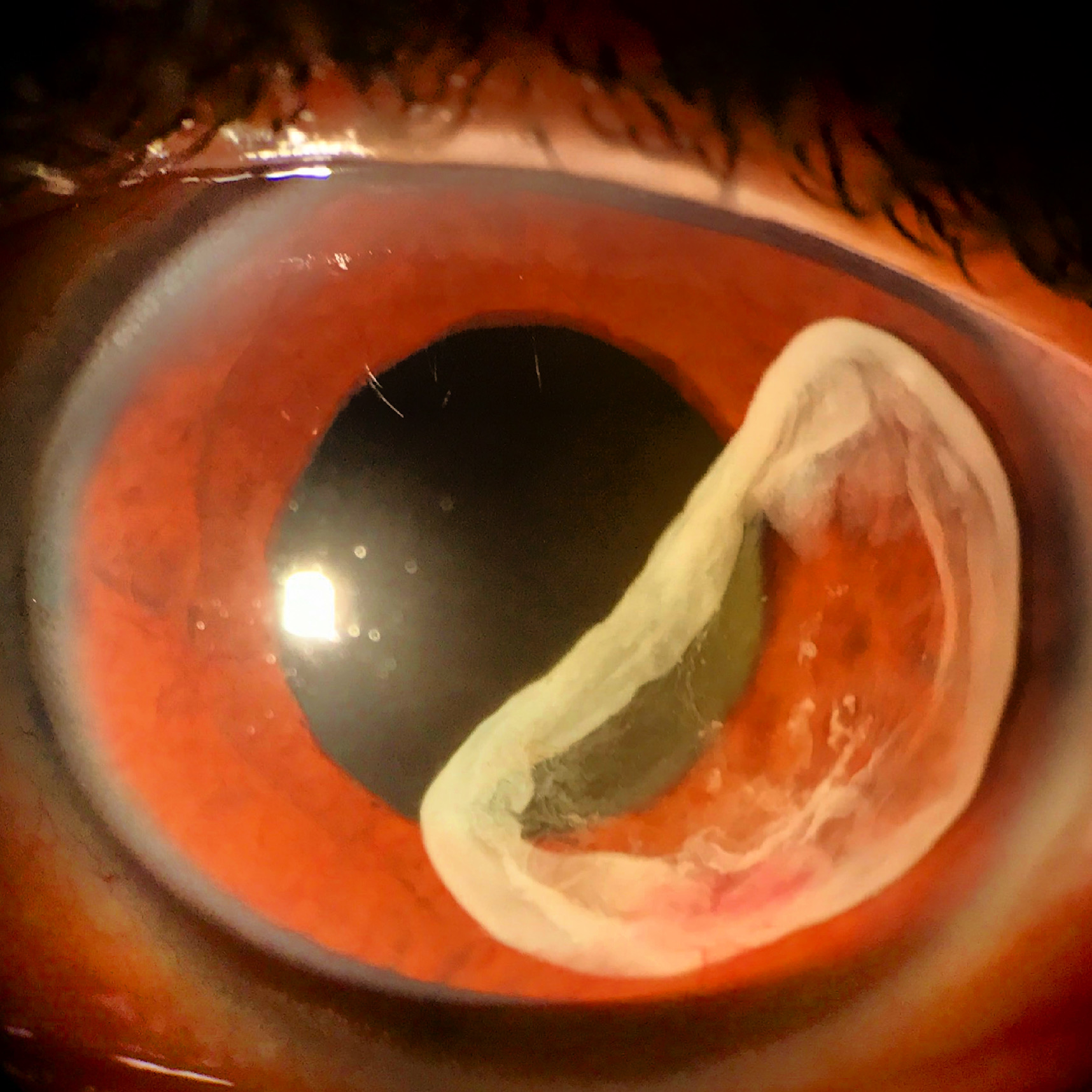
Enter the Dragon
A 68-year-old man presented with draconic dislocation of his right residual lens capsule and Soemmering ring, both prolapsing out from the pupil. He had undergone pars plana vitrectomy, scleral buckle, and a sutured IOL followed by IOL repositioning 10 years later.
Matthew Feng, MD
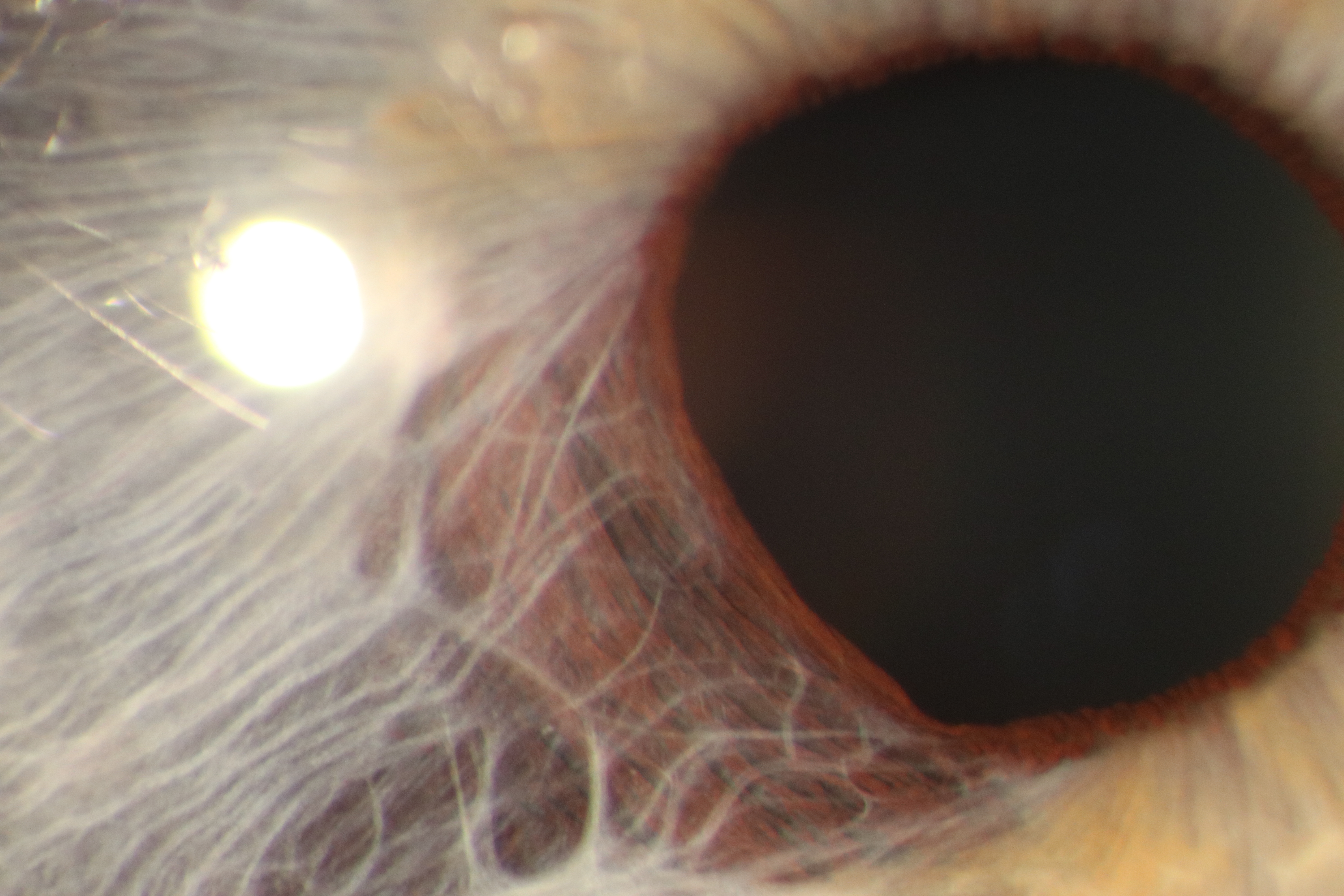
Postuveal Iridoschisis
This is a photograph of a patient with postuveal iridoschisis resulting from chronic anterior uveitis with intermittent exacerbations.
Dmytro Martynov, MD
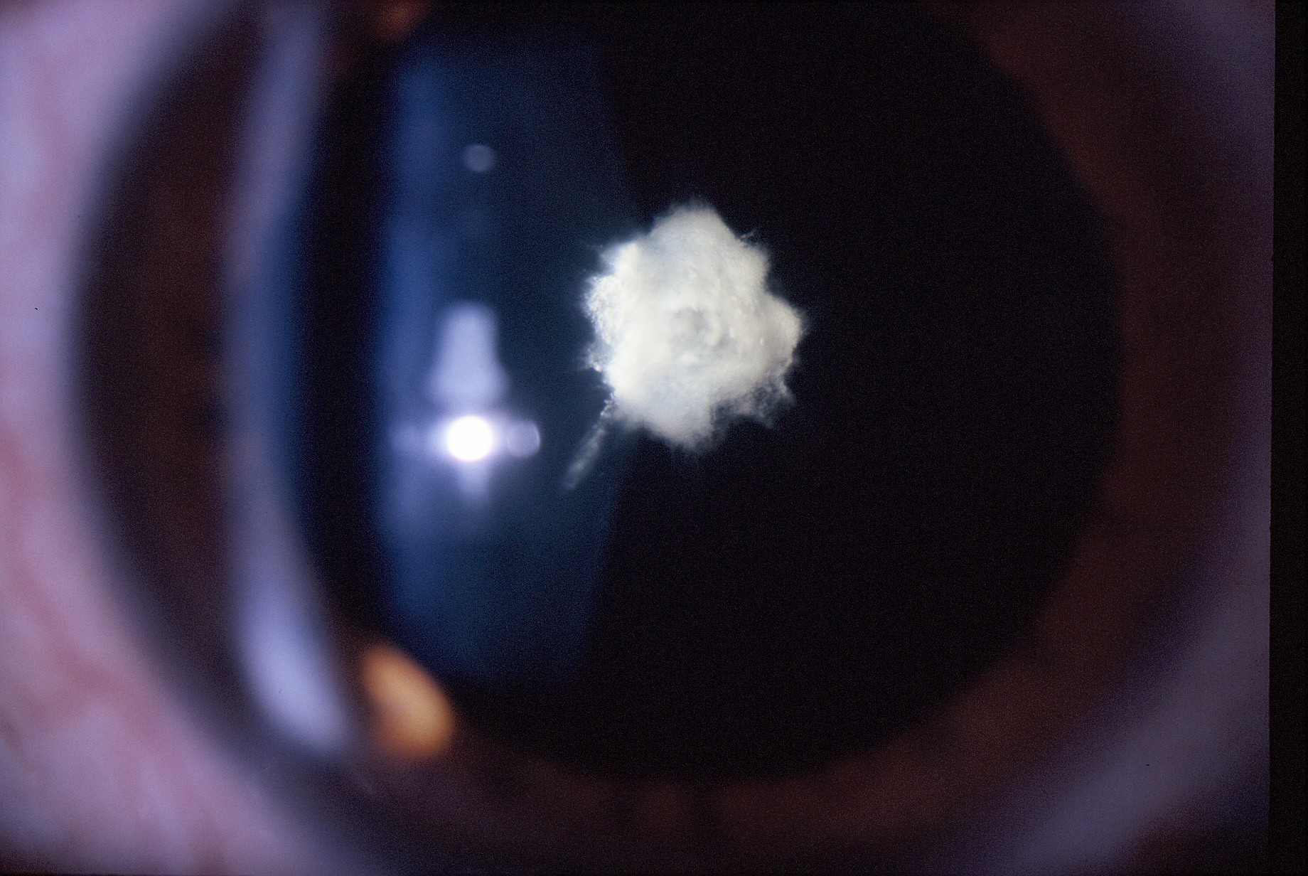
Ocular Popcorn
This photograph shows a posterior polar cataract that resembles popcorn.
C. Manuel Nicoli, MD
Trauma
Flap Bummer
This slit-lamp photograph was taken 1 day after an uneventful LASIK procedure using a corneal flap with a nasal hinge. The patient had unknowingly rubbed his eye in his sleep.
Anuja Desai, MS
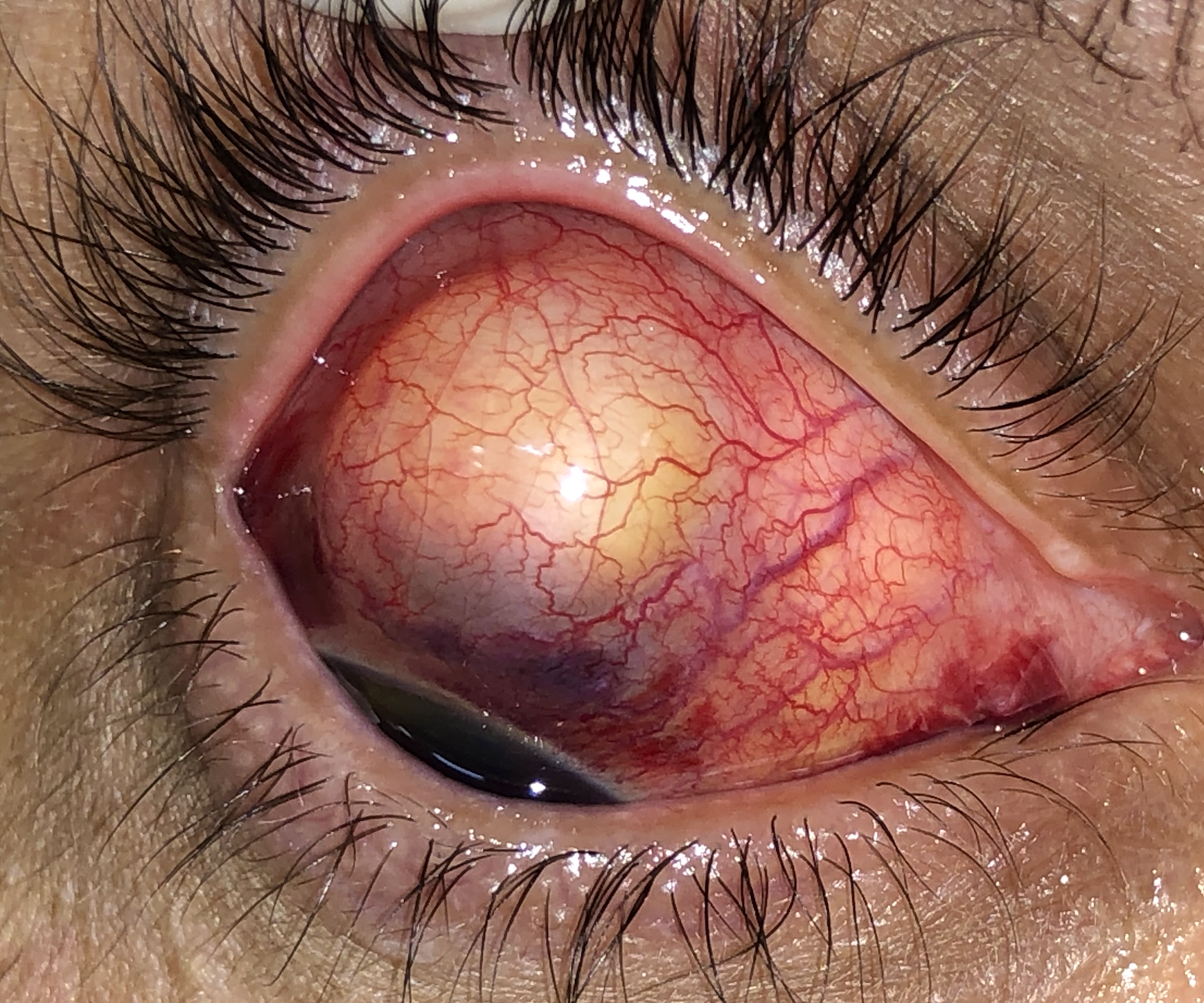
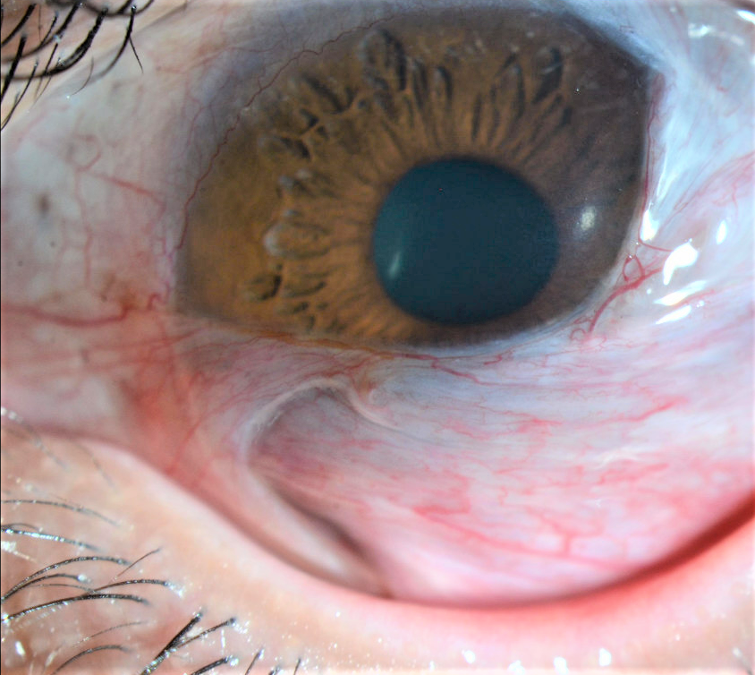
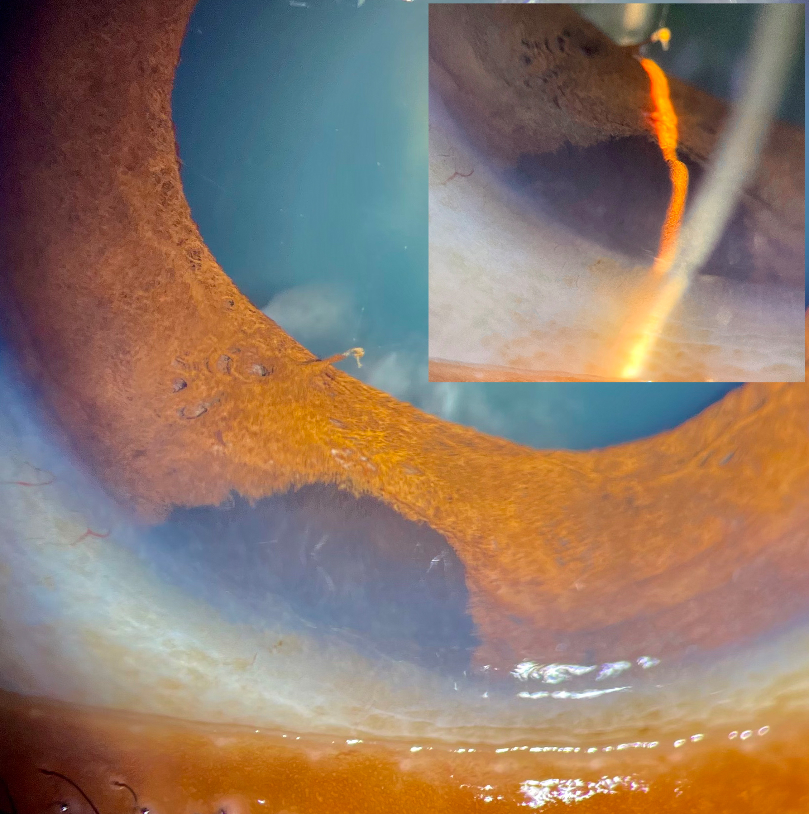
Traumatic Phacocele
This photograph shows a traumatic phacocele. In the image, the superior and nasal conjunctiva evinces a well-defined, elevated subconjunctiva with a yellow-orange mass underneath featuring the herniated lens.
Gabriel Castilho S. Barbosa, MD
Too Close for Comfort
This is a photograph of a glass fragment embedded in the cornea after a motor vehicle accident. The fragment extended into the anterior chamber and stopped just short of the anterior lens capsule.
Irving M. Raber, MD, and Thomas Tien, MD
Surgical Complications
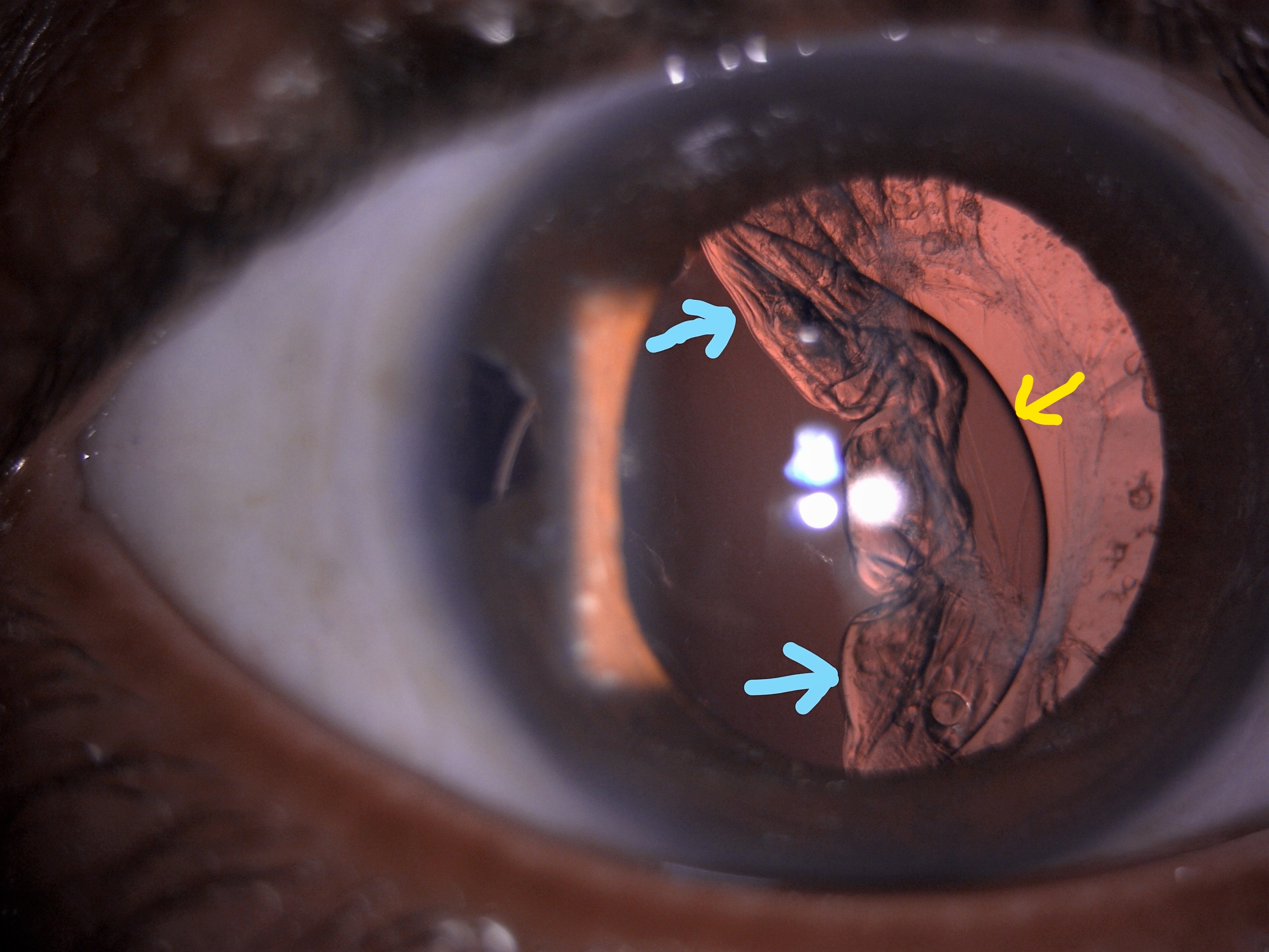
Decentered IOL With Crumpled Capsular Bag
This image shows a decentered rigid one-piece IOL temporally (yellow arrow), temporal zonular dehiscence, and a crumpled capsular bag (blue arrows) after an intraoperative complication.
Manas Nath, MBBS, DO, FAICO, FAEH
Lash in the Anterior Chamber
In this slit-lamp image, an eyelash in the anterior chamber protrudes out of a corneal sideport incision after phacoemulsification and IOL implantation.
Sashwanthi Mohan, DNB, MRCS
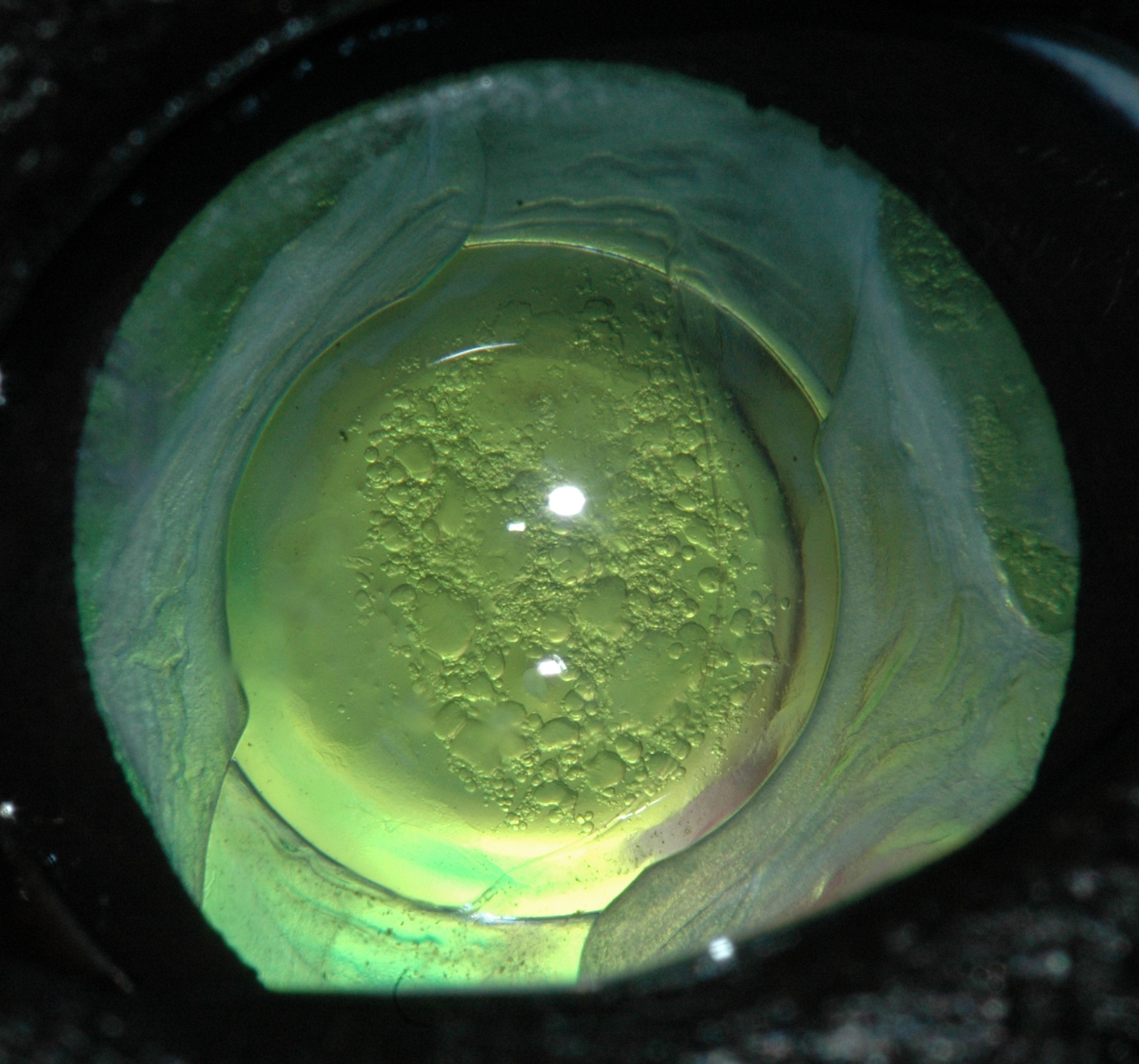
Vacuoles
This photograph shows an IOL, capsular opacity, and lens epithelial cell regrowth against the tapetal reflex after phacoemulsification in a dog.
John Mould, BA, BVSc, DVOphthal, FHEA, MRCVS