Sponsored By

Two surgeons share how the AL-Scan Optical Biometer and CEM-530 Specular Microscope help them optimize the outcomes of cataract and refractive surgeries with fast collection of key data.
AL-Scan Optical Biometer

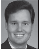
By Farrell “Toby” Tyson, MD
In the 2 years I have been using the state-of-the-art AL-Scan Optical Biometer (Nidek), it has become the biometer of choice for both my technicians and me. All of our biometers offer highly accurate axial length and the good average keratometry we need for IOL calculation formulas, while next-generation devices also take into account anterior chamber depth and white-to-white distance.
Our AL-Scan offers all of the metrics. In a 10-second test with the AL-Scan, we obtain six measurements for cataract surgery: axial length, corneal curvature radius, anterior chamber depth, central corneal thickness, white-to-white distance, and pupil size. That is with half the test time of our IOLMaster (Zeiss) and a quarter of the time required for our Lenstar (Haag-Streit). Obtaining calculations two to four times as fast as other biometers is beneficial in a busy clinic.
Any of the technicians in my practice can operate the AL-Scan because it is so easy to use and checks the accuracy of test results. Anyone who can operate an autorefractor can operate the AL-Scan. It automatically tracks movement of the eye in three dimensions and then captures images and data automatically when the eye is aligned. Technicians simply push a button, and the system takes tests automatically at the right time. I trust the AL-Scan results obtained by all of my technicians.
With the AL-Scan, technicians do not have to tell patients to stay perfectly still, staring straight ahead without blinking for a long time (an especially difficult task for elderly patients with cataracts). The fast, easy test instills confidence in our patients. Technicians can obtain quality data in a single reading and move on, rather than dragging out the process with repeated readings that make patients wonder why the process is taking so long or if the technician is testing correctly.
In addition, the AL-Scan’s flexibility allows surgeons to customize testing. For example, the keratometer uses two separate rings of data points, so I can choose the radius I prefer. A central reading of 2.5 mm mimics the IOLMaster, but a larger radius offers results similar to a manual keratometer. Anterior segment imaging includes sectional lens images, pupil images, and double mire rings projected onto the cornea. The system also reduces noise for cataract measurements and aids in toric IOL alignment.
From a business perspective, the AL-Scan is very cost effective. Because acquisition is accomplished quickly, the system requires less technician time. The time we gain allows us to see more patients or extend the duration of in-office examinations. It makes us more productive and boosts our ability to fully optimize cataract surgery. We can give patients the exemplary outcomes we always strive to achieve, helping ensure that patients are happier and require fewer enhancements.
CEM-530 Specular Microscope

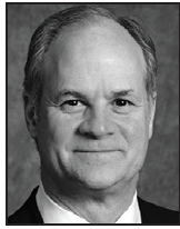
By Daniel S. Durrie, MD
Knowing the status of the endothelial cells before performing surgery on the eye is important. I image and analyze the endothelial cells for all new surgical patients, as well as anyone who has not had the analysis for 2 years or more. With many years of experience taking endothelial photographs and participating in the development of specular microscopes, I knew immediately that the CEM-530 Specular Microscope (Nidek) was exceptional. Because the technology is so easy to use, I am able to use it for all of my patients. Now, after just a few years with the CEM-530, I find it virtually indispensable.
The CEM-530’s imaging and analysis provide key information about the endothelial cell layer and corneal thickness. The system has excellent conventional central and peripheral specular microscopy, as well as paracentral images captured at eight points, yielding clear, highly detailed images for evaluation. We can look at any variation in size and shape of the endothelial cells and detect signs of stress on the endothelium. If there is little or no mycotic activity, cells have a tendency to die off, and surrounding cells get larger. When I see enlarged cells, I know there is stress, and I can show this evidence to my patients.
The cell images correspond to the software’s analysis, which helps me visualize the endothelial cells in trace, photo, area, and apex modes. It notes if the cell count is normal (2,500-3,000 cells/mm2) and displays histograms for variation in both shape and size. Users can even select an area of focus for the analysis. The CEM-530 performs and displays the entire analysis in just a few seconds.
Some of these features are available in technologies we have used in the past, but it has been difficult to get pictures and manually count cells. The CEM-530 is extremely easy to use. It automatically focuses, aligns, and measures the eye. When we got it, my technicians did back flips. We easily teach new technicians to use this technology, so no special technician is needed for the examination.
The CEM-530’s accurate central pachymetry stands out as well. Other technologies can have weaker results on patients with prior surgery, but we do not have that problem with this device.
Ultimately, information from the CEM-530 helps guide surgery and optimize postoperative outcomes. In fact, I will not do refractive surgery without axial length, topographic analysis, and an understanding of the health of the patient’s endothelium. The CEM-530 helps me identify patients who have lower cell counts or early guttata lesions that may be an early sign of Fuch’s dystrophy. The cell count is essential as well. If the endothelial cell count is 1,200 versus 2,500, I can adjust my technique and inflammation control to improve outcomes. Surprisingly, the CEM-530 has also revealed a lot of contact lens-related damage in my 20 to 40-year-old patients, which makes me steer them toward LASIK to get them out of contact lenses.
I tell colleagues that when they go to the next conference, they should look at the CEM-530’s capabilities and ease of use. Once they see how easy it is to use, they realize that they can have more endothelial evaluation in a very fast, affordable way and avoid surprises after surgery. They do not want to overlook guttata or endothelial dystrophy that cannot be seen in a screening slit-lamp evaluation.
In my own self-contained practice, the CEM-530 also has become part of our long-term documentation of vision and health of the eyes. Once people come to us for surgery, we continue to take care of them. Refractive surgery patients return for evaluation every 2 years. This approach to using the CEM-530 may not be for everyone. Because our practice does not take insurance or Medicare, we can perform the tests we want without a code or known diagnosis. There is a movement now within the insurance and Medicare world for physicians to perform unbilled tests that enhance outcomes. In situations like these, it is good to know that the CEM-530 is not an expensive test, and it has no film or prolonged costs. Quality analysis of the endothelial cells is also cost effective in helping us avoid what we dread most: patients who are unhappy with their surgical outcomes.
