

CHRISTOPHER E. STARR, MD
One of my most memorable patients was the famous and influential US artist and printmaker Eldzier Cortor (1916–2015). In addition to bilateral advanced nuclear sclerotic cataracts, Mr. Cortor also had 3.00 D of regular with-the-rule corneal astigmatism. At his preoperative examination, I was thrilled to explain to him that, in addition to removing the blur caused by dense cataracts, I could simultaneously remove the blur caused by corneal astigmatism. I assumed this would be revelatory news to him, but without a pause, he looked me straight in the eye and resolutely said, “Please do not alter my astigmatism in any way! It has shaped the unique painting style that I am known for, and it is very important to me to keep it intact.” Many of Mr. Cortor’s iconic paintings, some of which hang in the Smithsonian, feature beautifully exaggerated elongations of the female human form.
In that moment, he taught me a simple yet important lesson: Even though refractive surgeons can fully correct high astigmatism and ametropia, doing so is not the correct approach for all patients. This case highlights the importance of informed consent, effective patient education, managing patient expectations, and accounting for each individual’s unique needs. With this encounter in mind, I invited Vincent P. de Luise, MD, FACS, a world expert on the subject, to discuss the historical impact and influence of vision disorders on several iconic artists and the art they created.

VINCENT P. DE LUISE, MD, FACS
I would argue that sight informs people’s lives more than any other sense. How do changes in vision affect that reality? How do they affect people’s perception? How do these changes affect the creative spark and style of artists? Perception is ultimately an individual response to sensory stimuli; however, several universal neuroaesthetic principles underpin perception, specifically as it relates to beauty. These principles include symmetry, contrast, isolation, recursion (self-repetition), and peak shift, and they are cross-culturally perceived.1
DOES ASTIGMATISM CHANGE ARTISTIC STYLE?
The longstanding dialectic as to whether the elongations evident in the paintings of El Greco (Doménikos Theotokópoulos, 1541–1614) were a result of astigmatism or artistic style has been argued most compellingly in favor of style.2-4 Mild to moderate degrees of astigmatism blur vision more than they distort images. According to the self-correction hypothesis, if El Greco viewed a circle—even if he saw it in his mind’s eye as an oval—he would have drawn it as a circle.2,3 Some of El Greco’s works show distortion in meridians orthogonal to one another. This is not possible with regular astigmatism, which would cause distortion in only one meridian.
The hypnotic elongations of Alberto Giacometti (1901–1966) and Amedeo Modigliani (1884–1920) and the horizontal stretching evident in several portraits by Hans Holbein the Elder (1460–1524) are almost certainly stylistic.4 High degrees of astigmatism, however, can lead to an elongation of figures, as seen in some of Cortor’s works.
THE EFFECTS OF CATARACTS ON VISION
Cataracts, especially nuclear sclerotic cataracts, are the most common change in vision that can gradually affect an artist’s perception and style. The Venetian master Titian (Tiziano Vecelli, 1488–1576) lived well into his 80s. An increasing brunescence, a muddy brownish murkiness, is evident in the color palette of his works during his final decades of life, and it contrasts with the brilliantly colored works of his younger years. This change could certainly be a function of the artist’s style. An equally convincing argument, however, is that the brunescence in his late works was a result of progressive nuclear sclerosis, not an unusual occurrence in someone who lives into their ninth decade.
Several other artists, most notably Claude Monet (1840–1926), Mary Cassatt (1844–1926), and Honoré Daumier (1808–1879), had documented visually significant cataracts. They all eventually underwent cataract surgery, without success for Daumier and Cassatt.
The consequences of maturing cataracts on the color perception and style of a painter are most vividly observed in the trajectory of the late paintings of Monet.2,3,5,6 He began to notice changes in his vision around 1900. In 1913, he travelled to London to consult the German ophthalmologist and physiologist Richard Liebreich, who was the chair of ophthalmology at St. Thomas’ Hospital in London.7 Liebreich had a deep interest in art and had published an article on the effect of eye disease on the paintings of Joseph Mallord William Turner (1775–1851).7 Liebreich recommended surgery, but Monet demurred.
By 1915, Monet had begun labelling his paint tubes and putting them in a specific order to ensure that he knew which color he wished to use7 (Figure 1). A cache of correspondence between Monet and French President Georges Clemenceau discusses the commission of Monet to create a series of large canvases depicting waterlilies in a pond. In these letters, Monet laments his loss of vision and the change in his ability to perceive the intensity of colors.
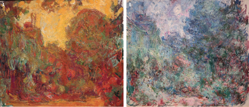
Figure 1. The Artist’s House Seen From the Rose Garden, Claude Monet, 1922. This work was created before the artist underwent cataract surgery on his right eye (A). The Artist’s House Seen From the Rose Garden, Claude Monet, 1924. This work was created after the artist underwent cataract surgery on his right eye (B).
Preoperative records from a 1922 examination by the artist’s ophthalmic surgeon, Charles Coutela, confirm advanced cataracts.6-8 Monet’s thick, postoperative Coke bottle glasses, newly invented by the Zeiss Company (now known as Carl Zeiss Meditec) in 1923, the year in which the cataract in Monet’s right eye was extracted, still exist. Monet complained bitterly of cyanopsia (a blue tint to the vision in his right eye) and peripheral distortion (an optical phenomenon inherent in aphakic glasses).7 The spectacle lens of the operated right eye was eventually tinted amber, and the lens of the unoperated left eye was occluded to prevent disabling aneisokonia.6,8 Two years elapsed before Monet came to accept his visual outcome (he never had the left cataract removed), and he completed the commission of the Grandes Décorations of waterlilies.7,8
FURTHER EXAMPLES
Goya. The trajectory of the artwork of the great Spanish master Francisco Goya y Lucientes (1746–1828) is marked by an abrupt change in style in 1792, immediately following a sudden bout of vision loss, vertigo, hearing loss, tinnitus, and aural hallucinations. Goya’s previously bright and colorful paintings morphed into a monochromatic series of bleak and sarcastic aqua-tinted etchings as well as works known as the Black Paintings. An overarching medical diagnosis to explain Goya’s midlife crisis is still debated; possibilities suggested include lead poisoning as well as cinchonism from the treatment for malaria.
Degas. Advanced glaucoma, macular degeneration, and inherited maculopathies can reduce central vision, potentially influencing artistic perception and style. This may explain the gradual change in the vision and choice of artistic technique by the French Impressionist artist Edgar Degas (1834–1917).8 As a result of a putative maculopathy (or a hereditary optic neuropathy), Degas gradually transitioned from painting in oils, which requires precise lines and edges, to pastels, which are more forgiving.8 Later in life, Degas sculpted in wax. By 1910, he had transitioned largely to photography and the photographic technology of gelatin silver print.9 Degas’ camera became his paint brush.
O’Keeffe. Renowned for her contribution to modern art, Georgia O’Keeffe (1887–1986) enjoyed an exceptionally long life and artistic career. In the mid-1970s, as a consequence of a loss of central vision from progressive macular degeneration, O’Keeffe stopped painting and transitioned to pottery.2,3
Bacon. The abstract expressionist Francis Bacon (1902–1992) was famed for his dark and disturbing paintings, many of which depict deformed figures. Bacon declared that his aim was to give a “visual shock” to viewers of his works of art.10 The raw, unsettling faces in a number of his portraits may be a result of a rare cortical perceptual abnormality called dysmorphia.11 This diagnosis is supported by Bacon's own detailed description of the phenomenon. The term he used was dynamic distortion, and he described dramatic and sudden changes in image magnitude and pattern during periods of close fixation (francis-bacon.com/artworks/paintings/1960s).10,11
Close. Chuck Close (1940–present) is a visionary artist who uses a multitude of inventive techniques to create powerful prints, paintings, and collages, including more than 100 immense dynamic neopointillist self-portraits.12 Close has been dyslexic since childhood, and he has prosopagnosia (ie, an inability to recognize faces). In 1988, he sustained a spinal artery stroke that rendered him paraplegic.
Close’s predominant and well-recognized artistic theme involves magnified images of himself, friends, and family using a painstaking photo-grid process of transferring each area of a photograph to an enormous canvas (www.pacegallery.com/artists/chuck-close).12 He also uses pixelated forms and computer-generated Iris printing (Iris Graphics), and he has revived daguerreotype technology.
Close continues to create monumental works of art, notwithstanding the enormous neurological challenges he faces. His work is a brilliant and courageous testimony to the creative urge that remains vital in him—an artistic passion to continue on, displayed by all of the artists discussed in this article.
READ IT NOW
Related Content From theCRST Archives
Tobias H. Neuhann, MD, FEBOS-CR, recalls an experience with an art professor who was unhappy with the dramatic change in his color vision after cataract surgery.
1. Ramachandran VJ, Hirstein W. The science of art: a neurological theory of aesthetic experience. J Consciousness Studies. 1999;6:15-51.
2. Marmor MF, Ravin JG. The Eye of the Artist. Mosby; 1996.
3. Marmor MF, Ravin JG. The Artist’s Eyes: Vision and the History of Art. Abrams; 2009.
4. Trevor-Roper PD. The World Through Blunted Sight: An Inquiry Into the Influence of Defective Vision on Art and Character. 2nd ed. Independent Pub Group; 1997.
5. Livingstone M, Hubel DH. Vision and Art: The Biology of Seeing. Abrams; 2014.
6. Ravin JG. Monet’s cataracts. JAMA. 1985;254(3):394-399.
7. Gruener A. The effect of cataracts and cataract surgery on Claude Monet. Br J Gen Pract. 2015;65(634):254-255.
8. Marmor MF. Ophthalmology and art: simulation of Monet’s cataracts and Degas’ retinal disease. Arch Ophthalmol. 2006;124(12):1764-1769.
9. Daniel MR. Edgar Degas, Photographer. Metropolitan Museum of Art: Yale UP; 1999.
10. Zeki S, Ishizu T. The "visual shock" of Francis Bacon: an essay in neuroesthetics. Front Hum Neurosci. 2013;7:850.
11. Safran AB, Sanda N, Sahel JA. A neurological disorder presumably underlies painter Francis Bacon distorted world depiction. Front Hum Neurosci. 2014;8:581.
12. Hylton WS. The mysterious metamorphosis of Chuck Close. The New York Times Magazine. July 13, 2016. Accessed June 9, 2021. https://www.nytimes.com/2016/07/17/magazine/the-mysterious-metamorphosis-of-chuck-close.html