
To provide patients with good quality of vision, astigmatism correction is essential. In my daily practice, I make a toric calculation for all of my crystalline lens surgeries. Toric IOLs represent more than 80% of my IOL implantations. Improvements in technology, including the availability of image-guided systems and total keratometry measurement on the IOLMaster 700 (Carl Zeiss Meditec), today give us security and simplicity in astigmatism correction (Figure).
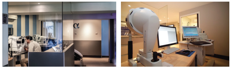
Figure. Images of some of the diagnostics devices used at Ophthalmologic Center Iridis.
Three elements must be considered to achieve the best final result in astigmatism correction:
Element No. 1: Surgically induced astigmatism (SIA); the best way to minimize SIA is to use a temporal approach with a microincision and a microincision IOL—one that will result in a final incision length of less than 2 mm;
Element No. 2: Axis and IOL power calculation; and
Element No. 3: Postoperative IOL rotation.
MINIMIZE SIA
Residual astigmatism diminishes the effect of a toric IOL, so it is essential to minimize or eliminate SIA. Surgeons have two options to achieve this: The first is to practice microincision cataract surgery (MICS), and the second is to use a slightly larger incision (generally 2.2 to 2.4 mm) and include the resulting SIA in the lens power calculations.
In my experience, it is better to perform MICS. In a personal study of 40 multifocal toric IOL implantations using bimanual MICS, the final mean SIA was -0.05 D @ 85°, and patients achieved mean outcomes of -0.04 D @ 96° OD and -0.08 D @ 79° OS (unpublished data).
AXIS AND IOL POSITIONING
To avoid mistakes in axis location due to cyclorotation, the eye should be marked before the patient is lying flat. Use of marking instruments is effective but time-consuming. Considering that up to 80% of cataract surgery cases have some degree of astigmatism, it is not possible to achieve an efficient workflow using these methods. Image-guided technology is therefore more appropriate. A printout from the IOLMaster 700 can be used to identify the 180° axis.
The capsulorhexis should be circular and should cover the optic edge. It is possible to fixate the IOL directly in the rhexis, as with the Femtis IOL (Oculentis, not available in the United States), but, in my opinion, there are other easier solutions.
POSTOPERATIVE IOL ROTATION
The risk of postoperative IOL rotation is related to total IOL length in comparison with corneal diameter (CD). To avoid IOL rotation, it is essential to remember that CD is correlated with capsular bag size.
Several years ago, we performed a prospective study to determine how CD affected postoperative rotation of toric IOLs (unpublished data). We found better results with IOLs that were 11 mm in diameter implanted in eyes with CD up to 12 mm, and there was double the amount of rotation with 11.4 mm–diameter IOLs implanted in eyes with CD greater than 12 mm.* If this concept is true for microincision IOLs, we must consider whether a standard diameter toric IOL (13 mm) would be better for eyes with larger CDs. It is not clear that there is no more rotation with a standard toric IOL in a large capsular bag (in eyes with greater than 12.5 mm CD).
In our study, several factors were noted in cases in which unexpected lens rotation occurred:
- Large corneal diameter (greater than 12 mm);
- Vertical (ie, with-the-rule) astigmatism; and
- No preponderance of myopic versus hyperopic eyes.
A novel concept
Adapting the overall diameter of toric IOLs to the CD is a novel concept. If lens sizing were to be made available, it might be preferable to implant a toric IOL with an 11-mm overall diameter, such as the AT Torbi (Carl Zeiss Meditec, not available in the United States), in eyes with a CD of 12 mm or less, and to implant a lens with greater overall diameter, such as the Ankoris (PhysIOL, not available in the United States), with an 11.4-mm diameter, when the CD is greater than 12 mm. It would be important, however, to ensure that the larger-diameter lenses could still be used in MICS in order to avoid SIA. Additionally, it would be interesting to include the lens thickness in the calculation of the possible capsular bag size.
CONCLUSION
It is certain that we must develop new IOL designs and concepts in order to achieve efficacy in astigmatism correction. This will be the next frontier in astigmatism correction.
Find out what diagnostic devices I use preoperatively in the Table. Click here!