

About 12 years ago, I noticed that some of my most frustrated post-LASIK patients and cataract surgery patients had severe symptoms of dry eye. On slit-lamp examination, however, their epithelium did not stain, and, no matter what dry eye therapy I tried, their symptoms persisted.
Such dry eye symptoms that occur without any signs are what I now refer to as phantom dry eye—a clinical entity I had seen in the past. What made me take notice was that two affected patients came in wearing a pair of glasses that they claimed, “made their dry eye symptoms totally go away.” With the glasses off, they wanted to scratch their eyes out; with the glasses on, they were completely comfortable. They had both come to me from the same optometrist, Jeff Krall, OD, of Mitchell, South Dakota, so I decided to set up a meeting with him. As it turned out, Dr. Krall taught me something that forever changed my approach to dry eye treatment.
A NOVEL HYPOTHESIS
Dr. Krall informed me that many of these dry eye (pain with no stain) patients also experience headaches, neck and shoulder pain, and eyestrain (digital vision syndrome), all of which are caused by a similar mechanism: an imbalance between the peripheral visual tracking system and central fixation. Constant effort by the extraocular muscles to correct this misalignment results in overstimulation of efferent proprioceptive fibers that run from the extraocular muscle to the trigeminal ganglion, the largest cranial nerve. This overstimulation results in trigeminal dysphoria, causing painful stimuli in the head, eyes, neck, and shoulders.
Dr. Krall found that, when he put a small amount of contoured (progressive) prism in the glasses of these patients, their symptoms were reduced or eliminated. The contoured prism, which he patented, is effective because patients’ prismatic needs are often less at distance, more at intermediate, and even more at near.
I asked Dr. Krall how he determined the amount of prism needed to achieve binocular visual alignment, and he showed me the measurement device he used to determine the disparity between central and peripheral binocular vision. When I saw the rudimentary manual instrument he was using, I felt like I was looking at Chitty Chitty Bang Bang.
“Jeff,” I said, “optometry doesn’t even realize it holds the keys to relieving many of the symptoms we see in clinic every day: headaches, neck and shoulder pain, and phantom dry eye—all symptoms that millions of patients experience daily.” In my mind, I began to count all the years we had spent working on clarity of vision but ignoring comfort of vision. I went on to say, “What you are doing is helping patients tremendously. You need to develop an automated measurement device.” Dr. Krall said that he had considered developing a device but that the cost was prohibitive. He had estimated that it would cost $1 million to develop (innocently underestimating the cost by millions). But, as Dr. Krall knew, research, development, and innovation are my specialties, and I believed I could help him find the funding and support to develop a new device and category of vision care.
Elements of visual imbalance in eye care have been known for years. Although traditional prisms can treat prismatic needs at only one distance, a portion of the eye care community understood and respected their value.1 If we were going to truly help this frustrated patient population, we were going to need a device that measured prismatic needs at distance, intermediate, and near in an automated fashion so that the contoured prism could be customized for each patient’s specific binocular visual needs.
Over time, I grew to know Dr. Krall and learned that he has built his own airplanes from scratch, among many other things; he is a true modern-day MacGyver. His discovery that this spectrum of symptoms is related to a binocular eye misalignment, and his subsequent development and patenting of the contoured prism spectacle lens, blew me away, but they did not surprise me.
MAKING IT WORK
Dr. Krall and I decided to work together to solve this issue. We embarked on a journey to tell the story of symptomatic binocular central and peripheral eye misalignment, how it could be measured and then treated with a contoured prism, and how we needed to develop an automated measuring device. We approached many venture capitalists and other investors, and when we met with Andy and Davis Corley, they believed in us and raised the money to bring our technology to market.
Based on Dr. Krall’s guidance, engineers developed an automated device, which we named the neurolens Measurement Device (eyeBrain Medical). The device accurately and objectively measures the degree of eye misalignment to determine the contoured prism needed to resolve the disparity at distance and near (Figure). It is important to note that the measurement must be done binocularly. Using Purkinje image analysis and eye-tracking software, this device measures the proposed misalignment, provides automated measurements with calculations to the hundredth of a prism diopter, and delivers a recommended prescription range for the neurolens contoured prism lens design to treat patients’ often severe symptoms.
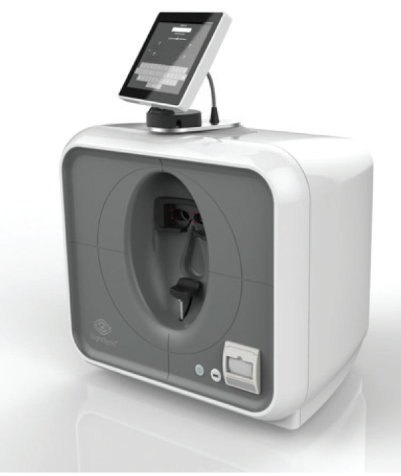
Figure. The neurolens Measurement Device measures the degree of eye misalignment to determine the contoured prism needed to resolve the disparity at distance and near.
This technology has been further refined and is now in use in a number of practices in the United States. I have personally witnessed multiple clinical situations in which the neurolens Measurement Device provided data to custom treat patients’ symptoms with contoured prism neurolenses.
CLINICAL EXAMPLES
Case No. 1. A contact lens–intolerant patient was extremely symptomatic after LASIK and was experiencing the same severe dry eye symptoms that he had before surgery with contact lenses. He showed no signs of dry eye on examination or testing, and no dry eye treatment helped relieve his symptoms. His problem now was that he couldn’t “take the LASIK off,” like he could remove his contacts preoperatively. After significant misalignment was detected with the neurolens Measurement Device, the patient was treated with neurolenses, and his symptoms are now gone.
This was the first patient who taught me about trigeminal dysphoria and how disruptive it can be. Myopic glasses are thin in the center and thick on the edge. This means that, with computer use and near work, the eyes turn in from the optical center of the glasses and get a base-in effect from the myopia-correcting lenses. Thus, many patients with this condition think they are contact lens–intolerant due to dry eye, and they stop wearing contacts and find some or total relief in their glasses due to the inherent base-in effect of myopic lenses in reading position.
This case, along with many subsequent phantom dry eye cases, made me realize why pharmaceutical companies have such a hard time achieving signs and symptoms endpoints in clinical trials for dry eye therapies. Any patients with symptomatic binocular vision misalignment should be excluded from pharmaceutical dry eye studies; otherwise, they may be the reason that endpoints are not achieved and a drug or other treatment fails to be approved.
Case No. 2. A contact lens–intolerant patient wore regular glasses and could not work past noon on the computer because of headaches and eyestrain. After being fit for neurolenses, her symptoms significantly improved, and she can now work a full day at her computer job.
Case No. 3. A mother of four was experiencing depression because of severe headaches. She had seen a headache specialist neurologist who treated her unsuccessfully with heavy headache drugs. She had lost her job, and her marriage was suffering significantly. After the neurolens Measurement Device measured her significant misalignment, she received neurolenses. She is now happy, and the whole family is doing much better.
As noted earlier, myopes, whose glasses are thick on the edge and thin in the center, experience some prismatic effect when their eyes turn in to read a computer or book. If this prismatic effect helps their binocular misalignment, they can be symptomatic when they switch to contacts. I now consider this possibility in all refractive surgery consultations that I would previously have labeled contact lens–intolerant. We can measure the binocular misalignment in these patients and educate them about their situation and the potential need for neurolenses postoperatively. They, in turn, can then make a better-educated decision about surgery.
CONCLUSION
In my practice and others’, the neurolens technology has significantly improved many patients’ quality of life. We have helped patients with severe dry eye or eyestrain after laser or implant vision correction who have previously been unable to find relief. The benefits also extend to the pediatric population (young individuals who experience headaches or eyestrain with reading that becomes misdiagnosed as attention deficit disorder).
Now that we have a way to accurately and objectively measure and treat eye misalignment, we can help the many patients who experience three or more symptoms while reading, using digital devices, or doing detail work. As we have restored quality of vision to many patients over the years, we now have a chance to diagnose eye misalignment and restore comfortable vision by recognizing and properly treating trigeminal dysphoria, whether surgery is involved or not.
1. Teitelbaum B, Pang Y, Krall J. Effectiveness of base in prism for presbyopes with convergence insufficiency. Optom Vis Sci. 2009;86(2):153-156.