

Retinal Vasculitis
Vasculitis is considered a specific disorder, in which components of the blood vessel are the center of the inflammatory focus. Fluorescein angiography (FA) has poor resolution of the deep retinal capillary plexus; optical coherence tomography angiography (OCTA) is not limited by leakage. OCTA provides microvascular morphological detail and information regarding capillary perfusion (Figure 1). In this case of Susac Syndrome, OCTA illustrated that areas of retinal capillary nonperfusion/hypoperfusion were more frequently observed in the deep than in the superficial capillary plexus (Figure 2). Capillary dropout is easily identified in FA, as are changes in macular capillary density in OCTA (Figure 3).
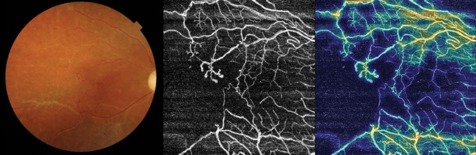
Figure 1. OCTA provides microvascular morphological detail and information regarding capillary perfusion.
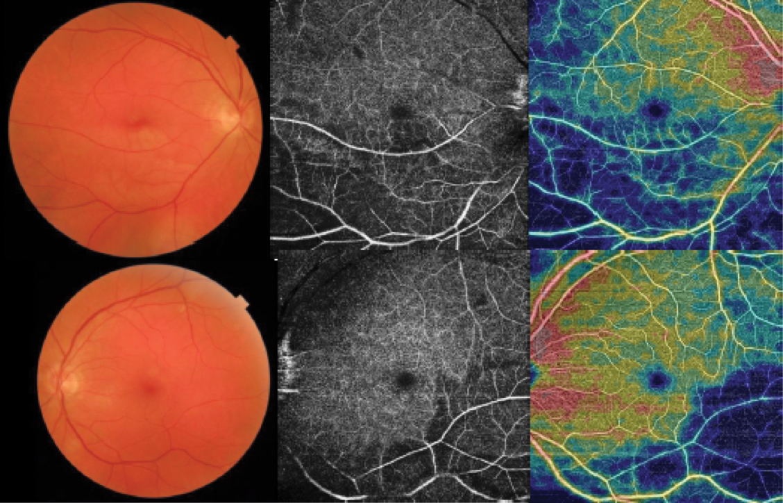
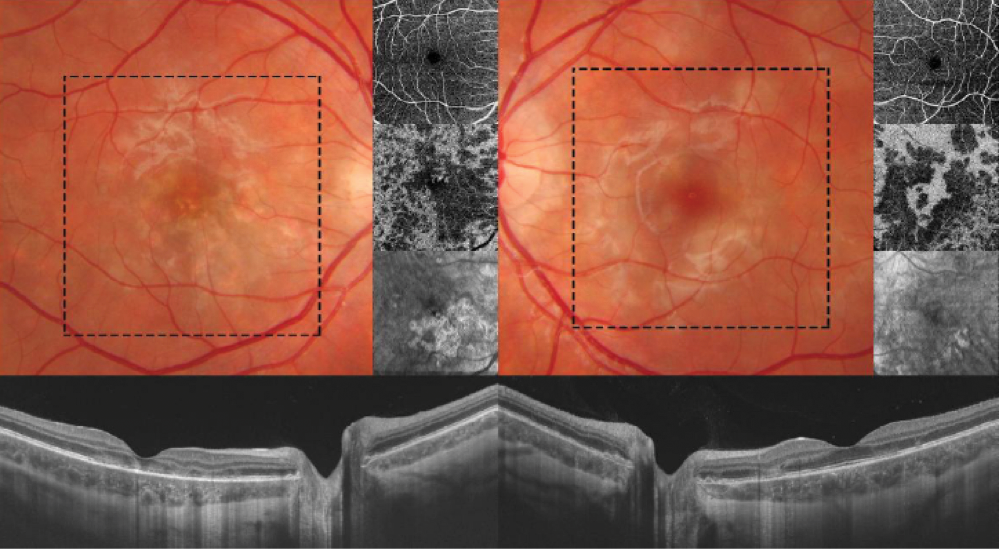
Figure 2. In a case of Susac Syndrome, areas of retinal capillary nonperfusion/ hypoperfusion are observed more frequently in the deep than in the superficial capillary plexus.
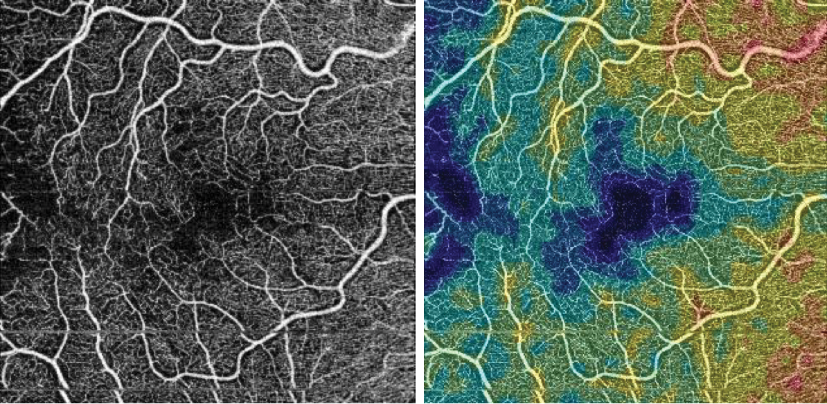
Figure 3. Capillary dropout identified in FA (left), and changes in macular capillary density identified in OCTA (right).
Topcon’s En Face Swept Source (SS)-OCT and OCTA enables the diagnosis of white dot syndromes. This, in turn, provides the information necessary to identify whether the primary inflammation is in the choriocapillaris, choroid, or ellipsoids layer. In cases of retinal vasculitis, OCTA shows that the ischemic changes, in the majority of cases, are in the deep capilar plexus. OCTA is also useful in cases of choroidal new vessels secondary to posterior uveitis.
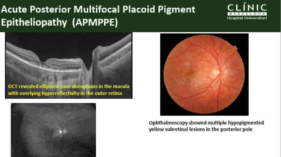
White dot syndromes include several disorders such as acute posterior multifocal placoid pigment epitheliopathy (APMPPE), multiple evanescent white dot syndrome (MEWDS), and punctate inner choroidopathy (PIC).
In this example of APMPPE, ophthalmoscopy showed multiple hypopigmented yellow subretinal lesions in the posterior pole, whereas SS-OCT revealed ellipsoid zone disruptions in the macula with overlying hyperreflectivity in the outer retina (Figure 4).
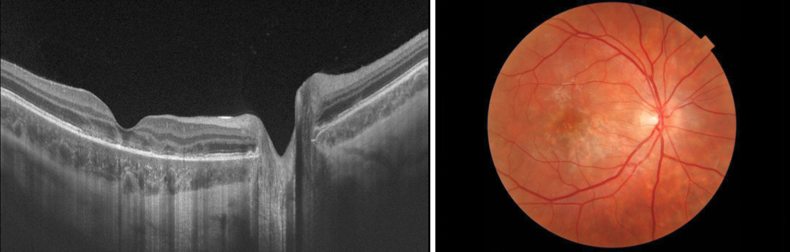
Figure 4. SS-OCT showing ellipsoid zone disruptions in the macula with overlying hyperreflectivity in the outer retina (left). Ophthalmoscopy showing multiple hypopigmented yellow subretinal lesions in the posterior pole (right).
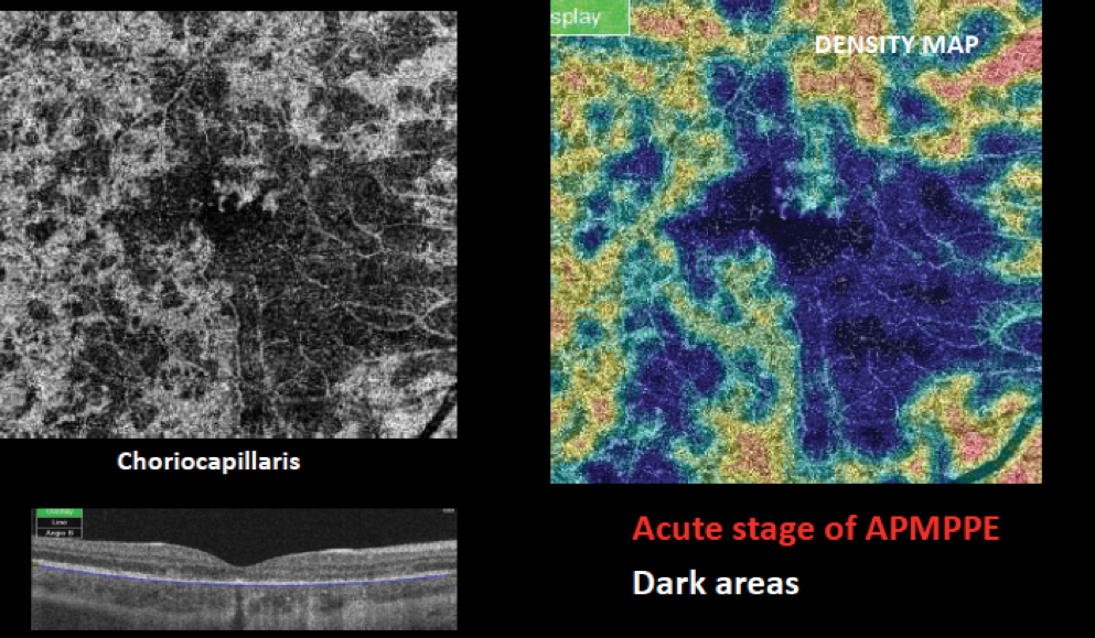
In the acute stage of the disease, these dark areas may result from decreased signal transmission or loss of blood flow — or sluggish blood flow — that may be below the limits of detection (Figure 5).

Figure 5. Dark areas resulting from a loss of blood flow in the acute stage of APMPPE.
OCTA provides new imaging evidence of the site and area of choriocapillary vascular pathology during the acute and later phases of APMPPE, including primary inflammatory choriocapillaropathy and progressive evidence of reduction in extent of sluggish flow areas. In APMPPE, changes in the outer retina are reversible.
In this example of MEWDS, OCTA imaging data localizes the pathologic process to the outer retina and the photoreceptors. OCTA imaging shows that choriocapillary circulation appears normal in the acute phase and suggests that MEWDS is an injury of the photoreceptors. En Face imaging illustrates the pathology as a pattern of hyperreflective dots (Figure 6).

Figure 6. En face OCT illustrating the pathology as a pattern of hyperreflective dots in an example of MEWDS.
In cases of serpiginous-like choroiditis, OCTA effectively documents progressive or recurrent choriocapillaris hypoperfusion via monitoring and follow-up of these patients.
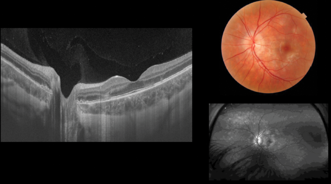
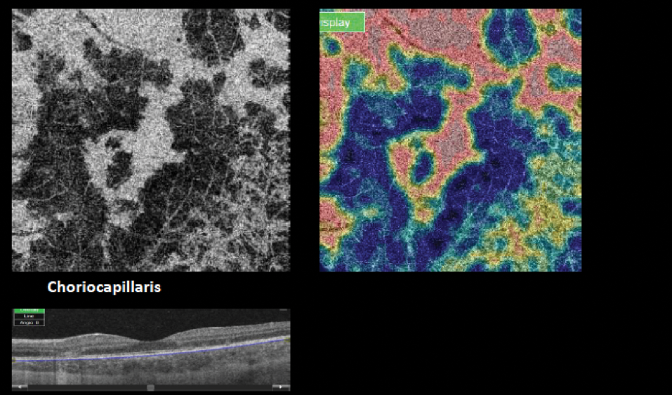
IMAGE SERIES
Acute Posterior Multifocal Placoid Pigment Epiteliopathy
This imaging represents a case of APMPPE with bilateral lesions. The series shows active and inactive lesions and evolution of the pathology from diagnosis through treatment with systemic steroids (Figures below).
In conclusion, our findings suggest that OCTA is an important imaging modality for managing posterior uveitis and retinal vasculitis together with other diagnostic tools.