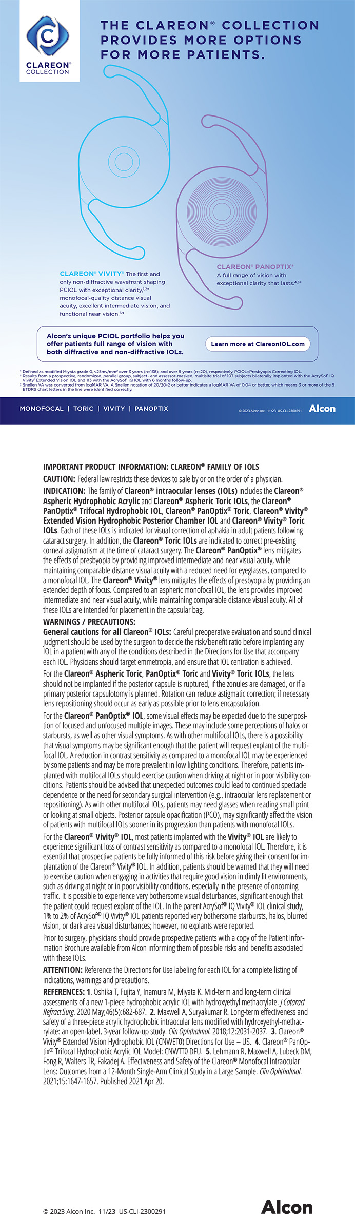
Clear corneal incisions have marked a breakthrough in phacoemulsification, because they reduce surgical time, speed patients' postoperative recovery, and reduce the rate of induced astigmatism. They also significantly decrease the incidence of the complications associated with scleral tunnels such as conjunctival manipulation and hyphema. Studies, however, have suggested a potentially increased risk of postoperative endophthalmitis with clear corneal incisions.1,2 For that reason, many ophthalmologists have become interested in strengthening the seal of these wounds. This article focuses on one such method, tissue adhesive.
CLEAR CORNEAL INCISIONS AND ENDOPHTHALMITIS
Recently, a multinational prospective study conducted by the ESCRS demonstrated that the risk of endophthalmitis following phacoemulsification was 0.38.1 Many peer-reviewed studies have compared the incidence of postoperative endophthalmitis with clear corneal versus scleral tunnel incisions. For example, a large, retrospective, comparative, case-controlled study evaluated the incidence of postoperative endophthalmitis in clear corneal incisions with or without sutures versus scleral tunnel incisions. The investigators compared 38 patients with culture-positive, acute, postcataract surgery endophthalmitis and 371 randomly selected control patients who underwent uncomplicated cataract surgery. They also compared the type of incision and use of a suture during cataract surgery between patients with endophthalmitis and controls. The researchers reported a threefold greater risk of endophthalmitis with clear corneal incisions than scleral tunnel incisions. The placement of a suture did not affect the incidence of endophthalmitis.2
In other studies, investigators have noted that self-sealing wounds were associated with the ingress of fluid into the anterior chamber in cases involving hypotony, minimal manipulation of the wound, and a postoperative fluctuation in IOP.3,4 Several other studies have demonstrated foreign bodies (including retained cotton fiber, cilia, and ointment) in the anterior chamber following clear corneal incisions.5-11 This research demonstrated the fragility of a clear corneal incision in the early postoperative period and the possible contamination of the anterior chamber by extraocular fluid entering through these wounds.
Nonetheless, a prospective case series involving 3,500 consecutive cataract surgeries performed by one surgeon using local anesthesia found different results. A 3- X2-mm beveled clear corneal incision was employed in each procedure, and no cases of endophthalmitis were reported.12
TISSUE ADHESIVES AND CLEAR CORNEAL INCISIONS
Tissue adhesives have been utilized for various purposes in ophthalmology, including corneal perforations, wound repair, and amniotic membrane transplantation. They have also been used as sealants for glaucoma and plastics procedures. Traditionally, ophthalmologists choose cyanoacrylate-based glues, but fibrin-based glues are gaining popularity as substitutes for sutures.13,14 Other new adhesives on the horizon include polyethyelene glycols, modified chondroitin sulfates, acrylic copolymers, and biodendrimers.
Many studies have demonstrated the efficacy of cyanoacrylate-based glue as a suture substitute in clear corneal incisions. In a prospective interventional case series, Meskin et al used 2-octyl cyanoacrylate with parabens (Liquid Bandage; Johnson & Johnson, Skillman, NJ) to seal the wound after clear corneal cataract surgery. In this series, a single surgeon created a 2.75-mm, triplanar, clear corneal incision in 51 patients. At the conclusion of each case, he sealed the wound with 2-octyl cyanoacrylate with parabens. All 51 eyes were found to have watertight wounds immediately after the application of the glue and at all postoperative visits. The most frequent complaint was a transient foreign-body sensation, which was reported by 31 patients (61). Other side effects included bulbar conjunctival hyperemia and the retention of glue on the second postoperative visit.15
Hermel et al conducted a similar study in bovine and rabbit eyes and demonstrated that the wounds in eyes with cyanoacrylic adhesives showed greater resistance in the early postoperative period than sutures. Seven days after the operation, however, the resistance of the incisions was similar among the groups.16
Shigemitsu et al compared the tensile strength of wounds that were not sutured, sutured using various techniques, or treated with various biotissue adhesives. They demonstrated that both cyanoacrylate and fibrin glue had tensile strength comparable to the sutures used in cataract surgery.17
Ritterband et al evaluated the effect of fluctuations in IOP on wounds that were sealed with cyanoacrylate glue. They made 3-mm clear corneal incisions in human donor globes. They used a transscleral cannula to alter the IOP while placing droplets of India ink on the wound. The eyes without glue were noted to leak with manual pressure at the wound's edge. None of the eyes with tissue adhesive showed an ingress of India ink with varying IOP or manual manipulation of the wound.18
Researchers have also evaluated the histological inflammatory activity with bioadhesives such as fibrin and cyanoacrylate versus unsutured valved closure and suturing. Alvarado et al demonstrated that fibrin bioadhesive induced a greater inflammatory response than other techniques at 1 week postoperatively, whereas cyanoacrylate adhesive caused less inflammatory activity than the suturing material. On day 30, both fibrin and cyanoacrylate adhesives had generated a slightly greater inflammatory response than sutured and unsutured techniques for closure. The study also showed that healing may be better with fibrin than cyanoacrylate glue in both the early and late postoperative period.19
Similarly, Henrick et al found that wounds sealed with organic tissue glue (Tisseel; Baxter Healthcare Corporation, Glendale, CA) had a higher incidence of inflammation than those closed with sutures. They found no difference in the wounds' integrity between these two methods of closure.20
Fibrin-based adhesives have been shown to have equal efficacy as cyanoacrylate glue with the added advantages of biocompatibility, the ability to be applied over larger surface areas, and an enhanced ability to secure amniotic membrane or conjunctival grafts. The potential disadvantages of human fibrin glue are the theoretical risk of viral transmission and the complexity of their preparation and application.21
Other glue materials such as Dermabond (2-octyl cyanoacrylate; Ethicon Inc., Somerville, NJ) have been shown to seal clear corneal incisions effectively such that their tensile strength is comparable to wounds closed by sutures.22
NOVEL TISSUE ADHESIVES
Many new adhesives are under investigation as alternate modalities to seal corneal wounds. In various studies, modified chondroitin sulfate aldehyde adhesive23 and acrylic copolymer tissue adhesive24 have proven successful.
Dendrimers are another promising class of tissue adhesives. Velazquez et al showed that, for both linear and stellate corneal wounds, biodendrimer adhesives provided a higher mean leakage pressure than closure with sutures.25
Dendritic macromers are also under investigation. Some macromers cross-link to form macroscopic gels, including hydrogels. Wathier et al evaluated several macromolecules. Their studies showed that hydrogel sealants produced from various macromolecules were effective in sealing corneal incisions (Figure 1). Their results also demonstrated that hydrogel sealants withstand higher pressures and stress placed on a wound than conventional self-sealed or sutured wounds. The hydrogel sealants offered the added advantages of easy preparation, reduced (four to six times) surgical time compared with conventional suturing, and minimized trauma to tissue26,27 (Figure 2).
CONCLUSION
Although no conclusive evidence exists to demonstrate a definitive relationship between clear corneal incisions and an increased risk of endophthalmitis, many ophthalmologists are investigating methods of sealing cataract incisions prophylactically. Traditionally, nylon sutures have been the most widely employed method of closing the wound. Tissue adhesives, however, have been quickly gaining in popularity as substitutes for sutures. Cyanoacrylate glue has been used most frequently, but other tissue adhesives, including human fibrin glue, are also employed. The disadvantages of cyanoacrylate and fibrin glues include a complex preparation process, foreign-body sensation, conjunctival hyperemia, and a possibly increased inflammatory reaction. Fibrin glue has the added theoretical risk of viral transmission. Other investigational bioadhesives may provide a safer, more biocompatible, and more effective alternative for closing corneal incisions after cataract surgery.
Bhairavi V. Kharod, MD, is a cornea and refractive surgery fellow at the Duke University Eye Center in Durham, North Carolina. She acknowledged no financial interest in the products or companies mentioned herein. Dr. Kharod may be reached at bhairavi.kharod@duke.edu.
Terry Kim, MD, is Associate Professor of Ophthalmology, Cornea and Refractive Surgery, Duke University Eye Center, Durham, North Carolina. He serves as a consultant for Hyperbranch Medical Technology, Inc. Dr. Kim may be reached at (919) 681-3568; terry.kim@duke.edu.