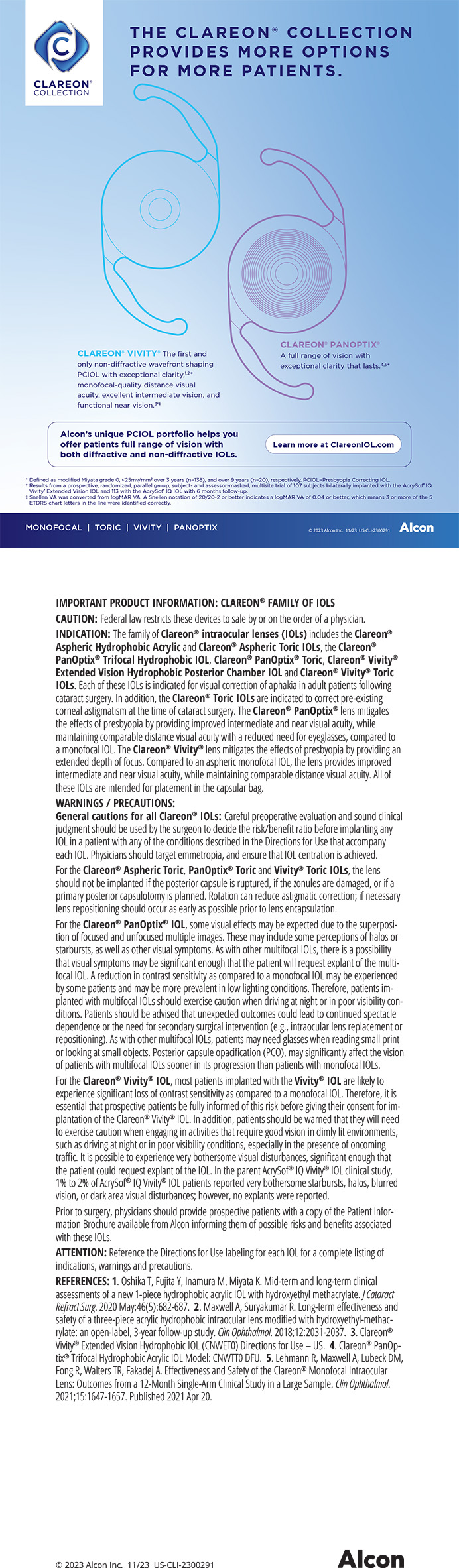
Many refractive surgeons must deal with a patient's dry eye during the weeks and months following a refractive procedure. Because I practice in a part of the US with extremely low humidity, pre- and postoperative dry eye is especially commonplace among my LASIK patients. Switching to the Intralase FS laser (Intralase Corp., Irvine, CA) from a mechanical microkeratome has decreased my incidence of induced postoperative dry eye in this population.
CONTRIBUTING FACTORS
Most published studies on the subject agree that, after LASIK, the incidence of dry eye is higher, particularly among women, long-term contact lens wearers, patients on certain systemic medications, older patients, and Asians.1-6 Prior to IntraLASIK, these findings were not reflected at my practice in Colorado, where about 50 of all patients treated with LASIK presented with some symptoms of dry eye during their recovery from refractive surgery.
The literature also shows reasonably close agreement on what contributes to dry eye after LASIK: the creation of the corneal flap and then laser ablation disrupt the corneal innervations, which contributes to a loss in corneal sensation.1 The decreased sensitivity can result in (1) a reduction in tear secretion, (2) the tear film's instability, (3) tear clearance, (4) a decrease in the number of conjunctival goblet cells, (5) an increase in tear osmolarity, and (6) punctate epitheliopathy.1-3 Furthermore, there is evidence that a superior hinge of the corneal flap can decrease corneal sensitivity, potentially leading to a reduction in the blink rate.4
In most patients, the cornea begins to return to normal by 6 months postoperatively, although one study showed that it can take up to 9 months for the cornea to return to its preoperative state.2 Anecdotally, I believe that refractive surgeons select for an increased number of dry eye patients in their LASIK population, because people who have a predisposition to dry eye and who are likely to be contact lens intolerant are more motivated to undergo the procedure than those who can tolerate contact lenses. Because of this tendency toward dry eye in LASIK patients, the standard of care at my practice has long been to insert temporary collagen plugs prior to surgery. This step helps to establish a more conducive environment for corneal healing postoperatively.
THE LASER FLAP DIFFERENCE
Although the aforementioned standard of care remains unaltered, the need for permanent punctal occlusion following LASIK has changed. With a mechanical microkeratome, my colleagues and I routinely used permanent punctal plugs in 20 of our LASIK patients. With the Intralase, that rate is down to approximately 5.
The first difference between mechanical and laser microkeratomes is how the flaps are created. With a mechanical microkeratome, there is a translation of the keratome head across the cornea under high pressure and with vibration as it makes the cut. There is sometimes macroscopic, and almost always some microscopic, damage to the corneal epithelium as a result. The Intralase procedure involves no movement across the cornea, only applanation.
The second difference is in the placement, thickness, and size of a flap created with the Intralase versus a mechanical microkeratome. The Intralase technology allows a smaller flap diameter, so there is less risk of corneal de-innervation. The creation of a well-centered flap is also easier with the laser. Using the patient's white-to-white measurement as my guide, the largest flap I create is 9.0mm in diameter for large corneas and 8.5mm for small corneas. My average Intralase flap size is 8.8mm. This contrasts to a 9.5-mm microkeratome flap, which is 15 larger in surface area. I am also able to create more consistent, thinner flaps with the Intralase laser. The deeper I cut into the cornea, the closer I get to the nerve bundles, thereby increasing the risk of impairing corneal sensitivity. As part of an unpublished multicenter clinical trial, my colleagues and I evaluated flap thickness using the Visante OCT. We compared flap thickness between microkeratomes at two sites and the Intralase at one site and found a more consistent correlation between programmed flap thickness and actual flap thickness with the Intralase (Figure 1).
In one study7 that looked at the average flap thickness (attempted vs achieved) with the Intralase laser, when the surgeon attempted a 100-µm flap, the actual mean thickness of the flap was 126µm, with a range of 89 to 165µm and a standard deviation of 14.1µm. This mean flap thickness was compared with the ranges seen in various clinical studies using mechanical microkeratomes.7 Table 1 shows the large standard deviation of flap thickness with microkeratomes, which is consistent with our own experience, as measured both by ultrasound and by the Visante OCT.
Any refractive surgery procedure somewhat exacerbates dry eye symptoms, but I have found the degree of irritation to be less in the 5 years (more than 6,000 cases) that I have used Intralase. In my practice, the two full-time optometrists who conduct the postoperative evaluations have reported on numerous occasions that they see less dry eye in the Intralase patients. One practical example of this low incidence of dry eye is that fluorescein staining is very common in eyes with flaps created by a mechanical microkeratome and almost nonexistent with the Intralase.
THE BEST CARE
When I bought my first Intralase laser in 2001, I had two offices. In one, I used a mechanical microkeratome for LASIK. In the other, I used the Intralase laser. Gradually, I concluded that providing my patients with the best possible care meant using the Intralase laser during all LASIK procedures. I closed the second satellite office primarily on this basis. Today, almost 100 of my LASIK cases are IntraLASIK. The only exceptions are in studies for which I am required to use a mechanical microkeratome and in patients who have a corneal scar or who previously underwent RK. There is no question in my mind that Intralase has improved the quality of my and my colleagues' LASIK practice, especially related to reducing dry eye issues.
Jon G. Dishler, MD, FACS, is in private practice in Greenwood Village, Colorado, and is Medical Director of the Dishler Laser Institute in Denver, Colorado. He is an investigator for Carl Zeiss Meditec, Inc., but acknowledged no financial interest in Intralase Corp. Dr. Dishler may be reached at jond@dishler.com.