Although it is a challenge to summarize more than 2,000 pages of trial transcripts during the course of the 12-day trial in Schiffer vs. Speaker et al, we have attempted to do so by presenting concise summaries of the trial testimony of each witness—complete with citations to the actual trial record—in the order in which they appeared during the trial. We hope that you find this article instructive and informative and that it sheds some light on our judicial system and the legal process, which has rendered this most recent landmark verdict. As always, we welcome your comments.
—Adam B. Krafczek, JD
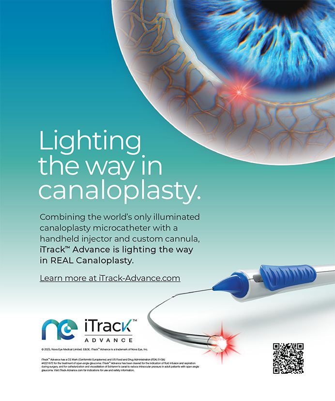
PLAINTIFF MARK SCHIFFER'S LEGAL CLAIMS AND ALLEGED DAMAGES
On October 6, 2000, Mark G. Speaker, MD, PhD, FACS, Medical Director of the Manhattan TLC Laser Eye Center at that time, performed LASIK surgery on Mark Schiffer. Subsequently, the patient claimed to have suffered impaired vision, which was particularly distorted in his left eye. Among Mr. Schiffer's claims is that his keratoconus was a condition that Dr. Speaker should have identified or anticipated prior to surgery and therefore disqualified him as a candidate for LASIK. Mr. Schiffer asserted that his impaired vision forced him to leave his position at Dresdner Kleinwort Wasserstein, an investment banking firm on Wall Street.
On January 23, 2003, Mr. Schiffer filed suit against Mark G. Speaker, MD; Laser and Corneal Surgery Associates, PC; TLC Laser Eye Center; Regina Zyszkowski; and Drs. Farkas, Kassalow, Resnick and Associates, PC, in the Supreme Court of the State of New York, County of New York, Index No. 101191-03. The Plaintiff claimed that the Defendants were negligent in the medical care and treatment rendered on behalf of Mr. Schiffer. His Complaint filed with the Court alleges the Defendants were negligent “in failing to use reasonable care; in failing to heed Plaintiff's condition, in departing from accepted standards in the procedures and treatment performed; in failing to follow appropriate practice; in failing to properly examine Plaintiff; in failing to properly treat Plaintiff's eyes; in failing to determine that Plaintiff was not a candidate for LASIK eye surgery, in performing LASIK eye surgery on Plaintiff when said procedure was contraindicated; and were otherwise negligent in their treatment of Plaintiff.”2
The Complaint further alleges the Defendants failed to provide Mr. Schiffer “with information that reasonably prudent medical practitioners should have provided under the circumstances, and failed to make Plaintiff aware of the risks and benefits of, and the alternatives to, the procedures employed.”3
The Defendants denied these allegations as set forth in Plaintiff's Complaint. The outcome of this lawsuit, tried in the Supreme Court of the State of New York, reportedly marks the largest award in a LASIK-related case to date, with the jury awarding the Plaintiff on July 27, 2005, the total amount of $7.25 million in damages.
THE TRIAL—Witnesses in Order of Their Appearance
Selig Percy Amoils, Plaintiff's Expert
Testified July 12 to 15, 2005
Direct Examination by Counsel for Plaintiff Mark Schiffer
Dr. Amoils is an ophthalmic surgeon who practices in Sandton, Johannesburg, South Africa. He is also licensed to practice medicine in England and in New York State.4 He completed his fellowship in ophthalmology in the US between 1966 and 1969, and he is a fellow of the AAO. He has written 35 peer-reviewed articles. In 2000, he published an article on iatrogenic keratectasia after LASIK.5 He has come across tremendous complications with LASIK surgery, and he has set up an organization in this country called Surgical Eyes and an ectasia study group under the Yahoo! help group.6
According to Dr. Amoils, the standard of care for LASIK surgery is the same worldwide. In June 2005, he was asked by Counsel for the Plaintiff to render an opinion on the quality of care received by Mr. Schiffer. Dr. Amoils reviewed the records of the Farkas Group; TLC Laser Eye Center; Susan Margolis, MD; Richard Braunstein, MD; the Massachusetts Eye and Ear Infirmary; and optometrist Michael Block. He also reviewed the sworn depositions that were taken prior to the trial. He asserted that Mark Schiffer had received poor treatment, that Mark Speaker, MD, was negligent, and that all of the Defendants had departed from the standard of care. He stated that this departure had caused Mr. Schiffer's injuries.7
Dr. Amoils said that Mr. Schiffer had forme fruste keratoconus preoperatively and that the danger of performing LASIK on patients with this ocular condition was well known in October 2000. He said that the preoperative evaluation of LASIK candidates should include topography and a slit-lamp examination but noted that a cornea with forme fruste keratoconus often appears to be normal. Dr. Amoils said that the FDA had warned against signs of keratoconus8 [end of day 1].
Dr. Amoils further testified that the treating optometrist's (Regina Zyszkowski, OD) preoperative screening of Mr. Schiffer departed from the standard of care when she failed to diagnose manifest, latent, forme fruste keratoconus. He said the most useful test for diagnosing this condition is topography. He described the topographic map of Mr. Schiffer's eyes from the Farkas Group dated September 29, 2000, as abnormal.9
When questioned by Counsel for the Plaintiff on Dr. Zyszkowski's testimony that colors can be arbitrary and that she relies on K values, Dr. Amoils stated that her remarks deviated from the standard of care, based on the published literature and his own research. Specifically, he said that inferior corneal steepening on the color scale of the preoperative topographic map of Mr. Schiffer's left eye reflected bulging of the inferior portion of the cornea. He faulted Dr. Zyszkowski for recommending LASIK for Mr. Schiffer and for not discussing the topography with Dr. Speaker, TLC, or Mr. Schiffer.10 Dr. Amoils testified that any retreatment of Mr. Schiffer's left eye would certainly have resulted in dandelion keratoconus or keratectasia, in which the condition worsens with each enhancement.11
After remarking on a lack of notes on the case byDr. Speaker, Dr. Amoils said that there was no indication that Dr. Speaker had reviewed the aforementioned topographic maps or those taken on October 6, 2000, prior to the LASIK procedure. Dr. Amoils testified that this behavior constituted gross negligence and a deviation from the standard of care on the part of Dr. Speaker. He added that the topography subsequently taken on October 6 by TLC was similar to the results of the topography taken by the Farkas Group on September 29 and confirmed the diagnosis of forme fruste keratoconus.12
Dr. Amoils referred to the pioneering work on corneal topography by Yaron Rabinowitz, MD, and Peter McDonnell, MD.13 He explained that Drs. Rabinowitz and McDonnell quantitatively evaluated the risk factors for keratoconus and described three diagnostic measures: (1) a central corneal power of greater than 47.00D; (2) a difference of 3.00D when comparing the inferior to superior cornea; and (3) asymmetry in the corneal powers of both of an individual's eyes.14
Applying these factors to Mr. Schiffer's preoperative topography taken on October 6, 2000 (Table 1),
Dr. Amoils noted that the central corneal power of the patient's left eye was 48.40D, a finding suggestive of forme fruste keratoconus. He said that there was an approximate 6.00D difference in corneal power in the inferior versus superior cornea, also significant for forme fruste keratoconus. He added that Mr. Schiffer's central corneal power measured 45.10D OD and 48.40D OS, a greater degree of asymmetry than the maximum 1.00D stipulated by Drs. Rabinowitz and McDonnell. He asserted that Dr. Speaker should have canceled the original surgery, repeated the topography, and measured the corneal thickness. The preoperative pachymetry of the central cornea measured 595µm, and a measurement with an Orbscan topographer (Bausch & Lomb, Rochester, NY) would have been appropriate.15
Dr. Amoils testified that LASIK performed in an eye with inferior steepening and asymmetric central corneal power would result in iatrogenic keratectasia, as it did in Mr. Schiffer's case. He said the complication had necessitated a corneal transplant in Mr. Schiffer's left eye. He cited study findings that the cornea's normal appearance on slit-lamp biomicroscopy does not rule out keratoconus.16 He stated that LASIK was “absolutely contraindicated” for Mr. Schiffer on the basis of his topography.17
If Mr. Schiffer's preoperative examination had been entirely normal aside from the October 2000 topography, Dr. Amoils said the patient would still definitely not have been a candidate for LASIK. He said his views were backed by the literature and the medical community.18 Dr. Amoils read into the record from an article by Douglas Koch, MD, a statement that surgeons should “be especially wary of performing LASIK in eyes with abnormal topography and have a low threshold for rejecting these patients for LASIK.”19,20
Referring to TLC's informed consent (dated September 1, 1999), which specified that LASIK candidates should not have certain ocular diseases, including keratoconus,
Dr. Amoils stated that Mr. Schiffer had not been adequately informed of his ocular problem and the risks of the procedure.21
According to Dr. Amoils, in the summer of 2001, Mr. Schiffer complained to Dr. Zyszkowski of a diminution of vision. She referred him to TLC for an enhancement, and he was seen by William
Tullo, OD, and Dr. Speaker on June 5, 2001. Dr. Tullo canceled the enhancement and ordered an Orbscan measurement, which was taken on June 7, 2001. Dr. Amoils stated that the cancellation was due to the patient's development of keratectasia and that the comanaged team had recognized that further LASIK surgery was contraindicated. Dr. Amoils said that Orbscan measurements on June 7, 2001, showed signs of abnormality in Mr. Schiffer's right eye and revealed a gross discrepancy in the corneal power of his left eye that is typical of keratectasia. Specifically, the posterior float was 75µm. He asserted that, based on its location, this inferior bulge could not have been produced by the laser. Central thinning was also evident.
Dr. Amoils said that the Orbscan measurement of Mr. Schiffer's right eye indicated an imminent progression to keratectasia.22
For his 10 days at the trial,
Dr. Amoils was compensated between $23,00023 and $29,000.24 “I have a huge practice,” he said. “It is costing me money to come here”25 [end of day 2].
Dr. Amoils testified that he had a reasonable degree of medical certainty that Mr. Schiffer will need a corneal graft in his right eye as well.26 He said the difference between the inferior and superior keratometry readings was approximately 6.00D on the September 29, 2000, topography.27 Specifically, the inferior value was 51.90D, and the superior value was approximately 45.18D.28
Dr. Amoils characterized Mr. Schiffer's vision prior to LASIK as good and said that patients usually do not regain their pre-LASIK vision. He said that Mr. Schiffer's case was distinct from that of the general patient population who receive corneal grafts in that he has post-LASIK keratoconus. He testified that the use of Intacs (Addition Technology, Inc., Des Plaines, IL) prior to the transplant necessitated a larger corneal graft.29
Drawing on his experience, Dr. Amoils said that keratectasia after LASIK is usually bilateral but more severe in one eye than its fellow. He said the condition is progressive.30 Mr. Schiffer's TLC chart dated June 5, 2001, from Plaintiff's Exhibit No. 2 contained handwritten notes that the enhancement should be cancelled and the left eye might have keratoconus. Dr. Amoils said the notes indicated that Mr. Schiffer has keratectasia bilaterally but to a greater degree in his left eye. He read similar findings on a topographic map dated May 2002 from Dr. Margolis' record; a handwritten diagnosis from Dr. Braunstein's records dated June 4, 2004; and a chart dated January 14, 2005, from Dr. Braunstein's office.31
Dr. Amoils stated that post-LASIK ectasia had been the subject of peer-reviewed articles prior to 2003 and that risk factors for the condition had been identified prior to that year. He disagreed with the possible defense that TLC's October 6, 2000, color topography showed a bowtie deformity, which might have been caused by contact lens warpage or a displaced apex.32
Cross Examination by Counsel for Dr. Speaker
Dr. Amoils acknowledged that Dr. Speaker has some standing in the US ophthalmic community and that he is an experienced corneal surgeon. He expressed familiarity with Dr. Speaker's publications.33 Dr. Amoils has not been affiliated with a hospital in New York, has no teaching privileges at any of the state's medical schools, and has not practiced medicine in the state. He has held the license for 20 years but has not used it technically. He disagreed with Dr. Rabinowitz's statement in his article13 that a comprehensive clinical examination remains the gold standard for the diagnosis of keratoconus and asserted that the gold standard is topography.34
Dr. Amoils first heard about Mr. Schiffer in June 2005, only 5 weeks prior to when this case went to trial. He spends 50% to 60% of his time performing surgery, generally 10 refractive cases, two retinal detachment surgeries, three to four cataract procedures, and three to four glaucoma operations per week. Dr. Amoils does not currently perform LASIK and last performed the procedure in 1994. His 10 refractive procedures per week are all PRK.35 He spends 40% to 50% of his time consulting.36 He has never testified in the area of medical malpractice in the US but has done so two or three times in South Africa.37
Dr. Amoils contended in 1999 that LASIK should never be performed in eyes with forme fruste keratoconus or in any eye with asymmetric topography with inferior steepening.38 He confirmed his earlier testimony that the topography taken at Dr. Zyszkowski's office showed corneal bulging. He said Mr. Schiffer had no clinical signs of keratoconus. He agreed that, in 2000, a physician could diagnose keratoconus or forme fruste keratoconus by looking at topography alone.39
In Mr. Schiffer's preoperative examination on September 15, 2000, he had myopia of -2.50 to -2.75D OD and a BCVA of 20/20+1 OD. Mr. Schiffer had myopia of -1.50 to -1.75D OS and a BCVA of between 20/25 and 20/20-2 OS. Dr. Amoils disagreed with the idea that visual acuity correctable to the 20/20 range is not suggestive of keratoconus.40
According to Dr. Amoils, in October 2000, Mr. Schiffer did not exhibit Rizutti's signs or a Charleaux sign. The mires did not pulsate during applanation tonometry, nor did he experience an acute rupture of Descemet's membrane, as some patients with keratoconus do.41
Dr. Amoils agreed that deep stromal stress lines may vanish if the examiner presses on the eye. He also acknowledged that, in keratoconus, there is normally a ring of iron deposits in the cornea and that these deposits also accumulate at the epithelium's base. Mr. Schiffer's October 2000 examination did not show a Fleischer's ring, scarring of Bowman's layer, or thinning of the cornea. Dr. Amoils disagreed with the contention that the pachymetry measurement was normal but acknowledged that the only reading was 590µm in the central cornea, an above-average finding. In addition, he concurred that corneal steepening in keratoconus produces clinical signs such as Munson's sign, which the Plaintiff did not have in October 2000.42
Dr. Amoils confirmed the existence of a global standard of care, both currently and in the year 2000. He said that LASIK had been accepted in some cities and countries before others and that, during that period, there was no global standard of care. A 1998 case report43 of a German patient was included on his list [in evidence] of 10 important publications relevant to the case.
Dr. Amoils maintained that its publication changed the standard of care for average US physicians,44 but he agreed that the AAO helps define the standard of care for the average ophthalmologist.45 He expressed unfamiliarity with an AAO-commissioned study of LASIK's safety and efficacy that was published in 2003.46,47
Dr. Amoils acknowledged that he had submitted a paper to Dr. Speaker for publication and that it had bothered him that it was rejected for publication. He had expressed some unhappiness over its rejection in 2000 by the AAO in a document he had written to Mr. Krouner (Plaintiff's Counsel).48
Cross Examination by Counsel for TLC Laser Eye Center
The October 2000 topography at TLC only reveals the curvature of the front surface of the cornea. Dr. Amoils concluded that this topography was abnormal based on measurements taken 3mm out and 3mm down from the corneal center. He judged the difference between these measurements according to Dr. Rabinowitz's work. Dr. Amoils confirmed that the contours shown on topography can change within 6 months, 1 year, and several years49 [end of day 3].
Medical records reflected that Dr. Braunstein's doctor/patient relationship with the Plaintiff began in January 2003. Dr. Braunstein ordered a wavefront examination approximately 6 weeks before scheduling corneal graft surgery.50 The examination (Tracey Technologies, Houston, TX) taken on January 14, 2005, of Mr. Schiffer's left eye included a Snellen letter E for higher-order aberrations 20/40.51 Dr. Amoils agreed that the wavefront studies ordered by Dr. Braunstein were considered reliable by the scientific community in 2005. He concurred that the use of the simulated Snellen letter was also accepted in the ophthalmological community in January 2000.52
Mark Schiffer, Plaintiff
Testified July 15, 2005
Direct Examination by Counsel for Plaintiff Mark Schiffer
Mr. Schiffer is 32 years old. He graduated from Yale University in 1995 with a B.A. in economics.53 He began matriculating at the Wharton School at the University of Pennsylvania in the fall of 1999 and graduated in the spring of 2001 with an MBA.54
The Plaintiff sought eye care in September 2000 while attending school in Philadelphia. He visited Dr. Zyszkowski at the Farkas Group's office in Long Island, New York. He was interested in LASIK, had a history of contact lens intolerance, and did not like wearing glasses. During the first consultation, Dr. Zyszkowski took his history. He expressed his interest in LASIK and underwent a LASIK screening. He discussed with Dr. Zyszkowski an orbital fracture he had sustained years ago, and she expressed uncertainty as to how the injury would affect his candidacy for LASIK. The Plaintiff said the injury had not affected his vision or his globe. He had been diagnosed with myopia and astigmatism and prescribed glasses while a teenager. He was examined, including at the slit lamp. His refraction and topography were measured. Dr. Zyszkowski said that topography confirmed his astigmatism, which could affect his candidacy for LASIK. He testified that there was no discussion about keratoconus or keratectasia. He received no indication that he had asymmetrical, inferior corneal steepening in his left eye.55
Dr. Zyszkowski said that a retinal spot in one of his eyes would need to be investigated further to determine his LASIK candidacy. She referred him to a retina specialist, Ken Carnevale, MD. He was thereafter told the retinal spot was not a contraindication to LASIK by Dr. Zyszkowski. Between leaving her office on September 29 and his LASIK surgery on October 6, he did not see Dr. Zyszkowski but spoke to her on the phone. She told him that, based on her conversation with TLC, the previous orbital fracture was not a contraindication for LASIK. She also said that his astigmatism was not a contraindication.56
She had originally wanted him to undergo LASIK with Eric Donnenfeld, MD, at TLC's Long Island office, due to its proximity to where the Plaintiff resided. She recommended he undergo the procedure in Manhattan with Dr. Speaker instead, because he used a laser that she believed was better for astigmatism.57
Mr. Schiffer recalled no discussion of the risks of LASIK between him and Dr. Zyszkowski, and he said she in no way indicated that he was at increased risk versus the general population. He arrived at the Manhattan office on October 6, 2000. A patient consultant helped him through the preoperative process. Mr. Schiffer made no direct payment to Dr. Zyszkowski or the Farkas Group. During the September visit, she had told him that, if he had LASIK, the fee for the initial consultation would be taken from what he paid TLC. Mr. Schiffer understood that she and her employer were responsible for his pre- and postoperative care.58
Mr. Schiffer did not recall receiving the informed consent prior to surgery but did recall signing it on that day at TLC. He said he did not discuss the contents of the document with Dr. Zyszkowski. Section three of the form listed LASIK indications and contraindications as well as perioperative care. Mr. Schiffer said he still did not know to what the term perioperative care refers. He acknowledged that the form stated, “Candidates must be free of certain eye diseases, including keratoconus, glaucoma, cataracts and retinal and optic nerve diseases.” Mr. Schiffer said he did not know in October 2000 that he had keratoconus. He said Dr. Zyszkowski told him nothing regarding whether he had the condition and neither did anyone at the Farkas Group, Dr. Speaker, or anyone from TLC. He had no reason to suspect he had keratoconus and had never heard of the condition at that time. He had no reason to expect he was at increased risk of injury from LASIK.59
He admitted signing and dating the informed consent and, as instructed, wrote on the form in his own handwriting that he understood that the procedure carried risks and no guarantees, although he said he was not told that he was at particular risk.60
On October 6, 2000, the day of the Plaintiff's surgery, a technician performed some tests on Mr. Schiffer before he met Dr. Speaker. One of the tests was topography. He did not recall being given a copy of this examination and did not discuss the results with anyone. No one discussed the results of the topography performed by Dr. Zyszkowski on September 29, 2000, with him on the surgical day, either.61
He recalled lying on the operating table with
Dr. Speaker behind him, from where he performed surgery and described the procedure. Dr. Speaker took no history from him and performed no tests before the surgery. The Plaintiff met with no optometrist on the morning of surgery.62
He underwent bilateral LASIK. Approximately 20 minutes elapsed between his entrance into and exit from the OR. No complication occurred during the procedure. Dr. Speaker and he did not discuss his candidacy for LASIK preoperatively. Neither did they speak about his topography. After surgery, he received a pair of dark sunglasses and an instruction sheet, and he left with his father.63
Mr. Schiffer met with Dr. Zyszkowski 1 day postoperatively and soon returned to the Wharton School of Business without incident with regard to his vision. Immediately postoperatively, he was pleased with his surgical result; he felt his UCVA was what it had been preoperatively with contact lenses. He had a number of follow-up visits, one of which was not with Dr. Zyszkowski but with Dr. Susan Resnick, a partner at the practice. Mr. Schiffer was pleased with his vision through the remainder of the year 2000, but he testified that it began to deteriorate in 2001 through the spring. At one of the follow-up visits, Dr. Zyszkowski recommended an enhancement. The visual deterioration consisted of regression to his preoperative visual acuity accompanied by some visual distortion he had not had previously.64
Dr. Zyszkowski referred the Plaintiff back to TLC. He visited TLC on June 5, 2001, and was examined by
Dr. Tullo, the clinical director. He recalled Dr. Tullo's performing a slit-lamp examination and topography.
Mr. Schiffer stated that no one performed a slit-lamp examination on the day of his original surgery and said he was unaware of any preoperative pachymetry readings on that day or in June 2001. Dr. Tullo informed him on June 5 that he might have keratoconus, a contraindication for the LASIK enhancement, which was canceled.
Dr. Tullo discussed keratoconus with him immediately after the examination and described the condition. He said he wanted the Plaintiff to undergo another test to confirm the diagnosis, specifically Orbscan topography. Mr. Schiffer testified he had never heard of this technology or been tested with it before June 5, 2001.65
Dr. Zyszkowski, Dr. Tullo, Mr. Schiffer, and Dr. Speaker met at the offices of TLC in New York. They discussed his possible keratoconus and need for an Orbscan. He was told that, if the diagnosis of keratoconus were confirmed, he would not be able to have an enhancement. He asked Dr. Speaker if he should not have undergone the original procedure. He testified that Dr. Speaker responded yes, in hindsight, but that the preoperative charts gave no indication that the problem would occur. He added that LASIK sometimes triggers the manifestation of keratoconus. Mr. Schiffer was told the prognosis of keratoconus, that the condition is not correctable, that it requires the use of hard contact lenses, and that it can progress to the point of requiring a corneal transplant.66
During his June 5 visit to TLC, an appointment was made for the Plaintiff to go to Cornell Medical Center for the Orbscan topography a few days later. The results of the Orbscan were communicated to Dr. Zyszkowski. He visited her in Long Island shortly after seeing her in New York. The results of the June 7, 2001, Orbscan confirmed the diagnosis of keratoconus (Table 2). He never spoke again to Dr. Speaker or Dr. Tullo. Mr. Schiffer and Dr. Zyszkowski discussed how to proceed with managing his left eye, which had keratoconus. She told him he would require a hard contact lens, and he understood he was no longer a LASIK candidate. He had no further contact with Dr. Zyszkowski, because he was unhappy with his care.67
He visited Dr. Margolis in Manhattan in April 2002. He accounted for the elapsed time between his last visit with Dr. Zyszkowski and his first with Dr. Margolis by saying that he did not require a corneal transplant at that time and that he knew he would not be able to tolerate a hard contact lens. He decided to cope with his deteriorating vision.68
During his visit to Dr. Margolis, he discussed with her his history. She diagnosed keratoconus in both of his eyes, but to a greater degree in his left. At TLC, Dr. Tullo had also said he thought Mr. Schiffer had keratoconus bilaterally, more so in his left eye. The Plaintiff had visited Dr. Margolis based on the recommendation of one of his mother's friends, whose sister has keratoconus. When he asked if Dr. Speaker had made an error operating on his eyes, she disclosed a personal relationship with him. The Plaintiff decided to visit another doctor.69
Mr. Schiffer left his job at Wasserstein in July 2002 and began working for his father's software company, Safe Banking. He said he departed because his visual difficulties were negatively affecting his career; he claimed he had been passed over for important projects and was working himself to exhaustion. Mr. Schiffer's initial responsibilities at Safe Banking were sales and marketing of the software products, office management, bookkeeping, and accounting.70 The Plaintiff said that his injury was the only reason he was not currently an investment banker. He admitted that he had said nothing about his vision's affecting his ability to perform his job to anyone when he left Wasserstein.71
Dr. Margolis had referred the Plaintiff to Dr. Block, who fitted him with piggyback contact lenses in 2002. According to Mr. Schiffer, the lenses were unsuccessful, and he had great difficulty putting them in his eyes and would often leave them out. When able to insert the lenses, he found them very uncomfortable, and they reddened and irritated his eyes. He further testified that the lenses had little effect on his visual distortion and he had trouble removing them. He used the lenses from the middle of 2002 to early 2004.72
Mr. Schiffer's treatment by Drs. Braunstein and Block overlapped. Dr. Braunstein practices at Columbia-Presbyterian Hospital in New York. He said the only possibility aside from the contact lenses was a corneal transplant, which he was reluctant to perform. He described a new Intacs procedure that he did not perform and referred the Plaintiff to Dimitri Azar, MD, at the Massachusetts Eye and Ear Infirmary, who implanted the segments. The Plaintiff hoped the surgery would reduce his discomfort with his vision. The procedure lasted approximately 1 hour and required him to miss a couple of days of work. According to Mr. Schiffer, he experienced no improvement, which he discussed with Dr. Braunstein, who prescribed eye drops to lower his IOP in an effort to improve the results. He testified that the drops did not help. The last resort was a transplant,73 which was scheduled for February 28, 2005, but was canceled, because the Plaintiff had the flu.74
The Intacs were removed in mid-February by Dr. Braunstein. This procedure caused the Plaintiff to miss a couple of days of work. The transplant was rescheduled for March 17, at which time he received eye-banked corneal tissue.75 He missed 1 week of work. Currently, he experiences no discomfort in his left eye. Since the transplant, he has experienced a lack of contrast sensitivity, however. The stitches from the surgery have not been removed, and, perhaps as a result, his vision is still blurry. His last visit with Dr. Braunstein was a couple of weeks prior to the trial.76 The Plaintiff remains under
Dr. Braunstein's care. The physician told him that he could expect a maximal cure 1 year postoperatively and that the stitches could stay in for that time. Mr. Schiffer said that his eye will never be normal again and that it will always be at risk of a ruptured globe due to disease and of rejecting the tissue.77
The Plaintiff characterized his pre-LASIK vision as normally blurry but correctable with contact lenses or spectacles. He described his current vision as severely distorted. He said he tries to avoid driving and stated that he cannot read signs. He said he does not recognize people at the distance normal people do and that his trouble seeing facial expressions negatively affects his interactions with people. He asked his wife that morning to verify if he had missed any spots while shaving.78
Mr. Schiffer plans to attend Rand Institute's doctoral program in public policy in September 200579 [end of day 4].
Cross Examination by Counsel for
Dr. Speaker
In 1993, Mr. Schiffer sustained an injury that ripped through his sinus cavity, fractured his left orbit, and required surgery with a titanium plate. His brain was herniated and contused. A nerve in the left side of his forehead was severed, resulting in a permanent loss of sensation and a partial loss of movement. He said the event did not permanently affect his earning potential. He admitted suing the Dix Hill Diner—where the injury took place—and others in 1995 and claiming $10 million in damages relating to his lifetime employment. The claim was based on his inability to achieve Phi Beta Kappa status as a college sophomore by maintaining a 4.0 GPA and his inability to participate in a Master's program at Yale simultaneously with his Bachelor's program.80
He admitted being notified in writing by TLC that the only way in which to avoid all surgical risks was not to have the procedure. He also admitted that he initialed this area of the informed consent. He read the form's general descriptions of LASIK and PRK. Prior to undergoing LASIK, he was aware of the alternatives to the procedure, including glasses, contact lenses, and PRK. He knew there were contraindications to LASIK. He declined monovision. He acknowledged that approximately three pages of the informed consent were devoted to complications and that he initialed the form. He was notified in writing of the refractive, flap-related, corneal healing-related, and other complications. The informed consent described the possibilities of foreign body sensation, pain, and discomfort postoperatively. The form also listed the complications of light sensitivity, blurred vision, ocular dryness, tearing, and fluctuations in vision. Persistent pain, although uncommon, was also included and noted to indicate a possible problem with the epithelium. Mr. Schiffer was further informed about the possible displacement of the corneal flap and potential infection, and he was instructed to contact the physician if the latter occurred.81
In addition, the informed consent indicated that steroid treatment and further surgery might be needed. It stated that his vision might not be fully restored and that both night vision and refractive problems were possible. He was informed that over- or undercorrections were possible and that imbalanced vision could be problematic.