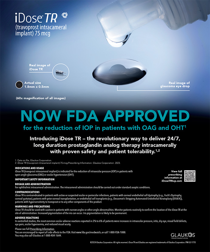
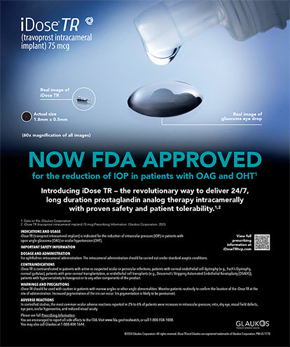
Cover Stories | Aug 2005
Preventing Pseudophakic CME
Prophylactic techniques and advice on managing the complication when it occurs.
Kensaku Miyake, MD
Thankfully, improved surgical techniques, methods of fixating the IOL, biomaterials, and anti-inflammatory drugs have dramatically reduced the incidence of cystoid macular edema (CME) after cataract surgery. This article describes the theoretical pathogenesis of CME, routine methods for preventing the condition, and how to manage the complication when it occurs.
THE PATHOGENESIS OF CME
Figure 1 summarizes my hypothesis on the development of CME following cataract and IOL surgery.1,2 According to my theory, prostaglandins and other cytokines are synthesized intra- and postoperatively from the anterior uvea and lens epithelial cells in response to intraocular surgical trauma. These mediators induce a disruption of the blood-aqueous barrier, followed by a disruption of the blood-retinal barrier concurrent with a manifestation of CME. The larger the magnitude of the blood-aqueous barrier's disruption, the larger the blood-retinal barrier's disruption and the greater the likelihood of CME.
Treatment with NSAIDs suppresses the synthesis of prostaglandins caused by surgical trauma to the intraocular tissues. It also alleviates the disruption in the blood-aqueous and blood-retinal barriers and decreases CME. According to this theory, factors such as older age, systemic vasculopathies, and inflammation predispose patients to CME as well as to disruptions of the blood-aqueous and blood-retinal barriers.
DETECTING CME
It is generally easy to diagnose CME with fluorescein angiography. The complication may or may not be symptomatic, and its peak incidence is approximately 5 weeks postoperatively. So-called clinical CME refers to a symptomatic decrease in vision that typically occurs 2 or 3 months postoperatively1,2 (Figure 2).
PREVENTING AND TREATING CME
Administering topical NSAIDs preoperatively and for 3 months postoperatively is an effective means of preventing CME.1,4 Tables 1 and 2 show the relation of a disrupted blood-aqueous barrier and the incidence of CME in a randomized study my colleagues and I conducted using small-incision surgery and foldable, acrylic IOLs.5 Our findings indicate that topical NSAIDs (such as diclofenac eye drops) reduce the incidence of CME by approximately 5% and normalize the blood-aqueous barrier within 1 to 2 months. In patients who are predisposed to developing angiographic CME, I would recommend the use of topical NSAIDs for 6 months or longer postoperatively.
Research indicates that NSAID eye drops are also effective for the treatment of clinical CME.4 Alternative therapies include triamcinolone acetate, Diamox (Wyeth Pharmaceuticals, Philadelphia, PA), and hyperbaric oxygenation.4,6,7 In cases of CME associated with intraoperative complications such as a rupture of the posterior lens capsule, vitreous loss, or abnormal IOL fixation, the etiological factor should be removed surgically.
CONCLUSION
Modern, small-incision cataract surgery and foldable, acrylic IOLs have helped to reduce the incidence of CME significantly. In patients who are predisposed to developing the complication, however, surgeons should consider prophylactic treatment with a variety of agents in addition to topical NSAIDs.
Kensaku Miyake, MD, is Director and Head, Shohzankai Medical Foundation, Miyake Eye Hospital, Nagoya, Japan. He is also Visiting Professor, Fujita Health University, Toyoake, Japan. Dr. Miyake may be reached at +81 52 915 8001; miyake@spice.or.jp.
1. Miyake K. Prevention of cystoid macular edema after lens extraction by topical indomethacin (I). A preliminary report. Albrecht Von Graefes Arch Klin Exp Ophthalmol. 1977;203:81-88.
2. Miyake K, Ibaraki N. Prostaglandins and cystoid macular edema. Surv Ophthalmol. 2002;47(suppl 1):S203-S218.
3. Miyake K, Ota I, Maekubo K, et al. Latanoprost accelerates disruption of the blood-aqueous barrier and the incidence of angiographic cystoid macular edema in early postoperative pseudophakias. Arch Ophthalmol. 1999;117:34-40.
4. Rossetti L, Chaudhuri J, Dickersin K. Medical prophylaxis and treatment of cystoid macular edema after cataract surgery. The results of a meta-analysis. Ophthalmology. 1998;105:397-405.
5. Miyake K, Masuda K, Shirato S, et al. Comparison of diclofenac and fluorometholone in preventing cystoid macular edema after small incision cataract surgery: a multicentered prospective trial. Jpn J Ophthalmol. 2000;44:58-67.
6. Cox SN, Hay E. Bird AC. Treatment of chronic macular edema with acetazolamide. Arch Ophthalmol. 1988;106:1190-1195.
7. Pfoff DS, Thom SR. Preliminary report on the effect of hyperbaric oxygen on cystoid macular edema. J Cataract Refract Surg. 1987;13:136-139.