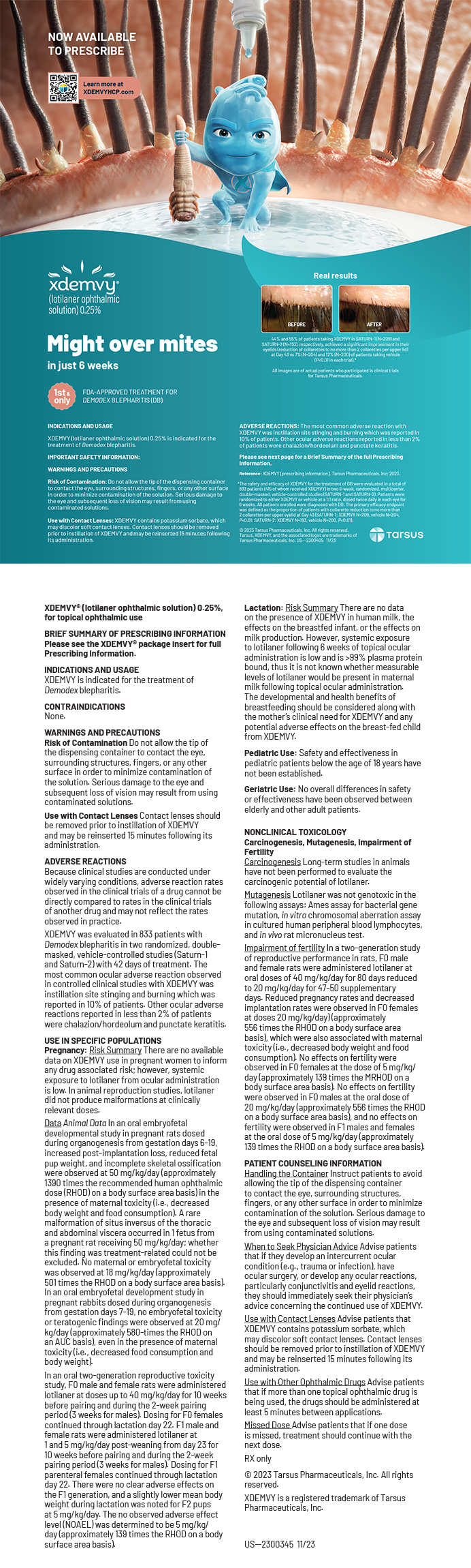
BY DANIEL H. CHANG, MD
Efficient removal of the nucleus is essential to minimizing corneal edema and delivering excellent early visual outcomes. To segment the nucleus, I use a horizontal phaco chop technique. This approach allows me to break up and remove the nucleus efficiently with minimal phaco energy.
I use a straight phaco tip with a 15º bevel to optimize my angle of nuclear engagement and holding power. I expose approximately 1 mm of the phaco tip past the end of the irrigation sleeve. After thorough hydrodissection and nuclear rotation, I enter the eye with the phaco probe and remove any anterior cortex within the capsular opening. I then engage the nucleus as close to the proximal edge of the capsulorhexis as possible. I use just enough phaco power to bury the tip. To prevent wound burn in this no-flow state of occlusion, I set the phaco machine on a micropulse setting, and I gently pulse the foot pedal until the tip is appropriately buried.
At this point, I advance the chopper underneath the distal edge of the capsulorhexis opposite the phaco tip (Figure). I use a Verges Phaco Chopper (Rhein Medical), which is ambidextrous and has a capsule-friendly olive tip for safety. To advance the chopper, I touch the tip to the nucleus centrally; then, I slide the instrument along the contour of nucleus and out under the capsular edge. To avoid chopping outside the bag, I never advance the chopper to the periphery without first contacting the lens nucleus centrally and sliding the instrument peripherally. When the chopper is positioned past the nuclear equator, I drop into foot position 1 (irrigation only), and I pull the chopper toward the phaco probe. For me, the vacuum is useful in maintaining nuclear stability as I advance the chopper, but I do not find it necessary during the actual chopping maneuver. By releasing vacuum, I am performing a purely mechanical maneuver, and there is minimal chance of the phaco tip’s inadvertently engaging the posterior capsule.
After creating two nuclear hemispheres, I rotate both the nucleus and the phaco tip slightly clockwise. I then engage the left hemisphere at approximately middepth and repeat the chopping procedure. I vary the size of my pieces inversely with the density of the nucleus— smaller pieces for denser nuclei and larger pieces for softer nuclei. I continue chopping until I reach the final nuclear fragments, at which time I watch carefully for surge and protect the posterior capsule with the chopper’s bulbous olive tip. After epinuclear removal, cortical cleanup, and careful placement of the IOL, I seal the wound and look forward to checking on a happy patient the next morning.
Daniel H. Chang, MD, is in private practice at Empire Eye and Laser Center in Bakersfield, California. He is a consultant to Abbott Medical Optics Inc. Dr. Chang may be reached at (661) 325-3937; dchang@empireeyeandlaser.com.
BY D. MICHAEL COLVARD, MD
My favorite chopping maneuver is a technique that I call “divide-and-conquer prechop.” I believe that it was first described by Jack Dodick, MD. After the capsulorhexis and hydrodissection, I place an ophthalmic viscosurgical device in the anterior chamber and introduce two specially designed choppers, either through a sideport and the phaco incision or through two sideports. I then slide the choppers under the anterior capsulotomy and direct them around the nucleus to a position approximately 180º from one another. I bring the choppers together centrally to create a bisecting crack in the nucleus, dividing it cleanly into two pieces. I then rotate the nucleus 90º and repeat the maneuvers, thus dividing the nucleus into quadrants. Once I have disassembled the nucleus, additional chopping is seldom necessary. If the need arises, however, I finish up with standard vertical chopping (Figures 1-4).
I like this prechopping technique because it combines the advantages of classic divide and conquer and intraoperative chopping without the disadvantages of either. The primary advantage of classic divide and conquer is that it allows the surgeon to disassemble the nucleus under very stable conditions. The chief disadvantages of the technique are that it is slow, places stress on the zonules, is associated with downward forces pushing on the nucleus, and requires more total phaco power to disassemble the nucleus than do well-performed chopping techniques. The primary advantage of intraoperative chopping techniques is their efficiency. No phaco energy or time is wasted making the grooves. The cracks are made as an integral part of the phaco process. Zonular stress, moreover, in the hands of an experienced surgeon, may also be reduced, because the nuclear material is lifted out of the capsular bag as intraoperative chopping is performed. The chief disadvantage is that, for optimal effectiveness, the technique requires higher vacuum levels and flow rates than are needed for divide and conquer. These heightened phaco parameters result in faster and less predictable movement of nuclear material and less stability of the anterior and posterior chamber volume.
Divide-and-conquer prechop quickly and easily divides the nucleus into quadrants without phaco energy and without placing stress on the capsule or the zonules. The compressive forces of the crossing choppers are isolated within the nucleus rather than transmitted to the supporting structures of the lens. The maneuver is performed with optimal visualization in a stable chamber filled with an ophthalmic viscosurgical device, and the nuclear quadrants are then brought into the pupillary plane without the need for high vacuum levels or high flow rates. I find this technique simple to perform, safe, and very reproducible. It is only for the very dense cataract with little or no cortex that I fall back on other techniques for nuclear disassembly. The specific choppers I use for this maneuver are Dodick-Colvard Cross Choppers (Rhein Medical).
D. Michael Colvard, MD, is a clinical professor of ophthalmology, Keck School of Medicine, University of Southern California, Los Angeles, and he is the director of the Colvard- Kandavel Eye Center in Encino, California. He acknowledged no financial interest in the product or company he mentioned. Dr. Colvard may be reached at (818) 906-2929; mike@mcolvard.com.
BY SUSAN M. MACDONALD, MD
Phaco chop is an advanced technique that requires the surgeon to possess a solid understanding of fluidics and power modulation. When chopping, he or she uses the phaco tip to grab and hold the nucleus. The “grab” is accomplished by the phaco needle’s being impaled into the nucleus.
The surgeon buries the phaco needle with a quick burst of energy. Next, moving the foot pedal to foot position 2 allows occlusion of the needle and causes vacuum to build. The vacuum will allow the surgeon to hold the nucleus and resist the movement of the chopper.
Diagonal quick chop is my primary technique. Under topical anesthesia, I start with a well-constructed wound and a generously sized capsulorhexis of at least 5 mm. A smaller one will make the surgery more difficult and increase the risk of complications. Equally important is thorough hydrodissection, resulting in a freely rotating nucleus. I use a J-cannula for subincisional hydrodissection and a straight cannula for the remaining areas. I make several gentle hydrodissections and do not move to the next step of the cataract procedure until the nucleus rotates easily.
I then pass the phaco tip through the main wound and place the chopper into the sideport 70º away from my main incision. I use a Seibel nucleus chopper (Ambler Surgical). Once the instruments are in place, I bury the phaco tip in the center of the nucleus, a step that can be accomplished with either longitudinal phacoemulsification or torsional power. The burst mode allows a short burst of energy that buries the tip without emulsifying too much nuclear material. The power and length of the burst can be adjusted depending on the density of the nucleus. I use a few short bursts of longitudinal phaco power (20%-60% ultrasonic power for 40 ms). If I am using torsional phacoemulsification, I adjust the amplitude to between 50% and 100% with a short burst, which limits movement of the phaco needle and allows me to bury it.
Next, I allow vacuum to build by shifting to foot position 2 (dynamic rise of 2, vacuum setting fixed at 350- 425 mm Hg). I place my second instrument ahead of the phaco tip, push the chopper down into the nucleus, and pull first toward the phaco needle then peripherally toward the paracentesis. The chop is initially down into the nucleus but then heads toward the needle and to the sideport incision. The needle holds the nucleus in place, creating gentle countertraction. I chop the nucleus into four to seven pieces (fewer for a soft nucleus, more for a hard one). Once the nucleus has cracked, I may continue in burst mode or change the power settings to 100% linear torsional burst with vacuum of 350 mm Hg. When I remove each of the pieces, I use a horizontal chop, taking my chopper to the periphery of the nuclear piece and pulling it against the needle.
I find this technique safe and easy to teach. It expends limited energy in the eye, and all of the phacoemulsification occurs in a safe zone.
Susan M. MacDonald, MD, is an assistant clinical professor at Tufts School of Medicine in Boston and director of comprehensive ophthalmology at the Lahey Clinic in Burlington and Peabody, Massachusetts. She acknowledged no financial interest in the products or companies she mentioned. Dr. MacDonald may be reached at (978) 538-4400; susan.m.macdonald@lahey.org.